HYPERPARATHYROIDISM AND. MINIMALLY INVASIVE ... Anatomy /
Embryology. • Physiology. • Pathology. • Surgery ... Clinical Presentation.
Symptom. %.
HYPERPARATHYROIDISM AND MINIMALLY INVASIVE RADIO-GUIDED PARATHYROIDECTOMY
•
INTRODUCTION • Anatomy / Embryology • Physiology • Pathology • Surgery
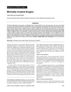
ANATOMY / EMBRYOLOGY The third branchial pouch gives rise to the inferior parathyroid glands (dark blue) in close association with the primordia of the the thymus gland (orange). As the thymus descends to the anterior mediastinum, parathyroids III follow along, ultimately coming into contact with the developing thyroid caudal to parathyroids IV (yellow). The parathyroid glands derived from pouch IV take a more direct route to come in contact with the thyroid, and become the more cephalad or superior glands. A portion of pouch IV (light blue) contributes a lateral C-cell component to the thyroid. The parathyroids usually (~80%) lie near the posterolateral capsule of the thyroid lobes.
Anatomy / Embryology The superior parathyroid glands are most commonly found about the middle third of the thyroid lobe, at the level of the cricothyroid junction, and near the point where the recurrent laryngeal nerve passes beneath the inferior pharyngeal constrictor to enter the larynx
Anatomy / Embryology The inferior glands are usually found near the lower pole of the thyroid lobe or below the lobe in the thyro-thymic ligament. They commonly lie below the inferior thyroid artery and anterior to the recurrent laryngeal nerve .
Anatomy / Embryology •
•
• Blood supply = inferior thyroid artery for both the superior & inferior thyroid glands
Aberrant parathyroid locations • • • •
Thymus gland (most common) Carotid sheath Vertebral body Thyroid gland
• Location identified by sestimibi
Anatomy • Superior Laryngeal nerve adjacent to the superior thyroid vascular pedicle, controls motor to the cricothyroid muscle, injury usually asymptomatic, but can cause loss of vocal projection & high pitch • Recurrent laryngeal nerve posterior to the inferior thyroid artery, motor for vocal cord abductors, unilateral injury causes hoarseness, bilateral injury causes airway occlusion (pt needs tracheostomy)
PHYSIOLOGY • Parthyroid Hormone (PTH) – Secreted by the Chief cells – Levels are inversely conrolled by [Ca2+ ]
• Effects: – – – – –
Tubular reabsorption of Ca2+ Osteoclastic resorption of bone Intestinal absorption of Ca2+ Synthesis of 1-25DHCC (active Vit. D) Excretion of phosphate
Incidence • HYPERPARATHYROIDISM – 1 : 1,000 prevalence – F:M 2:1 – Usually mild / asymptomatic – Primary assoc. w/ PRAD-1 oncogene
Etiology • Primary ( PTH, – Adenoma – Hyperplasia – Carcinoma
• Secondary
Ca 2+, renal cAMP, Phos) 90% (5% multiple) 10% assoc w/ MEN I & IIa < 0.1%
( PTH appropriate to low Ca 2+ )
– Chronic Renal Failure – Vitamin D Deficiency
• Tertiary – Continued excess PTH secretion following prolonged secondary hyperparathyroidism.
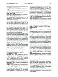
Parathyroid Adenoma : inferior rim of normal parathyoid tissue admixed with adipose tissue cells
Electrolytes • Hyperchloremic metabolic acidosis can occur in patients with primary metabolic acidosis • Renal failure – Ca PTH, Mag Na K Phos
Secondary Hyperparathyroidism • Decreased serum Ca & increased PTH • Associated with ESRD & vitamin D deficiency • Aluminum build up from ESRD increased osteomalacia • Tx - Dietary – Ca & Vit D supplements • Surgery only if symptomatic
Tertiary Hyperparathyroidism • Secondary hyperparathyroidism refractory to renal transplantation • Treated with surgery frequently
Signs / Symptoms • Asymptomatic (mild, < 2.99) • “Bones, stones, abdominal groans, psychic moans” Bones
Bone pain, #’s, arthralgia
Renal
Stones, polyuria
G.I.
Pain, duodenal ulcer, pancreatitis
Neuro.
Depression, apathy
Cardiac Hypertension, heart block
Clinical Presentation Symptom
%
Asymptomatic hypercalcaemia
50
Renal stones
28
Arthralgia
5
Peptic Ulcer
4
Hypertension
4
Bone disease / MEN 1 / others
9
Indications for Surgery Symptomatic hyperparathyroidism (stones, bone pain, peptic ulcers) Serum Ca 2+ >1.0mg/dl above normal Creatinine clearance < 30 % for age Renal stone on PFA Hypercalciuria ( >400mg/day) Bone marrow T-score 50%
Assay completion time
12 mins
Sensitivity 96% Specificity 100% Positive predictive value 97% of post-op calcium
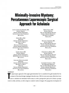
Intra-operative Gamma probe
Intra-operative Gamma probe “Minimally invasive parathyroidectomy facilitated by intraoperative nuclear mapping” Norman J, Surgery, 1997
15 patients with clearly a solitary adenoma on Sestemibi Average incision
2.4 cm
Mean operating time
24 minutes
97% of patients discharged within 2 hours of surgery Ex-vivo counts of 32% of background
Advantages of MIRP • • • • • • • • • •
Smaller incision 25 minutes Localization Pain Cost Haematoma Recurrent laryngeal nerve injury Tissue planes Contralateral structures Less post-op hypocalcaemia
Algorithm for MIRP PTH /
Calcium
Sestemibi scan Solitary adenoma
Negative or MGD
Unilateral exploration
Bilateral exploration
>50% iPTH