modification. This method was applied to identify ribose and phosphate moieties which could be important in the pre-tRNA recognition of E.coli RNase.
\.- 1993 Oxford University Press
Nucleic Acids Research, 1993, Vol. 21, No. 1
21-26
Modification interference approach to detect ribose moieties important for the optimal activity of a ribozyme Rajesh K.Gaur and Guido Krupp* Institut fur Aligemeine Mikrobiologie, Christian-Albrechts-Universitat, Am Botanischen Garten 9, W-2300 Kiel, Germany Received November 2, 1992; Revised and Accepted December 4, 1992
ABSTRACT A new approach for modification interference studies is presented. It involves the use of phosphorothioates as a handle to analyze any desired base or sugar modification. This method was applied to identify ribose and phosphate moieties which could be important in the pre-tRNA recognition of E.coli RNase P RNA (Ml RNA). The utility of this technique was confirmed by detecting the inhibitory effect of a deoxyribose in the 5"-flank (position -1). This site was already known to interfere with RNase P cleavage, if modified. We have analyzed pre-tRNATYr and pretRNAPhe and found different interference patterns for both tRNAs. Two unpaired regions were involved in both pre-tRNAs. Phosphorothioates interfered at the transition between acceptor- and D-arms. The results with deoxythymidines in the T-loop indicated that deoxyribose moieties or the extra methyl group in thymidine could interfere with RNase P cleavage. These data suggest that even in complete pre-tRNAs, only a few intact ribonucleotides are important in the substrate recognition by RNase P. We have demonstrated the potential of this new approach which offers many future applications in all fields involving nucleic acids, for example RNA processing, action of ribozymes, tRNA charging and studies related to DNA promoter recognition. INTRODUCTION Chemically synthesized RNAs which contain modified nucleotides at a few specific positions have been used to stabilize the RNAs and to elucidate the importance of these nucleotides in various studies. Prerequisites for such strategies are, first, the characterization of nuclease-resistant modifications, like phosphorothioates, or 2'-modified ribose moieties; second, the identification of the positions where modifications can be introduced into an oligonucleotide without loss of its activity. Unfortunately a serious drawback of this approach is the difficulty to predict all sites which can be modified without severely
*
To whom correspondence should be addressed
effecting the catalytic activity of the ribozyme. This is a cumbersome analysis even for small RNAs, as experienced by others, and it will be difficult to draw a final conclusion, unless all positions are analyzed independently (1-6). Moreover, for large RNAs this approach will be more tedious or even impossible; already transfer RNAs are refractory. A powerful alternative could be the modification interference analysis (7-11). However, in most of the cases this approach requires methods for the detection of modified sites in the nucleic acid which is impossible for many modified nucleosides, like m5C. We wish to report here a modification interference approach which can exploit phosphorothioates as a tool to analyze any kind of nucleoside modification in RNA and DNA. It is based on the presence of a low level (about 5-10%) of the modified nucleotide in enzymatically synthesized RNAs. The modification of interest is incorporated into an RNA transcript using an appropriate mixture of normal nucleoside triphosphates together with a modified NTPaS. As a result, each position with a modified base (or sugar) is next to a labile thio-substituted phosphodiester bond. In this fashion, any modification in sugar or base moieties can be combined with a scissile nucleic acid backbone; useful for both, RNA and DNA. As an example we analyzed which ribose moieties in pretRNAs should remain intact for efficient cleavage by RNase P. RNase P is a ribonucleoprotein which makes a single endonucleolytic cut in the pre-tRNA forming the mature 5'-terminus of the tRNA molecule (12). Protein and RNA components of RNase P are essential in vivo (13), however, in vitro the eubacterial RNA component acts as a true enzyme (ribozyme) and it is able to cleave tRNA precursors at the correct site (14,15). In our study we used the E. coli RNase P RNA (Ml RNA). Towards this end, we used dTTPcaS and dATPaiS to locate 2'-OH moieties which must remain intact for efficient pre-tRNA processing by RNase P. Obviously, one prerequisite of this analysis is the possibility to perform enzymatic RNA synthesis with the modified NTPsaS. However, only dATPaS and dTTPcaS were efficient substrates for T7 RNA polymerase (more details about the use of T7 RNA polymerase and 2'-deoxy- or 2'-O-methyl NTPsaS will be published elsewhere).
22 Nucleic Acids Research, 1993, Vol. 21, No. 1
Processing: of pre-tRNAs
MATERIALS AND METHODS Template preparations The plasmid p67YFO containing the gene for mature S.cerevisiae tRNAPhe (16) was a gift from Olke C. Uhlenbeck, Colorado, USA; plasmid pSU3 carrying the gene for E. coli pre-tRNATYr (17) was a gift from Leif Kirsebom, Uppsala, Sweden. The plasmids p67YFO and pSU3 were digested with Bst NI (NEBiolabs) and Fok I (NE-Biolabs) respectively.
Synthesis of pre-tRNA transcripts RNAs containing 2'-deoxynucleoside phosphorothioates were obtained by transcription reactions in which one of the NTPs was replaced by 0.9 mM dNTPcrS (Pharnacia, Uppsala) and 0.1 mM of the corresponding NTP. This resulted in 5-10% incorporation of the dNTPaS as evident from iodine-cleavage patterns. To obtain a similar modification level for ribonucleoside phosphorothioate containing pre-tRNAs, it was sufficient to use 0.1 mM NTPcaS and 0.9 mM of the corresponding NTP. The concentration of the other NTPs was 0.5 mM. In all reactions a dinucleotide, adenylyl-guanosine (ApG, Sigma) as an initiator has been used (a 4:1 ratio of the dinucleotide to GTP) to facilitate the subsequent [5'-32P]-end labeling of the pre-tRNAs, which otherwise would require a phosphatase treatment (20).
Preparation of Ml RNA The plasmid pDW27 (19), containing a gene for Ml RNA behind the T7 promotor was a kind gift of Norman Pace, Indiana University, Bloomington, IN. The plasmid was cleaved with Sna BI (Boehringer Mannheim) and transcribed with 0.5 mM NTPs as described earlier (20). A
The processing of 5 '-end labeled pre-tRNAs (about 200 fmoles) were carried out in 50 kl reactions as described previously (20). A 1:1 molar ratio of MI RNA to the substrate as the highest amount of enzyme and two serial tenfold dilutions were used. A control without Ml RNA was always included under identical conditions. After incubation for 1 h at 37°C the reaction products were applied on 8% denaturing polyacrylamide gels. The unprocessed substrates and products were visualized by autoradiography, excised and isolated by standard methods (18).
RNA analysis (i) Sequencing The 32P-labeled phosphorothioate transcripts were sequenced essentially according to the method reported by Schatz et al (21). The 5'-end labeled pre-tRNAs were dissolved in 10 ,ul of 10 mM HEPES (pH 7.2), incubated at 70°C for 3 min, chilled in ice, spun briefly and treated with 1 yl of 30 mM I2/ethanol solution (w/v). The above reaction mixture was left for 1 min at room temperature, excess reagent was removed by ethanol precipitation and the pellet was washed with 70% aqueous ethanol. The dry samples were dissolved in 4,d water, 6 ,ul urea/dye mixture (8 M urea, 0.03% xylene cyanol, bromophenol blue) and loaded directly on 8 or 20% denaturing polyacrylamide gels without prior heating.
(ii) Densitometer tracing An Ultra XL, enhanced Laser Densitometer (Pharmacia-LKB) was used for all densitometer analyses. The analyses of the extent of precursor tRNAs cleavage by RNase P and the autoradiograms of iodine cleavage patterns of uncleaved pre-tRNA were analyzed as described earlier (22).
B U +8/9
,
,_
a.
A*:** S
_
{1
X~~1)
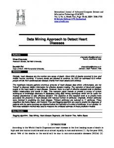
Figure 1. Modification interference with pre-tRNATYr and uridine caS triphosphate. Panel A Sequencing gel with phosphorothioate cleavage patterns. The [5'-32P]-labeled pre-tRNATYr was incubated with Ecoli RNase P RNA (Ml RNA). The uncleaved pre-tRNAs were gel-purified. We obtained cleavage of about 50% (1:1 molar ratio of Ml RNA to pre-tRNA; material analyzed in lane 1); 27% (lOfold molar excess of pre-tRNA; analysis in lane 2) and 2% (lOOfold molar excess of pre-tRNA; analysis in lane 3). The RNAs were treated with iodine and analyzed on a 20% denaturing sequencing gel. Lane C, control without Ml RNA. The left lanes always contain RNAs not treated with iodine while the material in the right lanes was treated with iodine to cleave the thio-phosphodiester bonds. Panel B Densitometer tracings of lane C (solid line) and of lane 1 (broken line). In both panels, we indicated the relevant uridine positions. In addition to U+8/9 (interference), also U-1 and U+54/55 (no interference) are marked for more convenient comparison of the results with deoxythymidine (Fig. 2).
Nucleic Acids Research, 1993, Vol. 21, No. 1 23 For each lane the average value was determined for four slightly shifted (800 itm steps), non-overlapping line tracings.
RESULTS Interference sites in pre-tRNATYr Throughout this report we describe the use of [5'-32P]-labeled phosphorothioate containing pre-tRNAs which were incubated without enzyme or with increasing amounts of MI RNA (the catalytic E.coli RNase P RNA), isolation of the uncleaved substrates followed by their iodine treatment and analysis on sequencing gels. All products were treated identically to avoid possible gel artifacts (23). We have varied the enzyme to substrate ratio from 1/100 to equimolar, and routinely obtained a cleavage efficiency of pre-tRNA by RNase P ranging from 2 to 50%, as determined by densitometer tracing. The interference sites were identified by comparing the 12/ethanol cleavage patterns of uncleaved pre-tRNAs (which were incubated with Ml RNA and isolated by gel electrophoresis) with the pre-tRNA incubated in the absence of Ml RNA. These comparisons were made by superimposing the densitometer traces of the gels obtained after iodine cleavage reactions of pre-tRNAs incubated in the presence or absence of MI RNA. We have analyzed E.coli pre-tRNATYr-UMPS and the results are shown in Fig. 1. The structure of pre-tRNATYr is shown in Fig. 5. It is evident from the densitometer tracings (Fig. 1B) that only the intensity of the band for U+8/9 was highly increased in the unprocessed substrates. This indicates that the corresponding thiophosphate interferes with RNase P cleavage; it was not possible to resolve the double band for both uridines, due to severe band compressions caused by base pairs formed between 5'-flank and acceptor stem. For the results shown in Fig. 2 we used pre-tRNATYr-dTMPS. In addition to thiophosphates, inhibitory effects of the 2'-deoxy groups were observed (possibly, also due to the extra methyl group in thymidines). These are evident by the accumulation of
A
dT + 54/55 and dT -1. In agreement with our observations, it is already well documented that the 2'-hydroxyl at the cleavage site (position -1) is required for the efficient cleavage of pretRNAs by RNase P (24,25). Mapping interfering sites in pre-tRNAPhe We have also analyzed a pre-tRNA with a short extra arm, the well characterized yeast tRNAPhe. Its cloverleaf structure is shown in Fig. 6. Here, already the ribonucleoside phosphorothioates at U + 54/55 and U + 68/69 interfered. Essentially the same interference pattern was obtained with 2'-deoxy-thymidines (data not shown). This suggests that no additional uridine positions have important 2'-hydroxyl groups. The possibilities to compare both tRNAs were limited. The cytidines at +68/69 in tRNATYr were not analyzed because dCTPaS was a poor substrate for T7 RNA polymerase (data not shown). In addition, position +9 is a uridine in tRNATyr whereas it is an adenosine in tRNAPhe. Therefore, we decided to analyze adenosines in tRNAPhe also. The interference analysis with pre-tRNAPhe-AMPS is shown in Fig. 3. In addition to minor effects, a strong interference was observed at adenosine +9. Similarly, in the analysis with dATPoS, dA+9 interfered (Fig. 4). In contrast to the ribonucleoside phosphorothioates (Fig. 3) the analysis with pretRNAPhe-dAMPS revealed a significant interference effect for dA+58 (Fig. 4). This suggests, the 2'-hydroxyl of the ribose at position +58 is important.
DISCUSSION In general, most of the modification interference approaches are based on either chemical modifications of a normal transcript (22,23,26,27) or the RNA synthesis can be performed with a mixture of normal and modified NTPs (10,11,20). The modification interference approach has been applied successfully, for phosphorothioates inserted during transcription reaction
B dT +54/55 Ii
I
dT +8/9 It
tI
II I
S~ ~ ~ ~ ~ ~d- _w
i?
IN
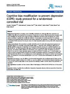
Figure 2 Pre-tRNATYr with 2'-deoxy-thymidine cS triphosphate. The samples were analyzed and presented as described in Fig. 1. This example shows the effects of increasing amounts of Ml RNA, no control without RNase P RNA is shown. The densitometer tracings in panel B compare the products obtained with the lowest amount of MI RNA (lane 3 in panel A; solid line in panel B) and the highest, lOOfold higher amount (lane 1 in A; broken line in B).
24 Nucleic Acids Research, 1993, Vol. 21, No. 1
R A --
A +9
IIi..7... .
a.
a
a
.
a
.>>nt8sm
_0
_A
_a o
.
_
_
'e
a AI?1
_
oo e_
a _a_
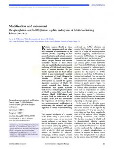
Figure 3 Pre-tRNAPhe with adenosine caS triphosphate. The analysis was essentially performed as in Fig. 1, but here an 8% sequencing gel was used. In addition to A+9 (interference), also A+58 (no interference) is marked for more convenient comparison of the results with deoxyadenosine (Fig. 4). H
A
dA +58
_ly, ,:
_.-
_a
dA +9
S
:w* .i _w
d '~
~
J iill ~ ~~~~~~~~~~~~~~~~~~~~~~~~~~~
Figure 4 Pre-tRNAPhC with 2'-deoxyadenosine aS triphosphate. The data are presented as described in Fig. 1. Here, only the products of two RNase P reactions are shown. Lane 1 with lOfold and lane 2 with lOOfold molar excess of pre-tRNA over MlRNA.
(10,11) and for some bases by chemical modification, like alkylated purines or uridines (22) or completely eliminated pyrimidines (27). However, the established methods have severe limitations as discussed in the following section and these have prompted us to develop a new approach. It is well established that in addition to the desired product, the chemical modification of nucleic acids results in the formation of other products which can even prevent Watson-Crick base pairing. This may obscure the interpretation
of the data obtained with chemically modified nucleic acids. As one example, the reaction of dimethyl sulfate with RNA yields the aniline-labile m7G, and in addition m1A and m3C which disrupt base pairs (28). Even worse, no detection system is available for the naturally occuring modified bases like m5C, m2G, m6A, inosin, ribo-thymidine in RNA or 2'-deoxy-uridine in DNA. In principle, the resistance against hydrolysis of the phosphodiester backbone next to 2'-modified nucleosides in RNA
Nucleic Acids Research, 1993, Vol. 21, No. 1 25 G
32pA
A U A C A C G C G U .G U A C G G * C A G G C C A G U A A A A A G C A U U A C C C C G *
A
-ffEJ
RNase P
C
U G
A C
G G
C C
RNase P
U *
*
*5*
G A A G G
G
C
A G
A.
C U
+69
U GA Uw A
GA CCGG* GC GU
cC A A AG
A ,G w C G C fi G*f C
GwFJ
CC U U C C
+I)
*''
/
C
CG C CC C
32pA
A
G G
+8
G
C C
A C U A C C
UJ
G
G
CA
A
G
AG
+93
+58
C 3" G C fi G A fi U A 3C A* U
A A
C U A
C
A C
U
G
can be used as a detection method (5,29). However, it will be difficult to detect the desired low percentage of 5-10% modification in an RNA and this will be impossible in DNA. These considerations, together with the recently developed complex procedure to study 2'-deoxy-uridines in protein-DNA interactions (30) highlight the inherent limitations of the current approaches and demand for a better method of detection. Obviously, all data obtained with the modification interference approach are only indirect. However, they are a useful first step in guiding future, more detailed and direct work with synthesized RNAs carrying the modification at defined positions only. We present an approach which is based on a phosphorothioate modification in the internucleotide phosphate backbone combined with the sugar or base modification of interest. This analysis is restricted to the Rp-diastereomer of the phosphorothioate since only the Sp diastereomer is a substrate for T7 RNA polymerase and is incorporated into the transcript with inversion of the configuration (31). We have used this method to identify the 2'-OH moieties in pre-tRNAPhe and pre-tRNATYr which could be important for efficient cleavage by RNase P. In order to distinguish between the thiophosphate interference and the interference due to sugar modification, we have compared the iodine cleavage patterns of pre-tRNA transcripts obtained with NTPaS with the iodine cleavage patterns of the corresponding pre-tRNA transcripts obtained with dNTPctS. The observed interfering sites are depicted in Figs. 5 and 6. In pre-tRNATYrUMPS, only thiophosphates at U + 8/9 interfered with RNase P cleavage whereas with pre-tRNATYr-dTMPS additional interference sites at -1 and +54/55 were identified. This suggests that 2'-hydroxyl at position -1, + 54/55 are important for efficient RNase P cleavage; also the possible importance of the extra methyl group in thymidine should be considered. It was already known that a deoxyribose at the RNase P cleavage site (position -1) is inhibitory (24) and we have also detected it with our approach. In this study it was not possible to decide if the interference at position +8/9 was only due to the modified phosphate backbone or due to both, backbone as well as sugar modification. In pre-tRNAPhe, thiophosphates at U+54/55,
ULA
A G
G
U
Figure 5 Cloverleaf model of pre-tRNA Yr. The [5'-32P]-labeled pre-tRNA is shown, obtained by transcriptions in the presence of ApG dinucleotide. An arrow indicates the RNase P cleavage site and the modification interference sites are marked by circles (phosphorothioates) and squares (2'-deoxyribonucleotides).
C
G G C U (G U 5 C C U _4+54 +-55
C U C G A" A G C
U G
*
U C U
U
G
UiC
GAlC A
U
AA
A
Figure 6 Cloverleaf model of pre-tRNAPhe.The structure and results are shown as described for Fig. 5.
U + 68/69 and A+9 were inhibitory. These interference sites were also observed with 2'-deoxy-nucleosides, but with 2'-deoxyadenosine an additional site was located at dA +58. The two pre-tRNAs gave different results, however, position +9 was always inhibitory. Three other interference sites, U+54/55 and A+58 were located in the T-loop which is an important contact area between substrate and enzyme, evident from base modification interference studies (22) and from the observation that a distorted T-loop can abolish RNase P cleavage completely (32). We had reported previously that a pre-tRNA with a short extra arm (pre-tRNAMet) has more contact points in the T-arm and base modifications were severely inhibitory, unlike a pre-tRNA with a long extra arm (pre-tRNAser) (22). These differences agree with the data presented here. The inhibition in the RNase P cleavage of pre-tRNAPhe (which has a short arm) was observed with both, thiophosphate and deoxyribose substitutions, whereas in pre-tRNATYr (which has a long extra arm) only the deoxyribose substitutions were inhibitory. In recent studies, additional important ribose moieties were identified near the cleavage site at positions -2, -1, +1 (25) and at the first cytidine (+74 in conventional tRNA numbering) in the 3'-terminal CCA-sequence (25). Small effects were also found in the 3'-terminal half of the acceptor stem, comparable to our interference data with U +68/69. In pre-tRNATYr, we also detected the inhibition of a 2'-deoxyribose in position -1, whereas an analysis of the other sites was not possible in our system. In this comparison, it is important to note that Perreault and Altman (25) used small, chemically synthesized model substrates which lacked the complete D-, anticodon- and extra arms, as well as all connecting nucleotides. All these elements are not absolutely essential for RNase P cleavage, but they are clearly important, as evident from reduced cleavage efficiencies of minimal substrates. In the T-loop of the model substrates, no effect was observed after introducing 2'-deoxyriboses in the conserved sequence GUUC (the positions from +53 to +56,
26 Nucleic Acids Research, 1993, Vol. 21, No. 1
refering to tRNA numbering); the position +58 was not analyzed by Perreault and Altman (25). In contrast to the findings with a minimal hairpin substrate, our data with fu-size pre-tRNAs suggest that the 2'-hydroxyls at +54/55 and +58 may be important and this could be caused by the different RNA structures. The hairpin substrates contain the acceptor- and Tstems in one continuous helix with twelve base pairs, whereas the two helices are separated in tRNAs. Very likely, the structure of the D-arm region effects the stacking interactions of the helices, moreover, their geometries will be more easily effected by a distorted T-loop. As a first step, the data reported here have pinpointed a number of positions where modified phosphates or ribose moieties can be expected to interfere with RNase P cleavage. Future studies with several chemically synthesized pre-tRNAs are needed to confirm these effects and they will reveal additional details. As a more general basis for further studies, we have shown that phosphorothioates can serve as a handle to perform modification interference analyses with otherwise refractory modified sugar moieties. With an increasing number of available modified NTPscaS and dNTPsaxS this approach can be useful in a variety of different fields.
ACKNOWLEDGEMENTS This work was supported by the Deutsche Forschungsgemeinschaft (Kr 817/3-1). R.K.G. is recipient of Alexandervon-Humboldt-Stiftung fellowship.
REFERENCES 1. Yang, J.-H., Perreault, J.P., Labuda, D., Usman, N., and Cedergren, R.
(1990) Biochemistry 29, 11156-11160. 2. Perreault, J.P., Wu, T., Cousineau, B., Ogilvie, K.K., and Cedergren, R. (1990) Nature 344, 565-567. 3. Perreault, J.P., Labuda, D., Usman, N., Yang, J.-H., and Cedergren, R. (1991) Biochemistry 30, 4020-4025. 4. Yang, J.-H., Usman, N., Chartrand, P., and Cedergren, R. (1992) Biochemistry 31, 5005-5009. 5. Paolella, G., Sproat, B.S., and Lamond, A.I. (1992) EMBO J. 11, 1913-1919. 6. Buzayan, J.M., van Tol, H., Feldstein, P.A., Bruening, G. (1990) Nucleic Acids Res. 18, 4447-4451. 7. Siebenlist, U., and Gilbert, W. (1980) Proc. Natt. Acad. Sci. USA 77, 122-126. 8. Conway, L., and Wickens, M. (1987) EMBO J. 6, 4177-4184. 9. Milligan, J.F., and Uhlenbeck, O.C. (1989) Biochemistry 28, 2849-2855. 10. Ruffner, D.E., and Uhlenbeck, O.C. (1990) Nucleic Acids Res. 18, 6025-6029. 11. Chowrira, B.M., and Burke, J.M. (1992) NucleicAcids Res. 20, 2835-2840. 12. Altman, S. (1989) In Adv. Enzymol. (Meister, A., ed) pp.1-36. John Wiley and Sons, New York. 13. Stark, B.C., Kole, R., Bowman, E.J., and Altman, S. (1978) Proc. Natl. Acad. Sci. USA 75, 3717-3721. 14. Guerrier-Takada, C., Gardiner, K., Marsh, T.L., Pace, N.R., and Altman, S. (1983) Cell 35, 849-857. 15. Gardiner, K., Marsh, T.L., and Pace, N.R. (1985) J. Biol. Chem. 260, 5415-5419. 16. Sampson, J.R., and Uhlenbeck, O.C. (1988) Proc. Natl. Acad. Sci. USA 85, 1033-1037. 17. Kirsebom, L., Baer, M.F., and Altman, S. (1988) J. Mol. Biol. 204, 879-888. 18. Krupp, G. (1991) In: Nucleic acid techniques in bacterial systemdtics (E. Stackebrandt and M. Goodfellow, eds.) pp. 95-114. John Wiley & Sons,
Chichester (England).
19. Waugh, D.S., Green, C.J., and Pace, N.R. (1989) Science 244, 1569-1571. 20. Kahle, D., Wehmeyer, U., Char, S., and Krupp, G. (1990) Nucleic Acids Res. 18, 837-844.
21. Schatz, D., Lebermann, R., and Eckstein, F. (1991) Proc. Natl. Acad. Sci. US4 88, 6132-6136. 22. Kahle, D., Wehmeyer, U., and Krupp, G. (1990) EMBO J. 9, 1929-1937. 23. Hegg, L.A., and Thurlow, D.L. (1990) NuceicAcidsRes. 18,2993-3000. 24. Forster, A.C., and Altman, S. (1990) Science 249, 783-786. 25. Perreault, J.P., and Altman, S. (1992) J. Mo. Biol. 226, 399-409. 26. Lang, K.M., and Keller, W. (1990) Mol. Cell. Biol. 10, 4942-4947. 27. Thurlow, D.L., Shilowski, D., and Marsh, T.L. (1991) NucleicAcids Res. 19, 885-891. 28. Ehresmann, C., Baudin, F., Mougel, M., Romby, P., Ebel, J.-P., and Ehresmann, B. (1987) Nucleic Acids Res. 15, 9109-9128. 29. Williams, D.M., Pieken, W.A., and Eckstein, F. (1992) Proc. Natl. Acad. Sci. US4 89, 918-921. 30. Pu, W.T., and Stuhi, K. (1992) Nucleic Acids Res. 20, 771-775. 31. Eckstein, F. (1985) Ann. Rev. Biochem. 54, 367-402. 32. Reilly, R.M., and RajBhandary, U.L. (1986) J. Biol. Chem. 261, 2928-2935.