1216
IEEE PHOTONICS TECHNOLOGY LETTERS, VOL. 23, NO. 17, SEPTEMBER 1, 2011
Modifying Plasmonic Spectral Properties of Engineered Silver Nanoblocks by Using Titanium Coating Li Wang, Wei Xiong, Yoshiaki Nishijima, Yukie Yokota, Kosei Ueno, Hiroaki Misawa, Gang Bi, and Jianrong Qiu
Abstract—Plasmonic spectral properties of silver nanoblocks fabricated by electron beam lithography are investigated. The extinction spectra of silver nanoblocks showed a red shift, broadening, and damping with the passage of storage time. In contrast, silver nanoblocks with titanium coating on the top did not show apparent spectral change even after storage for two months. Morphological change of the silver nanoblocks was characterized by scanning electron microscope. The present work clearly demonstrates that coating titanium on the top of the silver nanostructures is an effective way to prevent them from chemical corrosion and stabilize their plasmonic spectral properties. Index Terms—Nanotechnology, nanoblocks, surface plasmons.
protective
coating,
silver
I. INTRODUCTION
S
URFACE plasmons (SPs, also known as surface plasmon polaritons, SPPs), are surface electromagnetic waves that propagate in a direction parallel to the metal/dielectric (or metal/ vacuum) interface. SPs have drawn much attention in recent decades due to their promising applications in various fields of biosensor, imaging, solar cells, near-field probe, etc. Gold and silver nanostructures are most commonly used for generation of Manuscript received February 18, 2011; revised April 30, 2011; accepted May 28, 2011. Date of publication June 07, 2011; date of current version August 05, 2011. This work was supported by funding from the Ministry of Education, Culture, Sports, Science, and Technology of Japan: KAKENHI Grant-in-Aid for Scientific Research on the Priority Area “Strong Photon-Molecule Coupling Fields” [470 (19049001)], by Grants-in-Aid from Hokkaido Innovation through Nanotechnology Support (HINTS), and by National Natural Science Foundation of China (Grant 50872123 and Grant 51072054). L. Wang is with the State Key Laboratory of Silicon Materials, Zhejiang University, Hangzhou 310027, China (e-mail:
[email protected]). W. Xiong and G. Bi are with the Institute of Information and Electronics, Zhejiang University City College, Hangzhou 310015, China (e-mail:
[email protected];
[email protected]). Y. Nishijima, Y. Yokota, and H. Misawa are with the Research Institute for Electronic Science, Hokkaido University, Sapporo 001-0021, Japan (e-mail:
[email protected];
[email protected];
[email protected]. jp). K. Ueno is with the Research Institute for Electronic Science, Hokkaido University, Sapporo 001-0021, Japan. He is also with the PRESTO, Japan Science and Technology Agency, Kawaguchi 332-0012, Japan (e-mail: k-ueno@es. hokudai.ac.jp). J. Qiu is with the State Key Laboratory of Silicon Materials, Zhejiang University, Hangzhou 310027, China. He is also with the Institute of Optical Communication Materials, South China University of Technology, Guangzhou 510640, China (e-mail:
[email protected]). Color versions of one or more of the figures in this letter are available online at http://ieeexplore.ieee.org. Digital Object Identifier 10.1109/LPT.2011.2158640
SPPs. Compared with other noble metals, e.g., gold, silver has the following advantages. Firstly, the localized electric field enhancement of silver nanoparticles (NPs) is stronger than that of other noble metals [1]. Secondly, silver has its peak amplitude in vacuum at a higher frequency, which qualifies silver NPs to respond in the violet region of the visible spectrum, while the Mie resonances of gold and copper NPs typically appear in the green and red wavelength [2], [3]. Thirdly, the propagation length and the skin depth of the SPPs induced from the silver are longer than those of gold. Fourthly, silver is much cheaper than gold, which is very important for practical applications. However, the commercial applications of SPs based on silver nanostructures is not as popular as gold so far due to its lack of chemical stability under atmospheric conditions [7]. There have been many investigations about silver nanostructures protected by dielectric layer (e.g., silica) for applications to surface enhanced Raman scattering (SERS) [4], enhanced magneto-optics [5], lasers [6], etc. However, most of the silver nanostructures with protective layer focused on core-shell structures. In this investigation, we fabricated silver nanoblocks (NBs) with a protective layer of titanium coating on the top, and explored the effect of titanium thickness on protective action of preventing silver NBs from chemical corrosion. Titanium is chosen as the protective metal due to its excellent resistance to corrosion. It is almost as resistant as platinum, capable of withstanding attack by acids and moist chlorine in water [8]. II. EXPERIMENTAL Silver NBs with different aspect ratio ( ) defined as the ratio of the length to the width of the oblong NB in the horizontal plane were fabricated on conventional glass substrates by highresolution electron beam lithography (EBL) method [9]. The thickness of the nanostructures was measured by atomic force microscope (AFM, VN-8000, KEYENCE, Japan). Titanium of thickness 2.8 nm served as the adhesive layer of the silver and glass substrate. The real size of NBs with different in horizontal plane were nm nm nm nm (R2.2), nm nm (R3.2), nm (R1), nm nm (R5). The height of silver nm (R4.3), and NBs and the interval space between two neighboring NBs were set to be 40 nm and 200 nm, respectively. To study the stability of NBs, samples with and without titanium coating on the top were stored under the same conditions up to 2–3 months. The extinction spectra of silver NBs were measured and the surface morphology of silver NBs was characterized using
1041-1135/$26.00 © 2011 IEEE
WANG et al.: MODIFYING PLASMONIC SPECTRAL PROPERTIES OF ENGINEERED SILVER NANOBLOCKS
Fig. 1. Extinction spectra of silver NBs (R2.2, nm nm stored for different time under atmospheric conditions ( mode).
nm)
Fig. 2. Extinction spectra of mode LSPR bands of silver NBs (R2.2) with Ti coating on the top (Ti thickness of (a), (b), and (c) is 2.8, 5.6, and 10 nm, respectively) after storage for different time.
scanning electron microscope (SEM, JSM-6700FT, JEOL, Japan). Finite-difference time-domain (FDTD) simulation method (Lumerical) was adopted to simulate the extinction spectra of silver NBs without and with titanium coating of different thickness. To simulate the extinction spectra, several combinations of NB parameters were used. Refractive index of glass substrate was 1.5. The shape, size, thickness, and period of simulation model of silver NBs are in accordance with those of the real silver NB arrays. The cubic mesh accuracy was 1 nm. The optical constants for silver and titanium were taken from Johnson and Christy [10] and Palik [11], respectively. III. RESULTS AND DISCUSSION The extinction spectra of silver NBs were measured with light polarized parallel and perpendicular to the direction of the rectangular NBs’ elongation, which were corresponding to the longitudinal ( ) and transverse ( ) modes localized surface plasmon resonance (LSPR) bands of silver NBs, respectively. Fig. 1 shows the extinction spectra of silver NBs (R2.2) stored for different time under atmospheric conditions. We can see that the mode LSPR band shows a red shift, broadening, and damping with increasing storage time. Both of the and modes LSPR bands of silver NBs show the same trend. Fig. 2 shows the extinction spectra of mode LSPR bands of silver NBs (R2.2) with titanium coating on the top (titanium thickness of a, b, and c is 2.8, 5.6, and 10 nm, respectively) stored for different time. Compared with Fig. 1, it is obvious that
1217
Fig. 3. Peak shifts of mode LSPR bands of silver NBs (R2.2) without and with Ti coating of different thickness as a function of different storage time.
the red shift of LSPR band of silver NBs with titanium coating on the top is much smaller than that without titanium coating, and the red shift of the LSPR band of silver NBs with titanium coating of thickness 5.6 nm is smaller than that with titanium coating of thickness 2.8 nm. Since the titanium layer of thickness 5.6 nm is more protective than that of thickness 2.8 nm, we increase the titanium thickness to 10 nm to see if it is more protective with thicker titanium coating. From Fig. 2(b) and (c), we see that the red shift of LSPR band of silver NBs with titanium coating of thickness 10 nm is smaller than that with titanium coating of thickness 5.6 nm. Red shift almost disappears in Fig. 2(c). Moreover, for LSPR band of silver NBs with titanium coating, broadening and damping also can be neglected even with longer storage duration. Fig. 3 compares the peak shifts of mode LSPR bands of silver NBs (R2.2) without (0 nm) and with titanium coating (1, 2.8, 5.6, and 10 nm) after storage for different time (1 week, 2 weeks, 3 weeks, 1 month, 1.5 months, and 2 months). We find that the peak shift is very slight even after 2 months of storage when the titanium thickness is 10 nm. Hence, we can conclude that thicker titanium coating will be more protective for sliver NBs. The red shifts, broadening, and damping of the LSPR band with increasing storage time are considered to be due to structural breakage and sulfidation of silver NBs [7], [12]. It is well known that titanium has excellent resistance to corrosion. It can also serve as good adhesive layer metal as chromium. Here we use it as the cover layer as well as adhesive layer. After titanium coating, the optical properties of the silver NBs will change since the dielectric material contacting with the silver NBs changes from air to titanium. Therefore, we simulated the extinction spectra of silver NBs with titanium coating of different thicknesses by FDTD simulation method. The experimental (Exp.) and simulated (Sim.) results of R1 are shown in upper and lower parts in Fig. 4, respectively. We find that the simulated extinction spectra match well with the experimental result. The simulated result of the NBs with other matches well with the experimental result, too. The peak of experimental extinction spectra shifts a little compared to the simulated result, due to the size or thickness error during experimental process and the approximation of FDTD simulation method. Considering the effect of thickness of titanium coating on spectral properties of silver NBs, we compare the experimental (a) and simulated (b) extinction spectra of silver NBs without
1218
IEEE PHOTONICS TECHNOLOGY LETTERS, VOL. 23, NO. 17, SEPTEMBER 1, 2011
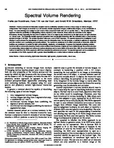
Fig. 6. SEM images of silver NBs (R5) (a) without Ti and (b) with Ti (Ti 2.8 nm) coating on the top, which were observed at the time when they were just fabricated and after storage for (a) two months and (b) three months under atmospheric conditions.
Fig. 4. Extinction spectra of silver NBs (R1, nm nm coated with titanium of different thicknesses (0, 2.8, 5.6, and 10 nm).
nm)
when they were just fabricated and after storage for 2 months [Fig. 6(a)] and 3 months [Fig. 6(b)] under atmospheric conditions. After two months of storage, the structural breakage of silver NBs without titanium coating can be observed clearly. In contrast, silver NBs with titanium coating almost show no apparent morphological change even after 3 months of storage. IV. CONCLUSION
Fig. 5. Peak shift of
mode LSPR band of as-made silver NBs with titanium coating of different thickness (0, 2.8, 5.6, and 10 nm) [(a) experimental data; (b) simulated data].
and with titanium coating. The peak shift of mode LSPR band of as-made silver NBs with titanium coating of different thickness (2.8, 5.6, 10 nm) compared with those of silver NBs without titanium coating (0 nm) is shown in Fig. 5. For the same aspect ratio, the experimental and simulated results both show that the peak shift of mode LSPR band becomes larger with increasing thickness of titanium coating basically. The experimental peak shift is not exactly the same as the simulated result, and the largest peak shift of experimental data is larger than that of simulated data, due to the size or thickness error during experimental process and the approximation of FDTD simulation method. We find that the largest experimental peak shift of mode LSPR bands as much as 120 nm occurs when the thickness of titanium coating is 10 nm compared with that without titanium coating. As we mentioned above, the peak shift of mode LSPR band becomes larger basically with increase of titanium thickness, the peak shift will be larger than 120 nm when the titanium thickness exceeds 10 nm. Since the peak shift is almost 0 nm even after 2 months of storage when the titanium thickness is 10 nm, which means titanium coating of thickness 10 nm is sufficient for protection of silver NBs, we suggest that the thickness of titanium coating should be around 10 nm. The morphology of silver NBs without and with titanium coating is also studied. Fig. 6 shows the SEM images of silver NBs (R5) without titanium [Fig. 6(a)] and with titanium [Fig. 6(b), thickness of titanium coating is 2.8 nm] coating on the top of silver NBs, which were observed at the time
We have studied the effect of titanium coating on the stability of plasmonic spectral properties of engineered silver NBs. The extinction spectra showed a red shift, broadening, and damping with increasing storage duration. The silver NBs coated with titanium of thickness 10 nm almost did not show any change in spectral properties even after 2 months of storage. The peak positions of experimental extinction spectra are in good agreement or close to those of simulated result. The optimal thickness of titanium coating to maintain the stability of plasmonic spectral properties of silver NBs is determined to be around 10 nm. REFERENCES [1] U. Kreibig and M. Vollmer, Optical Properties of Metal Clusters. Berlin, Heidelberg: Springer-Verlag, 1995, pp. 153–155. [2] G. Mie, “Beiträge zur optik trüber medien,” Ann. d. Physik, vol. 25, no. 3, pp. 377–445, 1908. [3] M. Chu, P. Sharma, C.-P. Chang, S. C. Liou, K.-T. Tsai, J.-K. Wang, Y.-L. Wang, and C.-H. Chen, “Probing surface plasmons in individual Ag nanoparticles in the ultra-violet spectral regime,” Nanotechnology, vol. 20, no. 23, p. 235705, Jun. 2009. [4] Y. Cui, B. Ren, J. Yao, R.-A. Gu, and Z. Tian, “Synthesis of Au bimetallic nanoparticles for immunoassay based on Ag surface-enhanced Raman spectroscopy,” J. Phys. Chem. B, vol. , 110, pp. 4002–4006, Feb. 2006. [5] L. Wang et al., “Plasmonics and enhanced magneto-optics in core-shell Co–Ag nanoparticles,” Nano Lett., vol. 11, pp. 1237–1240, Feb. 2011. [6] X. Meng, K. Fujita, S. Murai, T. Matoba, and K. Tanaka, “Plasmonically controlled lasing resonance with metallic-dielectric core-shell nanoparticles,” Nano Lett., vol. 11, pp. 1374–1378, Feb. 2011. [7] M. D. McMahon, R. Lopez, H. M. Meyer, III, L. C. Feldman, and R. F. Haglund Jr., “Rapid tarnishing of silver nanoparticles in ambient laboratory air,” Appl. Phys. B, vol. 80, pp. 915–921, Apr. 2005. [8] N. Casillas, S. Charlebois, W. H. Smyrl, and H. S. White, “Pitting corrosion of titanium,” J. Electrochem. Soc., vol. 141, pp. 636–642, Jan. 1994. [9] K. Ueno, V. Mizeikis, S. Juodkazis, K. Sasaki, and H. Misawa, “Optical properties of nanoengineered gold blocks,” Opt. Lett., vol. 30, no. 16, pp. 2158–2160, Aug. 2005. [10] P. B. Johnson and R. W. Christy, “Optical constants of the noble metals,” Phys. Rev. B, vol. 6, no. 12, pp. 4370–4379, Dec. 1972. [11] D. W. Lynch and W. R. Hunter, “Introduction to the data of several metals,” in “Subpart I: Metals” of Handbook of Optical Constants of Solids, E. D. Palik, Ed. San Diego, CA: Academic, 1998, vol. 3, pp. 240–249. [12] L. Wang et al., “Spectral properties and mechanism of instability of nanoengineered silver blocks,” Opt. Express, vol. 19, no. 11, pp. 10640–10646, May 2011.