PNAS PLUS
Modulation of synaptic function through the α-neurexin–specific ligand neurexophilin-1 Gesche Borna,1, Dorothee Breuera,1, Shaopeng Wanga,1, Astrid Rohlmanna, Philippe Coulonb, Puja Vakilia, Carsten Reissnera, Friedemann Kieferc, Martin Heined, Hans-Christian Papeb, and Markus Misslera,2 a Institute of Anatomy and Molecular Neurobiology, Westfälische Wilhelms-University, D-48149 Münster, Germany; bInstitute of Physiology 1, Westfälische Wilhelms-University, D-48149 Münster, Germany; cMammalian Cell Signalling Laboratory, Max Planck Institute for Molecular Biomedicine, D-48149 Münster, Germany; and dMolecular Physiology Group, Leibniz Institute of Neurobiology, D-39118 Magdeburg, Germany
Neurotransmission at different synapses is highly variable, and cell-adhesion molecules like α-neurexins (α-Nrxn) and their extracellular binding partners determine synapse function. Although α-Nrxn affect transmission at excitatory and inhibitory synapses, the contribution of neurexophilin-1 (Nxph1), an α-Nrxn ligand with restricted expression in subpopulations of inhibitory neurons, is unclear. To reveal its role, we investigated mice that either lack or overexpress Nxph1. We found that genetic deletion of Nxph1 impaired GABAB receptor (GABABR)-dependent short-term depression of inhibitory synapses in the nucleus reticularis thalami, a region where Nxph1 is normally expressed at high levels. To test the conclusion that Nxph1 supports presynaptic GABABR, we expressed Nxph1 ectopically at excitatory terminals in the neocortex, which normally do not contain this molecule but can be modulated by GABABR. We generated Nxph1-GFP transgenic mice under control of the Thy1.2 promoter and observed a reduced short-term facilitation at these excitatory synapses, representing an inverse phenotype to the knockout. Consistently, the diminished facilitation could be reversed by pharmacologically blocking GABABR with CGP-55845. Moreover, a complete rescue was achieved by additional blocking of postsynaptic GABAAR with intracellular picrotoxin or gabazine, suggesting that Nxph1 is able to recruit or stabilize both presynaptic GABABR and postsynaptic GABAAR. In support, immunoelectron microscopy validated the localization of ectopic Nxph1 at the synaptic cleft of excitatory synapses in transgenic mice and revealed an enrichment of GABAAR and GABABR subunits compared with wild-type animals. Thus, our data propose that Nxph1 plays an instructive role in synaptic short-term plasticity and the configuration with GABA receptors. synaptic transmission
not been studied in detail. Neurexophilins were discovered as a component of the latrotoxin receptor α-Nrxn (21) and comprise a family of four glycoproteins (Nxph1–4) that exhibit the characteristics of secreted, preproprotein-derived molecules (19, 22). Biochemical studies demonstrated that Nxph1 and Nxph3 interact with the second laminin-sex hormone-binding protein neurexin (LNS) domain of Nrxn (23). The LNS2 domain is present only in the extracellular sequences of α-Nrxn that contain six LNS domains, whereas β-Nrxn contain only a single LNS identical to the sixth domain of α-Nrxn (6). In situ hybridization data suggested that Nxph1 is present in select inhibitory interneurons of the adult brain (19) and in migratory interneuron precursors (24). Knock-in mice coexpressing lacZ with Nxph3, in turn, revealed expression in excitatory neurons of neocortical layer 6b and in the vestibulocerebellum (20). The localized expression of Nxph variants is contrary to the widespread distribution of α-Nrxn, raising the question of whether Nxph have modulatory functions at distinct subpopulations of synapses. We observed previously that deletion of Nxph1 and Nxph3 in mice has no major impact on postnatal survival (20, 23), unlike their cognate receptor α-Nrxn (9), but no information on their physiological roles is available yet. Here, we show that deletion of Nxph1 impairs GABAB-receptor (GABABR)–dependent short-term depression of inhibitory synapses in the nucleus reticularis thalami (NRT). In support, transgenic overexpression of Nxph1 at excitatory contacts in the neocortex demonstrates the ability to alter the molecular composition of synapses because functional GABAA and GABAB Significance
| thalamus | autism | neuroligin | ultrastructure
Communication between neurons via synapses is essential for information processing and cognitive function in our brains and is found impaired in many neuropsychiatric disorders. Synaptic transmission is remarkably variable in strength, and cell-adhesion molecules as neurexins and their binding partners are candidates to regulate neurotransmission. This study changes our understanding of how neurotransmission can be adapted to local demands by investigating the previously undescribed functions of neurexophilins, arguably the most elusive ligands of α-neurexins. Neurexophilins are expressed only in subpopulations of synapses, and their presence is able to change short-term plasticity and molecular composition at these terminals.
C
hemical synapses mediate signal transmission, integration, and plasticity. Synaptically transmitted signals differ between synapses of the same type and vary even at individual contacts of the same neuron (1), depending, for example, on their probability of release and history of activity (2, 3). Numerous studies have demonstrated that neurotransmission requires a plethora of synaptic molecules and signaling events (4). However, the mechanisms controlling the shaping of synapses with different properties are mostly unclear. We have addressed this problem by studying neurexins and their interaction partners (5, 6). Several aspects make neurexins candidates to couple local recognition/adhesion events to synaptic function: first, both extracellularly longer α-neurexins (α-Nrxn) and shorter β-neurexins (β-Nrxn) are able to induce functional synapses (7, 8); second, at least α-Nrxn are essential for synaptic transmission at excitatory and inhibitory terminals (9, 10); and third, α- and β-Nrxn are highly polymorphic, mostly presynaptic molecules (11, 12) that interact with transsynaptic binding partners like neuroligins (13, 14), LRRTMs (15, 16), or cerebellin/GluRδ2 (17, 18). In contrast to Nrxn that are expressed throughout the brain in virtually all excitatory and inhibitory neurons (12), the α-Nrxn–specific ligand neurexophilin (Nxph) is restricted to neuronal subpopulations (19, 20). The function of this ligand has www.pnas.org/cgi/doi/10.1073/pnas.1312112111
Author contributions: G.B., S.W., A.R., M.H., H.-C.P., and M.M. designed research; G.B., D.B., S.W., A.R., P.V., C.R., and F.K. performed research; P.C., C.R., and F.K. contributed new reagents/analytic tools; G.B., D.B., S.W., A.R., P.C., C.R., M.H., H.-C.P., and M.M. analyzed data; and M.M. wrote the paper. The authors declare no conflict of interest. *This Direct Submission article had a prearranged editor. Freely available online through the PNAS open access option. 1
G.B., D.B., and S.W. contributed equally to this work.
2
To whom correspondence should be addressed. E-mail:
[email protected].
This article contains supporting information online at www.pnas.org/lookup/suppl/doi:10. 1073/pnas.1312112111/-/DCSupplemental.
PNAS Early Edition | 1 of 10
NEUROSCIENCE
Edited* by Thomas C. Südhof, Stanford University School of Medicine, Stanford, CA, and approved February 25, 2014 (received for review June 27, 2013)
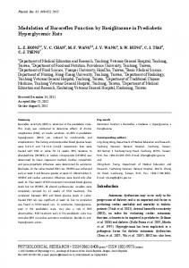
receptors become enriched and cause an impaired short-term facilitation. Results Nxph1 Functions at GABAergic Synapses in the Thalamus. To determine the physiological role of Nxph1 at synapses, we studied neurotransmission in Nxph1-deficient (KO) mice. We chose the NRT as a model system because expression of Nxph1 is normally high in the NRT (19). In addition, this region is composed of GABAergic neurons that sustain important brain functions, including sleep–wake regulation, cognition, and neuronal attention (25–27). Whole-cell patch-clamp recordings from Nxph1 KO neurons in the NRT (Fig. 1A) revealed only a small effect on spontaneous release because the frequency of miniature inhibitory postsynaptic currents (mIPSCs) was increased compared with wild type (WT) (Fig. 1 B and C). Amplitudes and kinetics such as decay times of mIPSCs were not different between genotypes (Fig. 1 D and E), and deletion of Nxph1 did not significantly alter electrically evoked inhibitory post synaptic currents (eIPSC amplitudes in WT: 418.5 ± 49.8 pA, n = 19 cells; in Nxph1 KO: 339.8 ± 54.1 pA, n = 14 cells; P = 0.30), although a tendency toward smaller responses in KO may exist. To test the hypothesis that Nxph1 might affect synaptic plasticity more strongly than basic transmission, we performed paired-pulse experiments
B
/ GAD65tg/+
-
VL
lateral
F
D
1.00
30
0.75 0.50 0.25
WT Nxph1 KO
1.5
***
1.0 0.5 0.0
0.00 0 1.0 2.0 3.0 4.0 5.0 interevent interval (s)
H1
G ppr (IPSC2/IPSC1)
1.2
* ** *
**
*
**
**
**
1.0
20 10
n.s. 50 40 30 20 10 0
0
**
WT Nxph1 KO
1.1
E
n.s.
100 pA 30 ms
Nxph1 KO
WT
25 pA 500 ms
W T N xp KO h1
C
IC dorsal
NXPH1 KO
decay time (ms)
+ +
cumulative probability
VB
mIPSC NRT
Cortex
NRT
+
WT
W T N xp KO h1
+
mIPSC NRT
amplitude (pA)
-/-
frequency (Hz)
Nxph1
W N T x KO ph1
A
to evoke short-term plasticity in the NRT (Fig. 1F). Short-term depression of eIPSCs, typical for NRT synapses (28), was impaired in KO neurons and replaced by facilitation at short interstimulus intervals, indicating that short-term depression as analyzed by paired-pulse ratio (ppr) depends on Nxph1 (Fig. 1G). This finding is supported by a second experimental protocol, a 20-Hz stimulation pulse that also yielded strong depression in NRT neurons of WT but not of KO mice (Fig. 1 H1, H2, and I), providing further evidence for a role of Nxph1 in synaptic plasticity. In contrast, basal neuronal cell properties such as membrane input resistance, resting potential, and capacitance were equal in both genotypes (Table S1). Current-clamp recordings of low-threshold spikes (LTS), a characteristic rebound response of NRT neurons (29, 30), also remained unchanged in the absence of Nxph1 (Fig. 1J and Table S1), suggesting that Nxph1 is not required for mediating intrinsic properties of the GABAergic NRT neurons. Because paired-pulse depression and spontaneous release at inhibitory terminals of NRT involves activation of GABABR (31), we probed the possibility that Nxph1 affects the function of these receptors by applying the antagonist CGP-55845 (Fig. 2A). We observed that mIPSC frequencies in KO were only weakly affected by (2S)-3-[[(1S)-1-(3,4-Dichlorophenyl)ethyl]amino-2hydroxypropyl](phenylmethyl)phosphinic acid hydrochloride
100 pA
H2
50 ms
50 100 150 200 inter-stimulus interval (ms)
0.9 0.8 WT
I
*
***
**
ppr 20 Hz stimulus train
1.00
Nxph1 KO
J
0.75 LTS
0.50
-60 mV
0.25 0.00
20 mV 100 ms
20 mV 100 ms A2/A1
A4/A1 WT
A10/A1 Nxph1 KO
2 of 10 | www.pnas.org/cgi/doi/10.1073/pnas.1312112111
Fig. 1. Nxph1 functions in subpopulations of inhibitory synapses. (A) Experimental setup for patchclamp recordings from neurons in the NRT (green indicates GAD65-driven GFP for reliable identification) and the VB. IC, internal capsule; VL, ventral posterolateral nucleus. Red arrows indicate GABAergic connections analyzed within the NRT and to the VB. (B) Representative mIPSC recordings from NRT neurons of WT (Upper traces) and Nxph1 KO (Lower traces). (C–E) mIPSC frequencies in KO neurons (green) show a leftward shift in the cumulative representation, and an increased average (Inset). Amplitudes (D) and decay times (E) of mIPSC are similar in both genotypes. (F) Averaged current traces from pairedpulse experiments in WT (black) and KO (green) to evoke inhibitory synaptic short-term depression. (G) Paired-pulse depression seen in WT at different interstimulus intervals is abolished in KO. (H and I) Short-term plasticity in NRT neurons evoked by a 20Hz stimulus train (H1, sample traces from WT; H2, sample traces from KO) reveals depression in WT. Ratios (ppr) were calculated between the 2nd, 4th, and 10th amplitude to the first amplitude, but depression (