arXiv:cond-mat/9911488v3 [cond-mat.stat-mech] 1 Mar 2000 ... Folding of the secondary structures proceeds through a well defined sequence of events. For.
arXiv:cond-mat/9911488v3 [cond-mat.stat-mech] 1 Mar 2000
Molecular dynamics of folding of secondary structures in Go-type models of proteins Trinh Xuan Hoang and Marek Cieplak Institute of Physics, Polish Academy of Sciences, Aleja Lotnikow, 02-668 Warsaw, Poland We consider six different secondary structures of proteins and construct two types of Go-type offlattice models: with the steric constraints and without. The basic aminoacid-aminoacid potential is Lennard Jones for the native contacts and a soft repulsion for the non-native contacts. The interactions are chosen to make the target secondary structure be the native state of the system. We provide a thorough equilibrium and kinetic characterization of the sequences through the molecular dynamics simulations with the Langevin noise. Models with the steric constraints are found to be better folders and to be more stable, especially in the case of the β-structures. Phononic spectra for vibrations around the native states have low frequency gaps that correlate with the thermodynamic stability. Folding of the secondary structures proceeds through a well defined sequence of events. For instance, α-helices fold from the ends first. The closer to the native state, the faster establishment of the contacts. Increasing the system size deteriorates the folding characteristics. We study the folding times as a function of viscous friction and find a regime of moderate friction with the linear dependence. We also consider folding when one end of a structure is pinned which imitates instantaneous conditions when a protein is being synthesized. We find that, under such circumstances, folding of helices is faster and of the β-sequences slower.
I. INTRODUCTION
Understanding of the statistical mechanics aspects of protein folding has been recently advanced through studies of coarse grained models in which aminoacids are represented by single beads. In particular, many valuable insights have been gained by considering such models on a lattice (see, e.g. ref.1–3 ). These toy models have allowed one to relate the folding process to the sequence dependent energy landscapes,4–6 to identify the folding pathways,7 and to study the issues of designability.8,9 They have also been used to demonstrate existence of tree like kinetic connectivities in the folding funnel10 that can be represented with the use of the disconnectivity graphs.11 More realistic coarse grained models, however, require an off-lattice setting. Recently, there have been a number of off-lattice studies, which focus on the kinetics of folding,12–16 as well as on the sequence design and determination of the interaction potentials.17,18 It has been also suggested that the geometry of the native structure of proteins itself, without a detailed information regarding the amino acid sequences, plays a decisive role in the folding process.19 The effective interactions between the beads in the coarse-grained models are difficult to derive from microscopic considerations and instead they may be chosen to reflect statistical properties of protein structures as collected in the protein data bank.20,21 The advantage of the lattice models is that they allow for an enumeration of conformations, at least for short chains, and thus for identification of the native state and determination of equilibrium properties of the system. However, the dynamics of these models are not related to any Newton’s equations (in the classical limit) since they have to be defined in terms of the discrete Monte Carlo steps made within a declared set of allowed moves.
Molecular dynamics (MD) simulations are a natural tool to study models set in a continuum space independent of whether they are coarse-grained or fully atomic. A decade ago, MD simulations of the microscopic representation of a polypeptide chain could explore time scales which were around nanoseconds and thus orders of magnitude too short to study the full duration of a typical folding.22 Currently, microsecond time scales have become accessible23 (in a special purpose computer) but these feats allow one to monitor individual trajectories in very restricted regions of the phase space. A detailed characterization is still restricted to up to 10ns long time scales.24 Thus such chemically realistic models cannot yet provide a sufficiently thorough equilibrium and kinetic characterizations of the system that are expected when setting up a model to be studied within the framework of statistical mechanics. Thus there is a need to study simplified coarse-grained continuum space models to understand the generic features of folding. These models must involve idealized potentials, for instance of the Lennard-Jones kind. These interactions may either be constrained to correspond to a target native state or they are not.12 The targeting may be facilitated by augmenting the model by an introduction of steric constraints,14,25 which takes into account the properties of the peptide bonds, but it can also be accomplished without such constraints.26 The dynamics of the simplified Lennard-Jones models are usually studied by the methods of MD. A novel and efficient variant of the MD technique has been recently proposed by He and Scheraga,25 in which one focuses the evolution on the torsional degrees of freedom. Some Monte Carlo studies for those models are also available.12,13,26 The applicability of the Monte Carlo methods to dynamical properties (as opposed to equillibrium) remains, however, untested. In this paper, we report on molecular dynamics stud-
ies of possibly the simplest models with the native states defined by target conformations – the off-lattice versions of the Go models.27 The interesting property of the Go model is that it essentially avoids the issue of the correct specification of the aminoacid-aminoacid interaction and yet it corresponds to a realistic conformation by defining the couplings in terms of the target conformation. Specifically, we consider the Lennard-Jones interactions between the beads and make them attractive for native contacts (two non-contiguous beads form a contact if they do not exceed a certain cutoff distance) and repulsive for non-native contacts. Such models are proteinlike also in the sense that they minimize the structural frustration. The object of our studies here is to investigate how viable are such Lennard-Jones-Go models in the context of the kinetics of protein folding. Our motivation for performing these studies follows from the expectation that such models may play a role similar to that of the Ising spin models in representing properties of the more complicated real life magnetic systems. Recently, there have been several related studies of the coarse-grained off-lattice Go models which, however, asked different questions than in this paper and were not based on the Lennard-Jones potentials. The study by Zhou and Karplus,28 employed a square well potential (also used before to analyze homopolymers29 ) which leads to a simplified discrete MD treatment. The authors have studied possible scenarios (with or without long-lasting intermediates) of the folding kinetics in a three-helix-bundle-like protein model as a function of the strength of the non-native contacts relative to the strength of the native ones. This kind of discrete MD have been also used by Dokholyan, Buldyrev, Stanley and Shakhnovich16 to identify a folding nucleus in a Gotype heteropolymer. (For a related full atom study of this issue for the CI2 protein see ref.30 ). Another study, by Hardin, Luthey-Schulten, and Wolynes31 used a detailed backbone representation and implemented an associative memory Hamiltionian in which the contact potentials are set to be the Gaussian functions. This study was aimed at understanding the kinetics of the secondary structures formation from the perspective of the energy landscape picture. Our partiality to the Lennard-Jones potentials is of a twofold nature. First, these potentials are well established in simulations of liquids and –essentially – continuum time stable MD codes are available. Second, their overall distance dependence seems qualitatively correct on a fundamental level. Real life conformations of proteins in the native state consist of interconnected secondary structures such as α-helices, β-hairpins (see, e.g. ref.32 ), and β-sheets. Understanding of the kinetics of protein folding should be first accomplished at the level of the secondary structures. This is, in fact, the task of this paper and we narrow it to two classes of the Go models: with and without the steric constraints and for each of these we consider six possible secondary structures with different numbers of monomers. The kinetics of folding are stud-
ied by the standard techniques of MD with a Langevin noise which controls the temperature of the systems and, at the same time, mimics the interactions of the protein fragments with the molecules of water. Our method of constructing the off-lattice Go models is outlined in Section II. In Section III, we determine the sizes of the native basins of the models through the shape distortion method33 instead of making assumptions about them, as it is done usually. In Section IV, we determine the phononic spectra of the models and discuss their relationship to the thermodynamic stability of the native structures. In Section V, we determine the folding times, the thermodynamic stability and other characteristic parameters for the models without the steric constraints and find that the β-structures have very low thermodynamic stability in this model. The folding times vary with the native conformation and the chain length. In particular, the α-helix folds faster than the β-hairpin of the same length. The latter also folds much slower than a similarly sized β-sheet with three strands. Experiments on folding indicate that the folding time for the hairpin is about 30 times slower that that for the α-helix.34 Our results do not yield the same rate but, at least, they show that the β-hairpin is the slower folder. A similar result has been obtained by Hardin, Luthey-Schulten, and Wolynes.31 Our studies of the three sizes of the helices confirm the general observation that folding properties deteriorate with the growth of the system size.35 In Section VI, we repeat the MD studies for the models with the steric constraints and find that these constraints substantially improve the thermodynamic stability, especially for the β-structures. At the same time, however, they raise the temperature of the onset of the glassy effects so the net result is that the αhelices remain good folders and the β-structures remain bad folders but their foldability is improved. Notice, however, that our models allow for good or adequate folding, depending on the system, without any additional dipoledipole interactions as introduced by He and Scheraga.25 In Section VII, we focus on the mechanisms of folding and discuss sequencing of events that takes place in folding of the secondary structures. In particular, we investigate characteristic time separations between the folding steps. In general, the time separations may depend on details of a model but the sequencing is expected to be model independent, i.e. it should proceed as predicted by our Go-type models. In Section VIII, we study the dependence of our results on the strength of viscous damping in the Langevin noise and find that it affects the folding time but it does not affect any of the other characteristic parameters except when the friction is set unrealistically low. Finally, in Section IX, we consider the processes of folding occurring when one end of the structure is fixed in space which imitates conditions found when a protein is being synthesized.36 We find that fixing one end of a helix accelerates folding and improves overall folding characteristics. The folding is found to originate at the unclamped end. On the other hand, folding of the β
structures becomes worse when one end is clamped. This paper sets the stage for studies of full proteins and kinetic interplays between various secondary structures. Results of such studies for the Go models of proteins will be presented in a separate paper.
II. MODELS
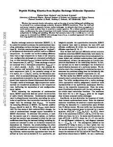
We represent a polypeptide in a simplified manner: by a chain of connected beads. The beads’ positions correspond to the locations of the Cα atoms. When the system is in its native state, the beads have no kinetic energy and the locations of the Cα atoms can be obtained from the Protein Data Bank. However, the secondary structures that we study are idealized and are not targeted to a specific real structure. Instead, they are just meant to be constructed in a way which is very close to typical α-helices and β-sheets found in the native states of real proteins. These secondary structures are stabilized primarily by the hydrogen bonds. The structures that we study are shown in Figure 1. These are: three α-helices, denoted as H10, H16 and H24, two β-hairpins, denoted as B10 and B16, and a β-sheet, B15. The numbers in the labels indicate the numbers of beads in the structures. All of the bonds between two consecutive beads along the chain of the helices, i.e. the peptide bonds, are assumed to have the same length of d0 = 3.8˚ A which is a typical real life value. As one proceeds along the axis of the helix (the z-coordinate) from one bead to another, the bead’s azimuthal angle is rotated by 100o and the azimuthal length is displaced by 1.5˚ A which again corresponds to a typical geometry. Each of the β-hairpins, B10 and B16, has two antiparallel strands which are connected by a turn. In the B15 β-sheet there are three strands and two turns. In the β-structures, the strands are not straight lines but have a zigzagged geometry as shown in Figure 1. The bond lengths between two connected beads are equal to 3.8˚ A but the displacement along the strand direction (the z axis in Figure 1) is 3.5˚ A. The distance between two opposite beads in two bonded strands, is set to 5˚ A, which is roughly equal to the hydrogen bond’s length. The turn region is constructed so that the bond length and the zigzag pattern match. The potentials of interactions between pairs of the beads are constructed in a way that ensures that the target structure coincides with the ground state of the system. i.e. with the native state. We pick the pair potentials to be of the Lennard-Jones type and select the parameters in a Go-type fashion27 so that significant attraction is associated with the native contacts and the non-native contacts are purely repulsive. In our model, we assume that a native contact is present if the distance between the two monomers in the designed structure is shorter than 7.5˚ A.
FIG. 1. Stereographic projections of the target structures studied in this paper: three α-helices H10, H16, H24, two β-hairpins B10, B16, and a β-sheet B15. The xz and yz planar projections for H10 and B10 are shown on the right hand side.
These Go-type couplings already stabilize the structures under studies but that stabilization will be found here not to be sufficiently adequate, especially in the case of the hairpins. We thus consider two classes of models: with and without additional steric constraints. The steric constraints add extra stability and they take into account the directional character of the peptide bonds in a more realistic manner. A. Go-type model with no steric constraints
This model is similar in spirit to that introduced by Iori, Marinari, and Parisi12 and to that studied by Li and Cieplak.26 The conceptual difference between these two papers is that the former is not constructed in reference to any predetermined target structure. For a conformation defined by the set of position vectors {ri }, i = 1, 2 . . . N , the potential energy is assumed to take the following form Ep ({ri }) = V BB + V N AT + V N ON .
(1)
The first term represents rigidity of the backbone potential, the second term corresponds to interactions in the native contacts and the third term to those in the nonnative contacts. N denotes the number of residues. The backbone potential takes the form of the sum over harmonic12 and anharmonic18 interactions
V BB =
N −1 X
[k1 (ri,i+1 − d0 )2 + k2 (ri,i+1 − d0 )4 ],
(2)
i=1
where ri,i+1 = |ri −ri+1 | is the distance between two consecutive beads; d0 = 3.8˚ A, k1 = ǫ and k2 = 100ǫ, where ǫ is the Lennard-Jones energy parameter corresponding to a native contact. The interaction between residues that form a native contact in the target structure is taken to be of the Lennard-Jones form: "� �12 � �6 # N AT X σij σij N AT , (3) − 4ǫ V = rij rij i 0.2m/τ , Tmin is equal to 0.3. The points are based on 200 trajectories. The inset shows the dependence at T = 0.3 in a non-logarithmic scale of γ, where for the higher values of γ, the points are fitted by a straight line.
D. Folding with one end fixed
Finally, we examine a special case, when one end of the chain is fixed during folding. This is relevant to the process of protein synthesis: one end can be thought of as being momentarily glued to the surface of a ribosome until the whole protein is constructed by adding new segments.36 In the synthesis process, the protein folds as it is being produced and its length extends. At each instant, however, one end of the protein can be considered pinned. Figure 16 shows that the helix H16 with the end bead fixed has a considerably lower value of Tmin , compared to no clamping, and it generally folds faster at low temperatures. pinned folds faster below Tmin and is characterized by a significantly lowered Tmin . We have also found that Tf is not affected by the pinning. Thus the helix becomes a much better folder. On the other hand, as seen in Figure 16, the contact sequencing becomes somewhat disturbed. Folding starts at the unclamped end. After the initial stage, many contacts get established almost simultaneously in an avalanche-like process. Then there is a large gap in time before the last contact, near the clamped end, comes into place. Analyzing the phononic spectrum of the pinned helix H16 yields ω1 τ ≈ 1.964 for the first excited mode which is almost the same as for H16 without the pinning.
FIG. 16. The order of contact appearance together with the average times for the contacts to be established for the first time for sequence H16 with one end monomer fixed. The fixed monomer is shown enlarged. The results are averaged over 1000 trajectories at Tmin . The inset compares the temperature dependencies of the median folding time for the situation in which the end monomer is (solid line) or is not (broken line) clamped.
VIII. CONCLUSIONS
In summary, we have studied the Go-type models of off-lattice secondary structures of proteins. We have provided a systematic characterization of both equilibrium and kinetic properties of these models. Models with the steric constraints were endowed with better thermodynamic stability. The stability can be assessed from the phononic spectra. The folding times strongly depend on the system size and on the geometry of the native state. The α-helices, which are stabilized merely by the local contacts appear to have much better folding properties than the β-structures. However, it should be noted that for both kinds of the structures none of the models is a very good folder – Tf is always smaller than Tmin , – although the choice of the native basin size δc has been made consistently by the shape distortion technique. We have checked that if one uses a somewhat larger δc then this results in an increase in the effective values of both
Tf and Tmin and thus their ordering remains unchanged. The kinetic criterion for good foldability does not appear to be compatible with the thermodynamic criterion provided by Klimov and Thirumalai43 in the case of the βsequences. We speculate that this may be related to the low compactness level of their native structures. Folding of the secondary structures at Tmin proceeds, on average, through a well defined sequence of events. That sequencing may become more complicated in proteins when several secondary structures compete when folding. We intend to explore this issue later. IX. ACKNOWLEDGMENTS
The idea of this project arose in discussions with Jayanth R. Banavar, whose subsequent interest and encouragement were vital for its completion. Many discussions with M. S. Li are also appreciated. We also thank M. Geller for pointing out to us that the folding process may be affected by the pinning. This work was supported by KBN (Grant No. 2P03B-025-13).
1
H. S. Chan, K. A. Dill, Phys. Today 46(2), 24 (1993). K. A. Dill, S. Bromberg, S. Yue, K. Fiebig, K. M. Yee, D. P. Thomas, and H. S. Chan, Protein Sci. 4, 561 (1995). 3 M. Cieplak, M. Henkel, J. Karbowski, and J. R. Banavar, Phys. Rev. Lett. 80, 3654 (1998). 4 H. S. Chan, and K. A. Dill, J. Chem. Phys. 100, 9238 (1994). 5 P. G. Wolynes, Proc. Natl. Acad. Sci. USA 93, 14249 (1996). 6 A. Sali, E. Shakhnovich, and M. Karplus, Nature 369, 248 (1994). 7 H. S. Chan, and K. A. Dill, Proteins: Struct., Funct., Genet. 30, 2 (1998). 8 H. Li, R. Helling, C. Tang, and N. Wingren, Science 273, 666 (1996). 9 M. Vendruscolo, A. Maritan, and J. R. Banavar, Phys. Rev. Lett. 78, 3967 (1997). 10 P. Garstecki, T. X. Hoang, and M. Cieplak, Phys. Rev. E 60, 3219 (1999). 11 O. M. Becker, and M. Karplus, J. Chem. Phys. 106, 1495 (1997). 12 G. Iori, E. Marinari, and G. Parisi, J. Phys. A 24, 5349 (1991). 13 A. Irback, C. Peterson, F. Potthast, and O. Sommelius, J. Chem. Phys. 107, 273 (1997). 14 T. Veitshans, D. Klimov, and D. Thirumalai, Folding Des. 2, 1 (1997). 15 D. K. Klimov, and D. Thirumalai, Phys. Rev. Lett. 79, 317 (1997). 16 N. V. Dokholyan, S. V. Buldyrev, H. E. Stanley, and E. I. Shakhnovich, Folding Des. 3, 577 (1998). N. V. Dokholyan, S. V. Buldyrev, H. E. Stanley, and E. I. Shakhnovich, condmat 9812284 (1998). 2
17
A. Irback, C. Peterson, and F. Potthast, Phys. Rev. E. 55, 860 (1997). 18 C. Clementi, A. Maritan, and J. R. Banavar, Phys. Rev. Lett. 81, 3287 (1998). 19 C. Micheletti, J. R. Banavar, A. Maritan, and F. Seno, Phys. Rev. Lett. 82, 3372 (1999). 20 S. Miyazawa, and R. L. Jernigan, Macromolecules 18, 534 (1985). 21 A. Kolinski, A. Godzik, and J. Skolnick, J. Chem. Phys. 103, 4312 (1995). 22 M. Karplus, Physics Today, 40, 68 (1987). 23 Y. Duan, and P. A. Kollman, Science 282, 740 (1998). 24 E. M. Boczko and C.L. Brooks III. Science 269, 393 (1995). 25 S. He, and H. A. Scheraga, J Chem Phys 108, 287 (1998). 26 M. S. Li, and M. Cieplak, Phys. Rev. E 59, 970 (1999). 27 N. Go, and H. Abe, Biopolymers 20, 991 (1981). 28 Y. Zhou, and M. Karplus. Nature 401, 400 (1999). 29 Y. Zhou, C. K. Hall, and M. Karplus. Phys. Rev. Lett. 77, 2822 (1996). 30 A. Li and V. Daggett. Proc. Natl. Acad. Sci. USA 91, 10430 (1994). 31 C. Hardin, Z. Luthey-Schulten, and P. G. Wolynes, Proteins: Struct., Funct., Genet. 34, 281 (1999). 32 F. Blanco, M. Ramirez-Alvarado, and L. Serrano. Curr. Opin. Struct. Biol. 8, 107 (1998). 33 M. S. Li, and M. Cieplak, J. Phys. A 32, 5577 (1999). 34 V. Munoz, P. A. Thompson, J. Hofrichter, and W. A. Eaton, Nature 390, 196 (1997). 35 M. Cieplak, T. X. Hoang, and M. S. Li, Phys. Rev. Lett. 83, 1684 (1999). 36 L. Stryer, Biochemistry, (W. H. Freeman and Company, New York, 1995). 37 G. S. Grest, K. Kremer, Phys. Rev. A 33, 3628 (1986). 38 M. P. Allen, and D. J. Tildesley, Computer simulation of liquids, (Oxford University Press, New York, 1987). 39 J. Callaway, Quantum Theory of the Solid State, (Academic Press, New York and London, 1974). 40 W. H. Press, B. P. Flannery, S. A. Teukolsky, and W. T. Vetterling, Numerical Recipes in FORTRAN, (Cambridge University Press, Cambridge, 1993). 41 D. Thirumalai, J. Physique I 5, 1457 (1995). 42 C. J. Camacho, and D. Thirumalai, Proc. Natl. Acad. Sci. USA 90, 6369 (1993). 43 D. K. Klimov, and D. Thirumalai, Phys. Rev. Lett. 76, 4070 (1996). 44 N. D. Socci, and J. N. Onuchic, J. Chem. Phys. 101, 1519 (1994). See also M. Cieplak, and J. R. Banavar, Folding Des. 2, 235 (1997). 45 R. Unger, and J. Moult, J. Mol. Biol. 259, 988 (1996). 46 A. M. Gutin, V. I. Abkevich, and E. I. Shakhnovich, Phys. Rev. Lett. 77, 5433 (1996).