Mar 12, 2001 - Molecular Dynamics Simulation of Continuous Current Flow through a Model Biological. Membrane Channel. Paul S. Crozier and Richard L.
VOLUME 86, NUMBER 11
PHYSICAL REVIEW LETTERS
12 MARCH 2001
Molecular Dynamics Simulation of Continuous Current Flow through a Model Biological Membrane Channel Paul S. Crozier and Richard L. Rowley Department of Chemical Engineering, Brigham Young University, Provo, Utah 84602
Nathan B. Holladay and Douglas Henderson Department of Chemistry and Biochemistry, Brigham Young University, Provo, Utah 84602
David D. Busath Department of Zoology and Center for Neuroscience, Brigham Young University, Provo, Utah 84602 (Received 22 August 2000) The conductance of sodium ions through a simplified channel-membrane system immersed in a reservoir of 1M NaCl in SPC兾E water is examined by molecular dynamics simulation. An applied external potential of 1.1 V drives the ions and water through a channel of length 25 Å producing a current of 19.6 pA, in reasonable agreement with experimental findings. The stream of ions and water molecules flows continuously because of the constant applied field and periodic boundary conditions. We also examine the potential profile across the simulation cell, the average density distributions of the various species in the reservoir and radially in the channel, and the ion velocity in the channel. DOI: 10.1103/PhysRevLett.86.2467
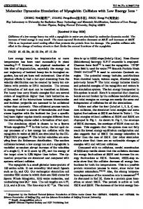
Ion channels are proteins that control the passage of ions across cell membranes. They are responsible for such important functions in biological cells as the generation of action potentials in nerves and muscles, the regulation of hormone release from endocrine cells, and the initiation of the cortical reaction to prevent polyspermy in oocytes [1]; despite this, the microscopic mechanisms for the function of such channels are imperfectly understood. Ion channels are among the simplest proteins to which statistical mechanics may be applied. Additionally, the electrolyte in the channel is an example of an inhomogeneous fluid, a topic of considerable interest in statistical mechanics. As a result, in recent years there has been increasing interest in the application of physics to membrane channels and, more generally, biophysics. Roux and Karplus [2] have reviewed earlier simulations. Recent investigations of channels have included equilibrium studies of the mechanism for channel selectivity using a continuum model for water [3,4], the so-called primitive model (PM) of an electrolyte, and a hard sphere model for water [5], the so-called solvent primitive model (SPM). Recent nonequilibrium investigations of channel conductance, the topic of interest here, can be grouped into three classes. In the first group, a Debye-Hückel or PoissonPlanck-Nernst level treatment is employed [6]. Thus, the PM is used and the molecular nature of the water is again ignored. In another type of study, the molecular nature of the water is included but the channel is approximated as a cylinder of infinite length [7,8]. As a result, the process of entry of ions and water into the channel cannot be studied. In the third class, the channel is finite in length, but the contribution of the water molecules is included only indirectly using Brownian or Langevin dynamics [9,10]. The study of Chung is performed under open circuit conditions 0031-9007兾01兾86(11)兾2467(4)$15.00
PACS numbers: 87.10. +e, 68.05. – n, 87.15.–v, 87.16. – b
with closed circuit conditions mimicked by a recycling of the ions that pass through the channel. We consider a 1M saline bath with explicit Na1 and Cl2 ions and extended simple point charge (SPC兾E) water on either side of our model membrane. Under in situ physiological conditions, electrolyte concentrations are typically 0.15M. However, concentrations of 1M are common in ex situ experiments. An applied electric field of 1.1 V兾 55 Å, corresponding to what may be expected from a pair of planar electrodes at finite separation from the membrane, is applied to all the ions and the water molecules. Periodic boundary conditions are used to allow a continuous flow of ions and water molecules without repositioning or recycling. A Gaussian thermostat is used to prevent the system from heating due to particle transport across the membrane. Because this nonequilibrium molecular dynamics (MD) is performed under a constant uniformly applied field, we refer to our simulations as applied field MD. To our knowledge, this is the first application of this method in studies of membranes. To illustrate our method, the channel is modeled as a nearly cylindrical rigid atomic pore with polar walls and internal diameter 8.1 Å (similar to that of the nicotine acetycloline receptor channel [1]) embedded in a rigid uncharged membrane. We address such questions as the voltage profile in the bath and in the channel and its change during ion passages, the ion passage time and current as a function of the applied voltage. The model channel and membrane system that we use is simple. However, we intend to move rapidly to more sophisticated models. Ten independent runs of 10 ns each were performed for 100 ns of total simulation time. Each run was started from a structured lattice of 600 SPC兾E water [11] molecules, 8 Na1 ions, and 8 Cl2 ions. Since we have an explicit © 2001 The American Physical Society
2467
VOLUME 86, NUMBER 11
PHYSICAL REVIEW LETTERS
molecular model for water, the polar properties of water are implicit in the model and the dielectric constant that we use is unity. The ions were distributed randomly and uniquely for each run. The Lennard-Jones (LJ) parameters for ion-ion and ion-water interactions are taken from the work of Bopp et al. [12] and Smith and Dang [13,14] and are summarized in Table I of Spohr [15]. For each run, an equilibration period of 100 ps was performed, followed by the 10 ns period of data collection. A fourth-order Gear predictor-corrector integration scheme was employed, with a time step size of 2.5 fs. Each simulation cell consisted of a constant volume parallelopiped 25 Å 3 25 Å 3 55 Å in the x, y, and z directions, respectively, where the x and y directions are parallel to the membrane surface and the z direction is perpendicular to the membrane surface. Rigid membrane walls were fixed in space at z 苷 15 Å and at z 苷 40 Å. The walls consisted of uncharged LJ spheres with s 苷 2.5 Å and e兾k 苷 60 K. These spheres were placed on a 10 atom 3 10 atom square lattice at 2.5 Å intervals, with a 4 atom 3 4 atom section missing from the center, opening into the channel. The channel walls were also composed of LJ spheres with s 苷 2.5 Å and e兾k 苷 60 K. In addition to the LJ parameters, channel spheres were assigned partial charges to mimic the environment found in biological channels. Charges of 20.35e, 10.35e, 20.5e, and 10.5e (e being the magnitude of the electron charge) were given to the spheres in a repeating pattern around each ring. The channel was constructed of eleven 20-atom rings spaced at 2.5 Å intervals down the axis of the channel, beginning at z 苷 15 Å and ending at z 苷 40 Å (from one membrane surface to the other). The 20-atom rings were given a 10.625 Å center-to-center diameter, with the 20 members evenly distributed about the perimeter. Each repeating ring was rotated 9± relative to the previous ring in order to stagger them by one atom radius and produce a helical charge pattern. To assist in visualizing our model channel, three consecutive rings of channel wall atoms are plotted in Fig. 1. The magnitudes of the partial charges that were used are typical of those commonly used for the peptide units of a protein backbone [16]. The atom spacing is similar to that found in peptide units. The overall polarity of the channel is about what would be found in backbone-lined pores, such as gramicidin or a potassium channel. However, we emphasize that this model channel is only a qualitatively reasonable model. Force calculations were performed using the particleparticle兾particle-mesh methodology [17] according to the optimization recommendations of Deserno and Holm [18]. Coulombic real space interactions and LJ interactions were truncated at 10 Å. Reciprocal space contributions to the Coulombic interactions were treated according to the particle-mesh methodology with a seventh-order charge assignment function on a 16 3 16 3 64 mesh point grid, in the x, y, and z directions, respectively. An external potential corresponding to an electric field of 1.1 V兾55 Å in the z direction was applied throughout 2468
12 MARCH 2001
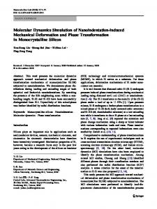
FIG. 1. Structure of the channel wall. Three consecutive 20atom rings are shown. From lightest to darkest, the spheres have partial charges of 10.5e, 10.35e, 20.35e, 20.5e. As the differences in shading of dark and light spheres may be difficult to see, one can start with a dark sphere and give it a specific negative charge. Proceeding counterclockwise, the next sphere in a given ring has a positive charge of the same magnitude, the third sphere has the other negative charge, and the fourth sphere has a positive charge of the same magnitude as that of the third sphere. Successive rings are displaced by 9±. The figure is for illustrative purposes only and is not drawn to scale; the exact specifications of the system are given in the text.
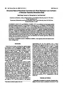
the simulation cell to mimic the potential gradient that drives ion transport through biological channels. As can be seen in the potential plot of Fig. 2, the fluid responds to this external field, neutralizing it in the reservoir region,
FIG. 2. Relative potential as a function of z in a straight line through the center of the channel and the reservoir region. Potentials were calculated by insertion of a test particle that sampled the electrostatic interactions from the average charge distribution over the duration of all of the simulations. The net potential drop across the cell remains constant at 1.1 V due to the imposed external electric field.
VOLUME 86, NUMBER 11
PHYSICAL REVIEW LETTERS
and amplifying it across the membrane so that the 1.1 V potential drop is felt mainly across the membrane. In analogy to an electrical circuit, the potential drop is found mainly across the channel (resistor), rather than across the reservoir (conducting wire). Periodic boundary conditions were applied in all three directions, maintaining a constant volume and enabling the simulations to be run continuously without particle insertions or deletions, even though a net current of Na1 ions was flowing in the positive z direction. During the 100 ns of data collection, 12 complete Na1 ion passages occurred, wherein a sodium ion entered the
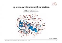
FIG. 3. Snapshot of the model system. The large dark spheres represent the Cl2 ions, the smaller light colored spheres represent the Na1 spheres, the smaller “boomerang” objects represent the SPC兾E water molecules, the two planes of smaller spheres represent the neutral membrane walls, and the rings are made up of small spheres (the darker spheres have a negative charge) that represent the atoms in the eleven 20-member rings comprising the channel structure. The figure is for illustrative purposes only and is not drawn to scale; the exact specifications of the system are given in the text.
12 MARCH 2001
channel at z 苷 15 Å and exited the channel at z 苷 40 Å. In addition, one partial passage occurred, in which a sodium ion traversed only 26% of the way through the channel within the allotted time. A Na1 could be found in the pore for 14.76 ns of the total 100 ns of data collection. A snapshot of the channel is given in Fig. 3. Although several Cl2 ions entered the channel at z 苷 40 Å and subsequently reexited at z 苷 40 Å after a few ps, there was no complete Cl2 ion passage and the total net fraction of Cl2 ion partial passages was therefore zero during the simulation period. For the given channel structure and 1.1 V applied voltage, there were 12.26 Na1 ion net passages within the allotted 100 ns. This yields a current of 19.6 pA and a conductance of 17.8 pS. The mean passage time is 1.2 ns, calculated by computing the total time in which there is a Na1 ion in the channel divided by the net number of passages. Since experimentally measured [1] channel conductances are of the same order of magnitude, the calculated current demonstrates the viability of the applied field MD method for studying channel conductance. The velocity of an ion in the channel, although fairly constant in the bulk of the channel, increases, up to a factor of 2, as the ion approaches the exit. Unlike present experimental measurements, MD simulations allow us to directly explore subtle atomic-level details of the channel conductance. For example, do the sodium ions prefer to travel down the center of the channel, or along the sides? Figure 4 shows the average density distribution in the radial direction of Na1 ions, O atoms, and
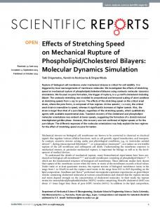
FIG. 4. Average density distribution of H atoms (dotted line), O atoms (solid line), and Na1 ions (dashed line) in the channel as a function of distance from the center of the channel. Sodium ion information comes from all 100 ns of property collection; hydrogen and oxygen atom distributions were collected during a 50 ps period while there was no ion in the channel. Densities are relative to the bulk density of each species.
2469
VOLUME 86, NUMBER 11
PHYSICAL REVIEW LETTERS
FIG. 5. Average density distribution of H atoms (dotted line, density divided by two), O atoms (solid line), Na1 ions (squares), and Cl2 ions (3) in the reservoir region as a function of z. Samples were taken from a 6 Å diameter cylinder that was parallel to the z axis and centered at the points farthest from the channel. Sampling took place at 50 fs intervals during all 100 ns of property collection. Note that the left axis corresponds to the hydrogen and oxygen traces and that the right axis corresponds to the ion traces. Please also note that because of the periodic boundary conditions, the negative z values of this plot are equivalent to those values plus the cell length, LZ 苷 55 Å (i.e., z 0 苷 z 1 55 Å).
H atoms inside the channel. The Na1 ions prefer to be about 1.1 Å from the center of the channel. In contrast, the Cl2 ions, whose density is too small to be seen, prefer to be at the channel center during their brief visits. On the other hand, the water molecules adhere to the walls of the channel, presumably due to the polarity of the channel atoms. For our model, there are few water molecules in the center of the channel. Note that in contrast to the water molecules and the Cl2 ions, the Na1 ion concentration in the channel reaches a level considerably in excess of the bulk concentration. As seen in Fig. 3, the water dipoles in the channel are oriented by the external field, as would be expected. Space does not allow a consideration of ion dehydration or water channel wall interactions here, but we speculate that the positioning of Cl2 ions near the center of the channel and their low conductance reflect the hydration of these ions within the pore. The structure of water around ions is undoubtedly peculiar to particle diameters and charges and the channel diameter. Average density distributions as a function of z, for a region with x and y far from the channel, for each atomic species in the reservoir region are given in Fig. 5. The Na1 ions tend to exhibit a higher concentration probability, with some layering, on the high-potential side of the membrane whereas the Cl2 ions tend to exhibit a higher concentration probability, again with some layering, on the low-potential 2470
12 MARCH 2001
side of the membrane. Some of the H atoms approach the membrane more closely than the O atoms because of their smaller size. Thus, something like a double layer forms on the membrane. There is rough electroneutrality in the center of the reservoir. Presumably, a more precise electroneutrality for the “bulk” fluid would be exhibited with a larger unit cell. We have demonstrated the feasibility of modeling an ion current through a membrane using molecular dynamics with closed circuit conditions. Sufficient ion passages are observed to permit a statistical reasonable estimate of the channel conductance. It will be interesting in the future to utilize this approach to study conductance/concentration relationships, conductance/voltage relationships, and channel permeation selectivity, as well as to study systems with a protein structure embedded in a lipid. This work was supported in part by the National Science Foundation (Grant No. CHE98-13729), by the donors of the Petroleum Research Foundation, administered by the American Chemical Society (Grant No. ACS-PRF 31573AC9), and by the National Institutes of Health (Grant No. AI23007).
[1] B. Hille, Ionic Channels of Excitable Membranes (Sinauer Associates, Sunderland, MA, 1992), 2nd ed. [2] B. Roux and M. Karplus, Ann. Rev. Biophys. Biomol. Struct. 23, 731 (1994). [3] W. Nonner, L. Catacuzzeno, and B. Eisenberg, Biophys. J. 79, 1976 (2000). [4] D. Boda, D. D. Busath, D. Henderson, and S. Sokołowski, J. Phys. Chem. B 104, 8903 (2000). [5] D. Golding, J.-P. Hansen, and S. Melchionna, Phys. Rev. Lett. 85, 1132 (2000). [6] U. Hollerbach, D. P. Chen, D. D. Busath, and B. Eisenberg, Langmuir 16, 5509 (2000). [7] R. M. Lynden-Bell and J. C. Rasaiah, J. Chem. Phys. 105, 9266 (1996). [8] C. Hartnig, C. Witschel, and E. Spohr, Ber. Bunsen-Ges. Phys. Chem. 102, 1689 (1998). [9] S.-H. Chung, T. W. Allen, M. Hoyles, and S. Kuyucak, Biophys. J. 77, 2517 (1999). [10] J. Hu, S. Goldman, C. G. Gray, and H. R. Guy, Mol. Phys. 98, 535 (2000). [11] H. J. C. Berendsen, J. R. Grigera, and T. P. Straatsma, J. Phys. Chem. 91, 6269 (1987). [12] P. Bopp, W. Dietz, and K. Heinzinger, Z. Naturforsch. 43A, 1424 (1979). [13] D. E. Smith and L. X. Dang, J. Chem. Phys. 100, 3757 (1994). [14] L. X. Dang, Chem. Phys. Lett. 200, 21 (1992). [15] E. Spohr, Electrochim. Acta 44, 1697 (1999). [16] B. R. Brooks, R. E. Bruccoleri, B. D. Olafson, D. J. States, S. Swaminathan, and M. Karplus, J. Comput. Chem. 4, 187 (1983). [17] R. W. Hockney and J. W. Eastwood, Computer Simulation Using Particles (IOP, Bristol, UK, 1988). [18] M. Deserno and C. Holm, J. Chem. Phys. 109, 7678 (1998); 109, 7694 (1998).