ORIGINAL RESEARCH ARTICLE published: 12 October 2012 doi: 10.3389/fnint.2012.00090
INTEGRATIVE NEUROSCIENCE
Monosynaptic functional connectivity in cerebral cortex during wakefulness and under graded levels of anesthesia Jeannette A. Vizuete 1 , Siveshigan Pillay 2 , Kamran Diba 3 , Kristina M. Ropella 1 and Anthony G. Hudetz 4* 1 2 3 4
Department Department Department Department
of Biomedical Engineering, Marquette University, Milwaukee, WI, USA of Biophysics, Medical College of Wisconsin, Milwaukee, WI, USA of Psychology, University of Wisconsin at Milwaukee, Milwaukee, WI, USA of Anesthesiology, Medical College of Wisconsin, Milwaukee, WI, USA
Edited by: John J. Foxe, Albert Einstein College of Medicine, USA Reviewed by: Pierre Megevand, Albert Einstein College of Medicine of Yeshiva University, USA Eugene F. Civillico, Food and Drug Administration, USA *Correspondence: Anthony G. Hudetz, Department of Anesthesiology, Medical College of Wisconsin, 8701 Watertown Plank Road, Milwaukee, WI 53226, USA. e-mail:
[email protected]
The balance between excitation and inhibition is considered to be of significant importance for neural computation and cognitive function. Excitatory and inhibitory functional connectivity in intact cortical neuronal networks in wakefulness and graded levels of anesthesia has not been systematically investigated. We compared monosynaptic excitatory and inhibitory spike transmission probabilities using pairwise cross-correlogram (CCG) analysis. Spikes were measured at 64 sites in the visual cortex of rats with chronically implanted microelectrode arrays during wakefulness and three levels of anesthesia produced by desflurane. Anesthesia decreased the number of active units, the number of functional connections, and the strength of excitatory connections. Connection probability (number of connections per number of active unit pairs) was unaffected until the deepest anesthesia level, at which a significant increase in the excitatory to inhibitory ratio of connection probabilities was observed. The results suggest that the excitatory–inhibitory balance is altered at an anesthetic depth associated with unconsciousness. Keywords: consciousness, cross-correlogram analysis, cortical monosynaptic connectivity, excitatory–inhibitory balance, connection strength
INTRODUCTION Local computations within neuronal networks constitute the foundation for information processing that ultimately leads to conscious experience and behavior (Buzsaki, 2006, 2007; Buzsaki et al., 2007). The balance between excitation and inhibition in local networks is also considered to be of significant importance for neural computation, cognitive function, and regulation of global firing activity (Bartho et al., 2004; Buzsaki, 2006, 2007). Parallel recording of extracellular activity using microelectrodes is the principal technique to investigate neuronal communication within localized areas. Accordingly, there has been a strong interest in reliable approaches to extract neuronal connectivity from multichannel unit recordings in both awake and anesthetized animals. How the derived neuronal interactions depend on the level of consciousness including waking, sleep, and anesthesia is a principal question that may shed light on the neuronal mechanisms underlying neuronal computations that support cognitive functions. To date, relatively little is known about the nature of anesthetic dose-dependent changes in functional interactions in intact neuronal networks. The modulation of neuronal communication by anesthetic agents is of particular interest because anesthetics can be applied to investigate the emergence and breakdown of consciousness in a controlled manner. Several studies suggest that the brain’s ability to process and integrate information across remote and local areas in the cerebral
Frontiers in Integrative Neuroscience
cortex gives rise to conscious experience (Tononi and Edelman, 1998; Alkire et al., 2008). We suggested that long-range functional communication within the cerebral cortex is disrupted during loss of consciousness as produced by various anesthetics (Hudetz, 2002; Hudetz et al., 2003; Imas et al., 2005a,b, 2006). Likewise, a loss of cortical effective connectivity has been demonstrated in humans at an anesthetic depth associated with unconsciousness (Lee et al., 2009; Ferrarelli et al., 2010; Langheim et al., 2011). Furthermore, a recent study using local field potential recordings, found a concentration-dependent effect of several anesthetics on intracortical functional connections, suggesting that anesthetics modulate neuronal communication in local circuits (Kreuzer et al., 2010). Thus, functional communication in neuronal networks may be a primary target of anesthetics. Anesthetic agents have been shown to exert graded suppressive effects on both spontaneous and evoked neuronal activity (Detsch et al., 2002; Villeneuve and Casanova, 2003; Hudetz et al., 2009; Sleigh et al., 2009). Moreover, most common anesthetics suppress excitatory and facilitate inhibitory synaptic transmission (Pearce et al., 1989; Pittson et al., 2004). Whereas the effect of anesthesia on single unit activity (UA) has been studied extensively, how the observed synaptic changes influence communication in the intact neuronal network remains unclear. Elucidation of the latter requires an estimation of functional neuronal connectivity from the simultaneous recording of a large number of active units, in vivo, across multiple states of arousal.
www.frontiersin.org
October 2012 | Volume 6 | Article 90 | 1
Vizuete et al.
Wakeful and anesthetized neuronal connectivity
Numerous techniques have been recently applied to estimate functional connectivity in intact neuronal networks (Brown et al., 2004; Kass et al., 2005). Putative monosynaptic connections can be identified in local networks of extracellularly recorded units by estimating spike transmission probabilities from crosscorrelogram (CCG) analyses (Csicsvari et al., 1998; Bartho et al., 2004; Fujisawa et al., 2008). Results showed that spike transmission probabilities were state-dependent in rat hippocampal cells: highest during exploration and rapid-eye movement (REM) sleep, as observed by the presence of theta waves, and lowest during sharp-wave bursts associated with slow-wave sleep (Csicsvari et al., 1998). Fujisawa et al. further demonstrated behavior-dependent changes in short-term functional connectivity as measured by monosynaptic interactions in the medial prefrontal cortex (Fujisawa et al., 2008). These studies demonstrate that the efficacy of spike transmission within a neural network may depend on brain state, and consequently, the animal’s level of consciousness. The studies conducted using CCG analysis have been mainly performed in intact cortical neuronal networks during wakefulness, sleep or deep anesthetic levels (McGaraughty and Reinis, 1993; Csicsvari et al., 1998; Bartho et al., 2004; Fujisawa et al., 2008; Fujiwara et al., 2008). However, deep anesthesia associated with nociceptive immobility (Rampil, 1994; Antognini and Kien, 1995) does not inform us about dose-dependent changes associated with the loss and return of consciousness (Gugino et al., 2001). To understand the critical changes in network function associated with loss of consciousness, there is a need to determine, in a controlled manner, how spike transmission probabilities are altered at multiple graded levels of anesthesia. In this study, we compare excitatory and inhibitory spike transmission probabilities in rat cerebral cortex during wakefulness and under graded levels of anesthesia.
RESULTS BEHAVIORAL OBSERVATIONS
Experiments were performed on seven rats at three levels of inhaled desflurane anesthesia (6, 4, and 2%) and wakefulness. At the 6% level, spontaneous movement was absent. As the anesthetic was withdrawn, rats exhibited a gradual increase in their level of alertness. At moderate depth of anesthesia (4%), they displayed sporadic and brief behaviors such as, temporary whisker twitching or chewing, but for the most part, they remained immobile. During light sedation (2%), most rats displayed head and limb movements, and postural changes that lasted for several seconds. Finally, during wakefulness (0%), rats displayed typical intermittent grooming and exploratory behaviors as well as quiet (absence of movement) alertness. The return of righting reflex suggested that consciousness was regained at 4% anesthetic concentration. UNIT ACTIVITY AND MONOSYNAPTIC CONNECTIONS
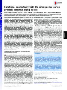
Spontaneous extracellular spikes were recorded using 64-contact multishank neural probes chronically implanted in the primary visual cortex (Figure 1A). Each electrode shank spanned the entire depth of the cortex, recording from eight equally spaced depths and eight equally spaced positions. Spikes were detected at
Frontiers in Integrative Neuroscience
approximately half of the electrode contacts (54 ± 16%). Spike sorting yielded one to three units from each electrode contact (Figure 1B). In seven rats during wakefulness, 434 active units with spike rates of at least 1 s−1 were recorded. The number and spike rate of units decreased with the anesthetic concentration (p < 0.05, linear trend, Table 1). Putative excitatory and inhibitory monosynaptic connections were identified by CCG analysis from the counts of correlated spiking between each possible pair of units at various time lags. Examples of CCG corresponding to excitatory, inhibitory, and reciprocal functional connections are illustrated in Figure 1C. The mappings of classified monosynaptic connections found between and within electrode contacts in wakefulness and at the deepest anesthesia level are illustrated in Figures 1D and 1E. In wakefulness, a total of 94 connections were found. This number represents approximately 0.5% of all possible unit pairs. The majority of connections were excitatory (ratio: 1.82 ± 0.71). Anesthesia reduced the number of all connections (p < 0.05, linear trend, Table 1). The CCG analysis also classifies the presynaptic unit as a putative pyramidal cell or interneuron depending on whether it forms an excitatory or inhibitory connection. Putative pyramidal cells fired at a lower rate (median: 3.76, 95% CI: 3.25–5.48) than interneurons (median: 6.27, 95% CI: 4.87–7.91) during wakefulness, and their spike rate distributions were significantly different (p < 0.01, K–S, data not shown). In addition, a significant difference (p < 0.05, M–W) between the spike rates of putative pyramidal cells and interneurons was present after one outlier was removed (>3 SD). The number of both cell types was reduced with deepening anesthesia (Table 1). SPATIAL DISTRIBUTION OF MONOSYNAPTIC CONNECTIONS
During wakefulness, most connections were short-range, within 200 um (Figure 2A), and most inhibitory and excitatory connections were confined to the same electrode contact at 73 and 64%, respectively (Figure 2B). This was similar at the deepest anesthetic level (6% desflurane, Figure 2E), where short-range excitatory and inhibitory connections were present at 81 and 69%, respectively. However, the number of long-range connections was noticeably smaller than in wakefulness (Figure 2D). During wakefulness, most excitatory connections projected from deeper to more superficial layers, whereas inhibitory connections were widespread, spanning nearly all cortical layers (Figure 2C). During anesthesia, the connections were limited to a shorter intralaminar span (Figure 2F). To investigate if a reduction in active units contributed to the paucity of long-range connections, we compared the statistical distribution of the Euclidean distance of all possible connections among the measured units in wakefulness and anesthesia (data not shown). We found that the distributions were essentially identical (p = 0.74, K–S) implying that the reduction in connection distances was not due to a reduction in the number of active units. CONNECTION PROBABILITY AND CONNECTION STRENGTH
An unbiased measure of functional connectivity is connection probability, defined as the number of observed monosynaptic connections relative to the number of all possible pairs of the
www.frontiersin.org
October 2012 | Volume 6 | Article 90 | 2
Vizuete et al.
Wakeful and anesthetized neuronal connectivity
FIGURE 1 | Schematic of electrode placement and examples of recorded units and connection types. (A) Electrode placement of the 64-contact neural probe in the rat primary visual cortex monocular region (V1M) in the right hemisphere. Each dot represents the approximate location of an electrode shank. Schematic is overlaid on a stereotaxic drawing obtained from the Paxinos rat brain atlas. (B) Example of recorded spike waveforms from 12 channels in one experiment. Color waveforms represent online sorting of units during acquisition. (C) Spike cross-correlograms for excitatory, inhibitory and reciprocal connections. Thresholds are represented for excitatory connections (blue line), inhibitory connections (red line), and jittered mean displayed as gray trace. Bin size
Frontiers in Integrative Neuroscience
is 1.3 ms. The gap in the center bin reflects the blanking period of spike sampling for connections observed within the same electrode contact. (D) Illustration of excitatory (blue) and inhibitory (red) connections superimposed on a map of electrode contacts during wakefulness (0% desflurane) and unconsciousness (6% desflurane) from all experiments. Presynaptic cell putatively defined as pyramidal cell (blue triangle), interneurons (red circle), or unclassified (gray square). In some cases multiple units are shown at the same contact. For greater clarity, connections between electrode contacts only are shown. (E) Number of classified within-contact excitatory and inhibitory (E,I) connections during wakefulness (left) and unconsciousness (right).
www.frontiersin.org
October 2012 | Volume 6 | Article 90 | 3
Vizuete et al.
Wakeful and anesthetized neuronal connectivity
Table 1 | Properties of classified units and connections used for CCG analysis from seven experiments. Desflurane concentration 0%
2%
4%
6%
Units*
434
309
346
271
Spike rate, 1/s*
5.5 (4.9, 6.5)
4.2 (3.6, 4.8)
3.3 (2.9, 3.7)
2.8 (2.4, 3.2)
ALL
CLASSIFIED Connections, all*
94
67
53
44
Excitatory
60 (64%)
39 (58%)
28 (53%)
31 (71%)
Inhibitory
34 (36%)
28 (42%)
25 (47%)
13 (30%)
90
47
50
42
Putative cells, all Pyramidal cell
58 (64%)
28 (60%)
27 (54%)
31 (74%)
Interneuron
32 (36%)
19 (40%)
23 (46%)
11 (26%)
Pyramidal spike rate, 1/s
3.8 (3.3, 5.5)**
3.5 (2.5, 6.2)
3.5 (2.2, 3.9)
2.6 (1.7, 3.4)
Interneuron spike rate, 1/s
6.3 (4.9, 7.9)
3.4 (1.6, 5.8)
4.0 (2.5, 5.2)
3.6 (1.4, 5.2)
Number in parentheses indicates percent of all connections and cells. Spike rates are median with 95% confidence intervals. A significant linear decrease in the number of units and unclassified spike rates with desflurane was present. A significant difference in the spike rates of putative pyramidal cells and interneurons was observed at wakefulness. *p < 0.05, linear trend; **p < 0.05, Mann–Whitney.
recorded units (Figure 3A). Anesthesia exerted a differential effect on excitatory and inhibitory connection probabilities, as indicated by a significant interaction term (p < 0.05, RM-ANOVA). This effect was due to a significantly higher probability of excitatory vs. inhibitory connections (ratio: 2.95, p < 0.01, T–K) at the 6% desflurane level. The higher excitatory to inhibitory connection probability at 6% resulted from a significant increase in the excitatory connection probability from the 4% concentration level (p < 0.05, Bonferroni). There was no difference in connection probability between wakefulness and the two lighter levels of anesthesia. We also examined connection strength, measured by the normalized height of the CCG peaks (Figure 3B). This quantity characterizes the efficacy of monosynaptic spike transmission. Anesthesia reduced excitatory connection strength in a dosedependent manner (p < 0.05, linear trend). There was no change in the strength of inhibitory connections. We considered the possibility that excitatory connection strength might decrease because of the reduced spike rate, reducing the height of the correlation peaks in the CCG. We examined this by constructing a correlation plot of the connection strength and the corresponding spike rate of each connected unit or unit pair (Figure 4). The results showed a very low correlation between connection strength and spike rate (source: R2 = 0.07, target: R2 = 0.05, combined: R2 = 0.08). To examine this question further, we sought to determine if a decrease in spike rate of either the presynaptic (source) or the postsynaptic (target) unit alone or both could alter the detectability of excitatory connections. To this end, we chose 13 classified excitatory connections of highly spiking source or target units, and decimated the number of spikes in the source, target or both units by 0, 50, 80, 90, or 95%. The CCG analysis was then repeated on all decimated data. The results showed that even at relatively low spike rates (