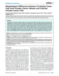
Pure ground-glass nodules (GGNs) appear as slight focal opacities on lung windows, and are invisible and contain no solid components on mediastinal ...
Xiang et al. Cancer Imaging 2014, 14:33 http://www.cancerimagingjournal.com/content/14/1/33
RESEARCH ARTICLE
Open Access
Morphological factors differentiating between early lung adenocarcinomas appearing as pure ground-glass nodules measuring ≤10 mm on thin-section computed tomography Wenjing Xiang1,2, Yanfen Xing2, Sen Jiang2, Gang Chen2, Haixia Mao2, Kanchan Labh2, Xiaoli Jia2 and Xiwen Sun2*
Abstract Background: We aimed to compare the morphological features of pure ground-glass nodules (GGNs; diameter, ≤10 mm) on thin-section computed tomography (TSCT) with their histopathological results in order to identify TSCT features differentiating between atypical adenomatous hyperplasia (AAH), adenocarcinoma in situ (AIS) and minimally invasive adenocarcinoma (MIA). Methods: Between January and December 2013, 205 pure GGNs with a diameter ≤10 mm on TSCT were pathologically confirmed as AAH (40), AIS (95) or MIA (70) lesions. The patients’ age and sex were recorded. The morphological features were evaluated, and maximum diameter and mean CT value were measured for each nodule. F test, Pearson χ2 test, Fisher exact test and multinomial logistic regression analysis were used to identify factors differentiating between AAH, AIS and MIA. Receiver operating characteristic (ROC) curve analysis was performed for maximum diameter and mean CT value. Results: F test, Pearson χ2 test and Fisher exact test revealed that maximum diameter (P 5 mm invasion). The term bronchioloalveolar carcinoma (BAC) is no longer used. In the new classification, AIS is equivalent to the formerly used term BAC. Pure ground-glass nodules (GGNs) appear as slight focal opacities on lung windows, and are invisible and contain no solid components on mediastinal windows. Persistent pure GGN is a common computed tomography (CT) finding in a variety of diseases, such as AAH, AIS, MIA, focal fibrosis and organizing pneumonia. It is well-known that GGNs containing a solid portion (mixed GGNs) are a sign of malignancy. Most studies [2-4] have investigated the solid components of mixed GGNs; however, few researchers have assessed pure GGNs. Pure GGNs are occasionally a sign of malignancy [5]. Lee et al. [6] compared the CT features of pure GGNs between patients with preinvasive lesions (AAH or AIS) and those with invasive adenocarcinomas, and found that a lesion size of