an increased risk of developing an adenocarcinoma (29, 30). Long-term gastric ... gastric cancer (4, 24). ... that is autochthonous to the stomachs of cats and dogs (23). Chen et ..... portion of the gastric antrum from each stomach was placed on.
Vol. 62, No. 11
INFECTION AND IMMUNITY, Nov. 1994, p. 4981-4989
0019-9567/94/$04.00+0
Recombinant Antigens Prepared from the Urease Subunits of Helicobacter spp.: Evidence of Protection in a Mouse Model of Gastric Infection RICHARD L. FERRERO,l* JEAN-MICHEL THIBERGE,' MICHEL HUERRE,2 AND AGNES LABIGNE' Unite des Enterobacteries (INSERM U389)' and Unite d'Histopathologie,2 Institut Pasteur, Paris 75724, France Received 6 July 1994/Returned for modification 15 August 1994/Accepted 30 August 1994
Urease is an important virulence factor for gastric Helicobacter spp. To elucidate the efficacy of individual urease subunits to act as mucosal immunogens, the genes encoding the respective urease subunits (UreA and UreB) of Helicobacter pylori and Helicobacter felis were cloned in an expression vector (pMAL) and expressed in Escherichia coli cells as translational fusion proteins. The recombinant UreA and UreB proteins were purified by affinity and anion-exchange chromatography techniques and had predicted molecular masses of approximately 68 and 103 kDa, respectively. Western blotting (immunoblotting) studies indicated that the urease components of the fusion proteins were strongly immunogenic and were specifically recognized by polyclonal rabbit anti-Helicobacter sp. sera. The fusion proteins (50 ,ug) were used, in combination with a mucosal adjuvant (cholera toxin), to orogastrically immunize mice against H. felis infection. Gastric tissues from H. felis-challenged mice were assessed by the biopsy urease test and by histology. In mice immunized with recombinant H. felis UreB, 60%o of animals (n = 7) were histologically negative for H. felis bacteria after challenge at 17 weeks. This compared with 25% (n = 8) for mice immunized with the heterologous H. pyloni UreB antigen. Neither the homologous nor the heterologous UreA subunit elicited protective responses against H.-felis infection in mice. The study demonstrated that a recombinant subunit antigen could induce an immunoprotective response against gastric Helicobacter infection.
Helicobacter pylori is a gram-negative spiral-shaped bacterium that colonizes the mucus layer associated with gastrictype epithelium in humans. The presence of the bacterium in the gastric mucosa is associated with chronic gastritis, often accompanied by an active inflammatory component, and promotes the formation of peptic ulceration in certain infected individuals (26, 27). Retrospective seroepidemiological studies have demonstrated that individuals infected with H. pylon have an increased risk of developing an adenocarcinoma (29, 30). Long-term gastric colonization with H. pyloni is thought to induce chronic atrophic gastritis (24), which is a precursor of gastric cancer (4, 24). It has therefore been proposed that eradication of the bacterium, particularly within those populations in which an H. pyloni infection is acquired at an early age, may reduce the cases of such a neoplasm (24). Several chemotherapeutic regimens for the treatment of H. pylori infection currently exist; nevertheless, the widespread treatment of individuals with antibiotics would be both unwise and impractical. Encouraging data supporting active immunization as a means of prophylaxis against H. pylori infection have emerged from experiments using a mouse model of gastric infection (2, 3, 7, 8). In this model, stomachs of mice were colonized by a close relative of H. pylori, Helicobacter felis (22), a bacterial species that is autochthonous to the stomachs of cats and dogs (23). Chen et al. (2, 3) and Czinn et al. (7) showed that it was possible to protect mice from such a colonization by orogastrically immunizing animals with sonicated extracts of H. felis, given in combination with cholera toxin (a mucosal adjuvant). Since these early studies were reported, attention has focused
on single antigens as possible candidates in an H. pylon vaccine; urease is one such antigen. During the initial stages of gastric colonization, urease activity plays a role in the protection of helicobacters from luminal acidity (9, 13). Urease is a conserved trait amongst gastric Helicobacter spp. Moreover, H. pylori urease was shown to be structurally (13, 18, 34) and functionally (13) similar to that of H. felis. By cloning the genes encoding the ureases of H. pylori (20) and H. felis (12), we showed that these enzymes, in contrast to other microbial ureases, consist of two subunits (designated UreA and UreB) which are highly conserved at the amino acid sequence level (12). Recently, it was shown that H. pyloni urease is a protective antigen in the H. felis-mouse model (8). The aims of the study were to develop recombinant antigens derived from the urease subunits of H. pyloni and H. felis and to assess the immunoprotective efficacies of these antigens in the H. felis-mouse model. Each of the structural genes encoding the respective urease subunits from H. pyloni and H. felis was independently cloned and overexpressed in Escherichia coli. The resulting recombinant urease antigens (which were fused to a 42-kDa maltose-binding protein [MBP] of E. coli) were purified in large quantities from E. coli cultures and were immunogenic yet enzymatically inactive. The findings demonstrated the feasibility of developing a recombinant vaccine against H. pylori infection.
MATERUILS AND METHODS Bacterial strains, plasmids, and growth conditions. H. felis
(ATCC 49179) was grown on a blood agar medium containing blood agar base no. 2 (Oxoid) supplemented with 10% lysed horse blood (BioMerieux) and an antibiotic supplement consisting of vancomycin (10 ,ug/ml), polymyxin B (25 ng/ml), trimethoprim (5 ,ug/ml), and amphotericin B (2.5 ,ig/ml).
* Corresponding author. Mailing address: Unite des Entdrobacteries (INSERM U389), Institut Pasteur, 28 Rue du Dr Roux, Paris 75724, France. Phone: 33-1-40 61 32 72. Fax: 33-1-45 68 88 37.
4981
4982
FERRERO ET AL.
Bacteria were cultured under microaerobic conditions at 37°C for 2 days (12). E. coli MC1061 cells were grown routinely at 37°C in Luria medium. The antibiotics carbenicillin (100 p,g/ml) and spectinomycin (100 p,g/ml) were added as required. DNA manipulations and analysis. All DNA manipulations and analyses, unless mentioned otherwise, were performed according to standard procedures (25). Restriction and modification enzymes were purchased from Amersham (Les Ulis, France). DNA fragments to be cloned were electroeluted from agarose gels and then purified by passage on Elutip minicolumns (Schleicher and Schull, Dassel, Germany). The PCR. Typical reaction samples contained 10 to 50 ng of denatured DNA; PCR buffer (50 mmol of KCl per liter in 10 mmol of Tris-HCl per liter [pH 8.3]); dATP, dGTP, dCTP, and dTTP (each at a final concentration of 1.25 mmol/liter); 2.5 mmol of MgCl2 per liter; 100 pmol of each primer; and 0.5 ,ul of Taq polymerase. The samples were subjected to 30 cycles of the following program: 2 min of denaturation at 94°C, 1 min of annealing at either 40 or 55°C (depending upon the level of stringency required), and extension for 2 min at 72°C. Cloning of amplification products in pAMP. Amplification products were cloned into the cohesive ends of the pAMP vector (Fig. 1) according to the protocol described by the manufacturer (CloneAmp System; Gibco BRL, Cergy Pontoise, France). Briefly, 60 ng of amplification product was directly mixed in a buffer (consisting of 50 mmol of KCI per liter, 1.5 mmol of MgCl2 per liter, and 0.1% [wt/vol] gelatin in 10 mmol of Tris-HCl per liter, pH 8.3) with 50 ng of the pAMP 1 vector DNA and 1 U of uracil DNA glycosylase. Ligation was performed for 30 min, at 37°C. Competent cells (200 RI) of E. coli MC1061 were transformed with 20 ,u1 of the ligation mixture. Purification of recombinant urease polypeptides. The urease polypeptides from Helicobacter spp. were overexpressed in E. coli cells with the expression vector pMAL-C2 (New England Biolabs Inc., Beverly, Mass.). The pMAL-C2 vector is under the control of an IPTG (isopropyl-o-D-thiogalactopyranoside)-inducible promoter (Ptac) and contains an open reading frame that encodes the synthesis of MalE (MBP). Cloning of sequences in phase with this open reading frame resulted in the production of MBP translational fusion proteins which, by virtue of the affinity between MBP and amylose, facilitated the purification of recombinant polypeptides. Large quantities of recombinant protein were purified according to the manufacturer's instructions. Briefly, fresh 500-ml volumes of Luria broth, containing carbenicillin (100 ,ug/ml) and 2% (wt/vol) glucose, were inoculated with overnight cultures (5 ml) of E. coli clones. The cultures were incubated at 37°C and shaken at 250 rpm, until the A600 was 0.5. Prior to adding 1 mmol (final concentration) of IPTG per liter to cultures, a 1.0-ml sample was taken (noninduced cells). Cultures were incubated for a further 4 h, at which time another 1.0-ml sample (induced cells) was taken. The noninduced and induced cell samples were later analyzed by sodium
dodecyl sulfate-polyacrylamide gel electrophoresis (SDS-PAGE). IPTG-induced cultures were spun at 7,000 rpm for 20 min in a Sorvall R-C5 centrifuge (Sorvall, Norwalk, Conn.) at 4°C. Pellets were resuspended in 50 ml of column buffer (200 mmol of NaCl per liter, 1 mmol of EDTA per liter in 10 mmol of Tris-HCl per liter [pH 7.4]), containing the following protease inhibitors (supplied by Boehringer, Mannheim, Germany): 2 ,umol of leupeptin per liter, 2 pumol of pepstatin per liter, and 1 mmol of phenylmethylsulfonyl fluoride per liter. Intact cells were lysed by passage through a French pressure cell (16,000 lb/in2). Cell debris was removed by centrifugation, and lysates were diluted in column buffer to give a final concentration of
INFECT. IMMUN. TAA ATG
ATG
(A)
-
pILL763
TAG
k.1-,Y , ureA X '
ureB
'I
I
mH (IB E-N*oACIUA....
I --P
I B E lH
.B
ERl l--
.E I
pILL920
pILL927
ATG
(B)
pILL205
_
|
TAG
TAA ATG
~~~~S, N/
D,, P.S
ureA
lureB TAG
ATC
ip)
pILL213 pILL219
(BH)VH
E
(B)B_)
pILL221
D (B
D
/
- -,,,,)
/
IH ,p
/
I
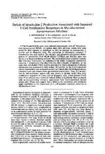
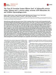
I%-/ 0771731i pILL222 FIG. 1. Plasmid constructions for the expression of recombinant urease antigens from H. pylon (A) and H. felis (B). The respective start codons (ATG) and stop codons (TAA or TAG) of the structural genes, ureA and ureB, are indicated. Numbers refer to the plasmid constructions (Table 2). Recombinant UreA and UreB constructions for H. pylon (pILL920 and pILL927, respectively), as well as H. felis UreA (pILL919 [not shown], as per pILL920) and UreB (pILL222), were constructed by PCR as described in Materials and Methods. Arrows indicate oligonucleotide primers and include the corresponding tails introduced as cloning sites. Plasmid vectors pILL570 (blackened box), pAMP-1 (stippled box), pUC18 (empty box), and pMAL (striped box) are shown. Restriction sites are indicated by abbreviations, as follows: H, HindIII; B, BamHI; E, EcoRI; P, PstI; S, Sau3A; D, DraI X, XmnI. Parentheses indicate sites present on the vectors. -11,
'Lol
2.5 mg of protein per ml, prior to chromatography on a column (2.6 by 20 cm) of amylose resin (New England Biolabs). The resin was washed with column buffer at 0.5 ml/min until the A280 returned to baseline levels. The MBP-fused recombinant proteins were eluted from the column by washing with column buffer containing 10 mmol of i-maltose per liter. Fractions containing the recombinant proteins were pooled and then dialyzed several times at 4°C against a low-salt buffer (containing 25 mmol of NaCl per liter in 20 mmol of Tris-HCl per liter [pH 8.0]). The pooled fractions were then loaded at a flow rate of 0.5 ml/min onto an anion-exchange column (1.6 by 10 cm) (HP-Sepharose; Pharmacia, Uppsala, Sweden), connected to a Hi-Load chromatography system (Pharmacia). Proteins were eluted from the column with a salt gradient (25 to 500 mmol of NaCl per liter). Fractions giving high readings
VOL. 62, 1994
RECOMBINANT UREASE SUBUNITS FROM HELICOBACTER spp.
atA280 were exhaustively dialyzed against distilled water at 4°C and analyzed by SDS-PAGE. Rabbit antisera. Polyclonal rabbit antisera were prepared against total cell extracts of H. pyloon 85P (11) and H. felis (ATCC 49179) (12). Polyclonal rabbit antisera against recombinant protein preparations of H. pyloon and H. felis urease subunits were produced by immunizing rabbits with 100 jig of purified recombinant protein in Freund's complete adjuvant. Four weeks later, rabbits were booster immunized with 100 jig of protein in Freund's incomplete adjuvant. At week 6, the animals were terminally bled and the sera were stored at
-200C. Protein analyses by SDS-PAGE and Western blotting (immunoblotting). Solubilized cell extracts were analyzed on slab gels, comprising a 4.5% acrylamide stacking gel and a 10% resolving gel, according to the procedure of Laemmli. Electrophoresis was performed at 200 V on a mini-slab gel apparatus (Bio-Rad Laboratories, Richmond, Calif.). Proteins were transferred to nitrocellulose paper in a Mini Trans-Blot transfer cell (Bio-Rad) set at 100 V for 1 h, with cooling. Nitrocellulose membranes were blocked with 5% (wt/vol) casein (BDH, Poole, England) in phosphate-buffered saline (PBS) with gentle shaking at room temperature, for 2 h (11). Membranes were reacted at 4°C overnight with antisera diluted in 1% casein prepared in PBS. Immunoreactants were detected with specific biotinylated secondary antibodies and streptavidin-peroxidase conjugate (Kirkegaard & Perry Laboratories, Inc., Gaithersburg, Md.). Reaction products were visualized on autoradiographic film (Hyperfilm, Amersham) by a chemiluminescence technique (ECL System, Amersham). Protein concentrations were determined by the Bradford assay (Sigma Chemical Co., St. Louis, Mo.). Animal experimentation. Six-week-old female Swiss specificpathogen-free mice were obtained (Centre d'Elevage R. Janvier, Le-Genest-St-Isle, France) and maintained on a commercial pellet diet with water ad libitum. To ensure that the specific-pathogen-free mice did not harbor urease-positive Helicobacter muridarum bacteria (13), six animals were randomly selected and the intestines and ceca of these animals were removed. The tissues were washed in saline, and mucus scrapings were then examined by phase-contrast microscopy for the presence of H. muridarum bacteria. The absence of H. muridarum from a random selection of animals suggested that the colony of mice was H. muridarum free. For all orogastric administrations, 100-,ul aliquots were delivered to mice with 1.0-ml disposable syringes, to which polyethylene catheters (Biotrol, Paris, France) were attached. Preparation of sonicated extracts of H. felis. H. felis bacteria were harvested in PBS and centrifuged at 5,000 rpm for 10 min in a Sorvall RC-5 centrifuge at 4°C. The pellets were washed twice and resuspended in PBS. Bacterial suspensions were sonicated as previously described (13) and were subjected to at least one freeze-thaw cycle. Protein determinations were carried out on the sonicates. Preparation of H. felis inocula for immunoprotection studies. To ensure virulent cultures of H. felis for protection studies, bacteria were reisolated from stomach biopsies of H. felis-infected mice. The isolates were passaged a minimum number of times in vitro. Stock cultures of these bacteria were stored at -80°C and were used, as required, to prepare fresh inocula for subsequent mouse protection studies. This procedure ensured the reproducibility of inocula used in successive experiments. Prior to inoculation of the animals, bacterial viability and motility were assessed by phase microscopy. Mouse protection studies. A total of 50 jig of recombinant antigen and 10 jig of cholera holotoxin (Sigma Chemical
4983
Corp.), both resuspended in HCO3, were administered orogastrically to mice at weeks 0, 1, 2, and 3. Mice immunized with sonicated H. felis extracts (containing 400 to 800 jig of total protein) were also given 10 jig of cholera toxin. At week 5, half of the mice from each group were challenged with 0.1 ml of an H. felis inoculum (approximately 108 bacteria per ml). The remainder of the mice received an additional booster immunization at week 15. At week 17, the latter were challenged with 0.1 ml of H. felis culture (approximately 106 bacteria per
ml).
Assessment of H. felis colonization of the mouse. Two weeks after receiving the challenge dose (i.e., weeks 7 and 19, respectively), mice were sacrificed by spinal dislocation. The stomachs were washed twice in sterile 0.8% NaCl, and a portion of the gastric antrum from each stomach was placed on the surfaces of agar plates (12 by 12 cm) containing a urea indicator medium (2% urea, 120 mg of Na2HPO4, 80 mg of KH2PO4, 1.2 mg of phenol red, 1.5 g of agar prepared in 100 ml). The remainder of each stomach was placed in formalsaline and stored until being processed for histology. Longitudinal sections (4 jim) of the stomachs were cut and stained by the Giemsa and hematoxylin-eosin techniques. The presence of H. felis bacteria in mouse gastric mucosa was assessed by the detection of urease activity (for up to 24 h) on the indicator medium, as well as by the screening of Giemsa-stained gastric sections that had been coded so as to eliminate observer bias. Bacterial colonization was defined as the presence of any H. felis bacteria in gastric sections, whilst mice were considered protected when no bacteria were seen in the sections. Mononuclear cell infiltrates were scored as follows: 0, no significant infiltration; 1, infiltration of low numbers of lymphocytes, limited to the submucosa and muscularis mucosa; 2, infiltration of moderate numbers of lymphocytes to the submucosa and muscularis mucosa, sometimes forming loose aggregates; and 3, infiltration of large numbers of lymphocytes, featuring nodular agglomerations of these cells. RESULTS Construction of recombinant plasmids. To clone the ureA genes of H. pyloni and H. felis, degenerate 36-mer primers were synthesized on the basis of the published urease sequences (primer set 1, Table 1). Purified DNA from E. coli clones harboring plasmids pILL763 and pILL205 (Table 2), which encoded the structural genes of H. pyloni and H. felis ureases, was used as template material in PCRs performed under nonstringent conditions. The amplification products were inserted into the plasmid vector pAMP (Fig. 1). Inserts were subsequently excised from the polylinker of the pAMP vector by double digestion with BamHI and PstI and then subcloned into the expression vector pMAL, chosen for the production of recombinant antigens (pILL919 and pILL920, respectively, Fig. 1). A product containing the ureB gene of H. pylori was amplified under stringent conditions with a set of 35-mer primers (set no. 2, Table 1). The purified amplification product (1,850 bp) was digested with EcoRI and PstI and then cloned directly into pMAL (pILL927, Fig. 1). H. felis ureB was cloned in a two-step procedure that allowed the production of both complete and truncated versions of the UreB subunit. Plasmid pILL213 (Fig. 1) was digested with the enzymes DraI, corresponding to amino acid residue number 219 of the UreB subunit (16), and HindIII. The resulting 1,350-bp fragment was purified and cloned into pMAL that had been digested with XmnI and HindIII (pILL219, Fig. 1). In order to produce a clone capable of synthesizing a complete
4984
FERRERO ET AL.
INFECT. IMMUN.
TABLE 1. The oligomeric primers used in PCR-based amplification of urease-encoding nucleotide sequences Primer set
Direction'
Nucleotide sequence (5'-*3')
1
Forw
2
Rev Forw
bCAU (CCNC) (AAR) (GAR) (YTN) (GAY) (AAR) (YTN) (ATG) (YTC) (YYT) (NCG) (NCG) (NSW) (DAT) (YTT) (YTT) (CAT) CUAb... CC GGA GAA 7TC AIT AGC AGA AAA GAA TAT GTI TCT ATG ...
EcoRId AC GTr CTG CAG
Rev
pStld 3
CIT ACG AAT AAC TIT TGT TGC TTG AGC
Forw
GGA TCC AAA AAG AIT TCA CG
Rev
GGA AGC !I C TGC AGG TGT GCT TCC CCA GTC
BamHId
HindIIId
Pstjd
aForw, forward; Rev, reverse. b
c
The 5' ends of these primers each had a series of four CAU and CUA codons, respectively, that were compatible with the pAMP vector. Degenerated nucleotides were synthesized according to the following code: Y, C or T; R, A or G; S, G or C; W, A or T; D, G or A or T; and N, G or A or C or
T. d
Restriction sites introduced in the amplified fragments.
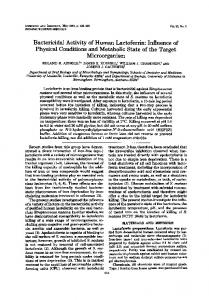
UreB protein, PCR primers (set 3, Table 1) that amplified a 685-bp fragment from the N-terminal portion of the ureB gene (excluding the ATG codon), which also overlapped the beginning of the insert in plasmid pILL219, were developed. The PCR-amplified material was purified and digested with BamHI and Hindlll and then cloned into pMAL (pILL221, Fig. 1). A 1,350-bp PstI-PstI fragment encoding the remaining portion of the ureB gene product was subsequently excised from pILL219 and cloned into a linearized preparation of pILL221 (pILL222, Fig. 1). 'Expression of Helicobacter urease polypeptides in E. coli. Recombinant urease proteins were purified from cell extracts of E. coli cells following chromatography on affinity (amylose resin) and anion-exchange (Q-Sepharose) gel media (Fig. 2). E. coli MC1061 cells transformed with recombinant plasmids encoding the respective ureA gene products of H. felis and H. pylori (pILL919 and pILL920, respectively) expressed fusion proteins with predicted molecular masses of approximately 68 kDa (Fig. 3). Two-liter cultures of these recombinant E. coli MC1061 cells typically yielded 30 mg of purified antigen. Similarly, the large UreB subunits of H. pyloni and H. felis ureases were expressed in E. coli (plasmids pILL927 and pILL222, respectively) and produced fusion proteins with predicted molecular masses of 103 kDa (Fig. 3). The yield in these cases was appreciably lower than that for the UreA preparations (approximately 15 mg was recovered from 2 liters of bacterial culture). Moreover, problems associated with the cleavage of the UreB polypeptides from the MBP portion of the fusion proteins were encountered (Fig. 3). These difficulties were attributed to the large sizes of the recombinant UreB polypeptides.
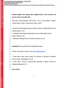
Western blot analyses of the UreA antigen preparations with rabbit polyclonal antisera raised against whole extracts of H. pyloni and H. felis bacteria demonstrated that the antigens retained antigenicity to the homologous as well as heterologous antisera (Fig. 4). The antisera did not recognize the MBP component alone. Cross-reactivity between the urease polypeptides of H. pyloni and H. felis was consistent with the high degrees of identity between the amino acid sequences of these proteins (12). Rabbit polyclonal antisera raised against purified recombinant UreB proteins prepared from H. pyloni and H. felis strongly reacted with the urease polypeptides present in recombinant UreB preparations (Fig. SA) as well as in whole-cell extracts of the bacteria (Fig. 5B). As reported previously (12), the UreB subunit of H. felis urease migrated slightly higher on SDS-PAGE gels than did that of H. pylori (Fig. 5B). Immunization of mice against gastric H. felis infection. Groups of mice were immunized four times (weeks 0 to 3) with the given antigen preparations. In a preliminary immunoprotection study, one-half of the mice from each group were challenged at week 5 with an H. felis inoculum containing 108 bacteria per ml (prepared as described in Materials and Methods). (i) Protection at week 5. Two weeks after challenge, 85% of stomach biopsy samples from the control group of animals immunized with H. felis sonicate preparations were urease negative and therefore appeared to have been protected from H. felis infection. This compared with 20% of those from the control group of mice given MBP alone. The proportion of urease-negative stomachs for those groups of mice given the recombinant urease subunits varied from 70% (for H. pylori
TABLE 2. Plasmids used Plasmid
Vector
Relevant phenotype or characteristic
pILL763 pILL199 pILL205 pILL919 pILL920 pILL927 pILL213 pILL219 pILL221 pILL222
pILL570 (Spr) pILL575 (Kmr) pILL570 pMAL-C2 (Apr)
9.5-kb fragment (Sau3A partial digest of H. pylori chromosome) 35-kb fragment (Sau3A partial digest of H. felis chromosome) 11-kb fragment (Sau3A partial digest of pILL199) 0.8-kb BamHI-PstI fragment containing PCR product encoding H. felis ureA gene 0.8-kb BamHI-PstI fragment containing PCR product encoding H. pyloni ureA gene 1.8-kb EcoRI-PstI PCR fragment encoding H. pylon ureB gene 2.2-kb fragment resulting from Sau3A partial digest of pILL205 1.4-kb DraI-HindIII fragment containing H. felis ureB (bp 657-1707) 0.7-kb BamHI-PstI PCR fragment encoding H. felis ureB (bp 4-667) 1.35-kb PstI-PstI fragment encoding H. felis ureB (bases 667-1707) from pILL219 cloned into linearized pILL221
pMAL-C2 pMAL-C2 pUC18 (Apr)
pMAL-C2 pMAL-C2 pMAL-C2
Reference
6
12 12
This study This study This study This study This study This This
study study
1 97 66
'^.r:
RECOMBINANT UREASE SUBUNITS FROM HELICOBACTER spp.
VOL. 62, 1994
2 3
4
5 6
__
45 _Wt _5_ 31
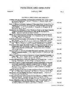
FIG. 2. Purification of H. pylon UreA-MBP recombinant protein by using the pMAL expression vector system. Extracts from the various stages of protein purification were electrophoresed on a resolving SDS-10% polyacrylamide gel. Following electrophoresis, the gel was stained with Coomassie blue. The extracts were as follows: lane 1, SDS-PAGE standard marker proteins (the molecular weights in thousands are shown on the side); lane 2, noninduced cells; lane 3, IPTG-induced cells; lane 4, French press lysate of induced cell extract; lane 5, eluate from amylose resin column; lane 6, eluate from anion-exchange column (first passage); and lane 7, eluate from anionexchange column (second passage).
UreB) to 30% (for H. pyloni UreA). Assessme: nt of coded histological slides for the presence of H. felis bactexria, however, indicated that the levels of protection in mice wer e lowerthan that observed by the biopsy urease test: for examp le, only 25% of gastric tissue from the control mice immunized with H. felis sonicate preparations was free of H. felis bacte-ria. Gastric
1 200 116 97 66
30
2 3 4 5
6
123)o
7
FIG. 3. Gel electrophoresis of purified recombinant urease preparations. Samples were resolved on an 8% polyacrylamiide gel in the following order: lane 1, standard marker proteins; lani 2, H. fpylo UreA-MBP; lane 3, H. pylon UreB-MBP; lane 4, MBP; Ile UreA-MBP; and lane 6, H. felis UreB-MBP. Protein degradation are products (small arrowhead) and unfused MBP (large ar indicated. The former were recognized by the homologous rabbit antiserum (Fig. 4). The numbers on the left refer to tthe molecular weights in thousands of standard marker proteins.
irowhead)
66
4985
1 2 3
U.~
45 30
'inti-H. pylor'I
anti-li-.
Jflis
FIG. 4. Recognition of UreA recombinant fusion proteins by polyclonal rabbit anti-Helicobacter sp. sera. Protein extracts of MBP (lanes 1), H. felis UreA-MBP (lanes 2), and H. pylon UreA-MBP (lanes 3) were separated on a 12% polyacrylamide gel. Western blotted proteins were reacted with rabbit polyclonal antisera (diluted 1:5,000) raised against whole-cell extracts of H. pylon and H. felis. Protein degradation products reacted with the homologous rabbit sera. The numbers on the left refer to the molecular weights in thousands of standard marker proteins.
tissue from these mice displayed relatively fewer H. felis bacteria and took a relatively long time to change the color of
the urease indicator medium (14). This suggested that a protective immune response had been induced in the mice but that the response was insufficient to protect against the large numbers of H. felis bacteria in the challenge inoculum. To test this hypothesis, mice were subsequently challenged at week 17 with an inoculum containing 100-fold fewer H. felis bacteria. (ii) Protection at week 17. The remaining mice from each group of animals were boosted at week 15. These mice were challenged at week 17 with an H. felis inoculum containing 106 bacteria per ml. Two weeks later, all stomach biopsies from the MBP-immunized mice were urease positive (Table 3). In contrast, the proportion of mouse stomachs that were urease negative varied from 50%, for H. pylon UreA-immunized animals, to 100%, for those immunized with H. felis UreB. The latter was comparable to the level of protection observed for the group of animals immunized with H. felis sonicated extracts. Histological evidence demonstrated that the UreB subunits of H. felis and H. pylori protected 60 and 25% of immunized animals, respectively. For mice immunized with sonicated extracts of H.
felis, histological analysis
of tissues
revealed that 85% of these animals had been protected from H. felis infection. Immunization of mice with recombinant H. pylori UreA did not protect the animals. Similarly, the stomachs of all H. felis UreA-immunized mice that had been challenged at week 5, and were not sacrificed until week 19, were
colonized with H. felis bacteria (Table 3).
The
urease
gastric biopsy
test,
compared with histological
sections, gave sensitivity and specificity analysis values of 63 and 95%, respectively. Thus, histology proved to be the more accurate predictor of H. felis infection in the of gastric tissue
mouse. Cellular immune response in immunized stomachs. In addition to the histological assessment of H. felis colonization,
4986
FERRERO ET AL.
INFECr. IMMUN.
TABLE 3. Protection of mice from H. felis infection following immunization with recombinant urease proteins
A
No. of mice colonized by H. felisa
1 2 3 4 97 68
46
4 _ #i _
~~~~a
-AM
31 anti-Uret13 II. plu/oi
ainti-UreB I I. fe/lis
B 1 2
Antigen
Ureaseb
Histologyc
MBP UreA H. pylori UreA H. felisd UreB H. pylori UreB H. felis H. felis sonicate
100 (10/10) 50 (4/8) 87.5 (7/8) 35 (3/8) 0 (0/7) 0 (0/8)
100 (10/10) 100 (8/8) 100 (8/8) 75 (6/8) 40 (2/7) 15 (1/8)
1 2 3 4
1 2
a Unless stated otherwise, mice had been immunized weekly (weeks 0 to 3), booster immunized at week 15, challenged with 105 H. felis bacteria per mouse at week 17, and sacrificed at week 19. bThe percentage of stomachs giving a positive urease biopsy test. Total numbers of mice are given in the parentheses. 'The percentage of stomachs with H. felis bacteria identified in histological sections of mouse gastric mucosa. Total numbers of mice are given in the parentheses. d Mice had been immunized weekly (weeks 0 to 3), challenged at week 5 (with 107 bacteria), and sacrificed at week 19.
coalesced to form lymphoid nodules that extended into the submucosal regions of the gastric epithelia (Fig. 7B and C). The mononuclear cell response appeared to be independent of the presence of bacteria. DISCUSSION
97 68 46
40,0
"m
.4110
31 anti--UreI3 I-1,
py/lori
ainti-U-reB II. fells
FIG. 5. Western blotting analyses with antisera raised against purified H. pylon and H. felis UreB recombinant proteins. (A) Nitrocellulose membranes were immunoblotted with antisera raised against the following purified recombinant protein extracts: lanes 1, biotinylated standard protein markers; lanes 2, H. felis UreB-MBP; lanes 3, MBP; and lanes 4, H. pylori UreB-MBP. (B) Recognition of UreB polypeptides in whole-cell extracts of H. felis (lanes 1) and H. pylori (lanes 2). Rabbit antiserum was diluted 1:5,000. The numbers on the left refer to the molecular weights in thousands of standard marker proteins.
gastric tissue was scored (from 0 to 3) for the presence of lymphocytic infiltrates (Fig. 6). In mice immunized with MBP alone, a mild chronic gastritis was seen with small numbers of lymphocytes restricted to the muscularis mucosa and to the submucosa of the gastric epithelium. In contrast, there were considerable numbers of mononuclear cells present in the gastric mucosae from animals immunized with either the recombinant urease polypeptides or H. felis sonicate preparations (Fig. 6 and 7A). These inflammatory cells sometimes
Individuals infected with H. pylon produce vast quantities of specific immunoglobulin G (IgG) antibodies in the serum (1, 31, 32), as well as IgA and IgG antibodies in the mucosal tissue (32). Despite the strong immune response, H. pylon bacteria remain firmly entrenched in the gastric mucosa. Consequently, immunization was for a long time dismissed as a method of prophylaxis against H. pylon infection. Impetus for the development of an anti-H. pylon vaccine, nevertheless, came from the results of several studies demonstrating the induction of protective mucosal immune responses against H. felis infection in mice. In the initial studies, sonicated H. felis extracts were used as the antigen (2, 3, 7); more recently, it was shown that H. pylon urease, purified either from the organism itself or
0
2.5
0
mouse
MBP
UreA H. pylori
UreB
H. felis
UreB H. pylori
H. felis sonicate
FIG. 6. Box plot representation of the distribution of mononuclear cell scores for the different immunization groups of mice. Mice had been immunized once per week (weeks 0 to 3), booster immunized at week 15, and challenged with an H. felis culture at week 17. Two weeks postchallenge, the mice were sacrificed. For each box plot figure, the highest point represents the 90th percentile while the lowest point represents the 10th percentile.
VOL. 62, 1994
A
RECOMBINANT UREASE SUBUNITS FROM HELICOBACTER spp.
4987
B
FIG. 7. Histological analysis of gastric tissues from immunized mice. Mice had been immunized once per week (weeks 0 to 3), booster immunized at week 15, and challenged with an H. felis culture at week 17. Two weeks postchallenge, the mice were sacrificed. Shown are mononuclear cells extending from the submucosa into the glandular region of the tissue (A). In a proportion of immunized animals, the lymphocytes coalesced to form lymphoid follicles in the subglandular region (B and C). Hematoxylin and eosin stain.
from recombinant E. coli cells, conferred protective immunity in mice (8). In order to determine whether this immunity might be conferred by one or more domains of the urease holoenzyme, recombinant urease subunit antigens from H. pylori and H. felis were expressed and characterized. An important aspect of the study was to compare the performance of heterologous and homologous Helicobacter antigens as mucosal immunogens in the H. felis-mouse model. The results from these studies allow us to propose the large urease subunit (UreB) as a potential component of a future H. pylon vaccine. The respective UreA and UreB subunits of H. pylori and H. felis ureases were overexpressed in E. coli cells and purified as MBP-fused proteins. Western blot analyses using anti-Helicobacter rabbit sera indicated that the urease recombinant proteins were strongly immunogenic (Fig. 4 and 5). Moreover, H. pyloni UreA and UreB recombinant proteins were recognized by sera from patients with confirmed cases of H. pylon disease (33). Purified MBP alone did not appear to cross-react with the rabbit antisera and so did not contribute significantly to the immunogenicity of the fusion proteins. In agreement with previous biochemical (13, 18, 34) and molecular (12) studies, immunological cross-reactivity between the recombinant urease subunits of H. pylori and H. felis was found. The cross-
reactivity appeared to be greatest when anti-H. felis sera were used in Western blot analyses (Fig. 4 and 5B). Though the UreB subunits of H. felis and H. pylori share an important number of immunogenic epitopes, the recombinant antigens derived from these proteins seemed to protect mice from gastric helicobacter infection to varying degrees. Hence, it is unlikely that a Helicobacter sp.-specific urease epitope might be sufficient to serve as a protective antigen in a vaccine. Given that heterologous H. pylori urease holoenzyme protected mice from gastric H. felis colonization (8), one might postulate that UreA, though not protective per se, may nonetheless be important in the presentation of immunoprotective domains. The H. felis inoculum used in the challenge procedure was found to be an important variable in immunoprotection studies. Amongst the different mouse protection trials, the bacterial densities of the H. felis inocula, as well as the methods of preparing the inocula, have varied greatly. This may, in part, account for the different levels of protection (varying from 35 to 85% protection) reported by the various studies (2, 3, 7, 8). By maintaining virulent cultures of H. felis, the quantity of bacteria needed to colonize the mouse was significantly reduced. Using this method, we have been able to colonize mice
4988
FERRERO ET AL.
with as little as 103 H. felis bacteria (14). This approach should ensure reproducibility between different immunoprotection trials. There have been several studies of the immune responses induced in the gastric mucosa of persons infected with H. pylori (5, 10, 19) and in animals experimentally infected with Helicobacter spp. (15-17, 22). In their original description of the H. felis-mouse model, Lee and colleagues (22) reported that, within the first 2 weeks of infection, H. felis-infected mice developed an acute inflammation composed predominantly of eosinophils and neutrophils. Moreover, lymphocytes did not become a predominating cell type until 8 weeks postinfection (22). The effect of mucosal immunization on gastric pathology has, thus far, not been investigated. In this study, pronounced lymphocytic infiltrations were observed in mice that had been immunized with either Helicobacter urease antigens or H. felis sonicated extracts (Fig. 6 and 7). A particularly interesting finding was the presence of follicular structures, resembling gut-associated lymphoid tissue, in the gastric mucosa of the immunized mice. Such structures were previously described for mice that had been experimentally infected for over 1 year with H. felis bacteria (15, 21). Though it may be argued that the lymphocytic gastritis seen in the immunized mice was induced in response to the bacterial challenge, this seems unlikely. As reported above, lymphocyte numbers appear to increase only during the chronic stages of the murine infection (15, 21, 22), and thus the 2-week postchallenge period would have been insufficient for the development of a bacterial gastritis. Furthermore, the MBP-immunized mice, which were all colonized with H. felis, had relatively low infiltration scores (Fig. 6). Mucosal immune responses normally require the uptake and presentation of antigens to lymphocytes at so-called inductor sites (28). The stimulated lymphocytes undergo differentiation and migrate to the given effector site, where specific IgAsecreting B cells proliferate and produce protective antibodies against the infectious agent (28). It is possible to speculate that the infiltrating lymphocytic cells seen in the stomachs of immunized mice may be involved in either antigen uptake or secretory IgA production. Further studies are required to address these questions as well as the types of clonal populations composing these mononuclear infiltrations. ACKNOWLEDGMENTS Financial support was provided, in part, by Galagen Incorp. (U.S.A.). R.L.F. was the recipient of a scholarship from the Direction Scientifique des Applications de la Recherche (from the Institut
Pasteur). We are indebted to Collette Coynault for her assistance in the early phases of the animal work and Nicole Wuscher for the histopathological processing of samples. We gratefully acknowledge Agnes Ullmann for her support and interest throughout. REFERENCES 1. Booth, L., G. Holdstock, H. MacBride, P. Hawtin, J. R. Gibson, A. Ireland, J. Bamforth, C. E. DuBoulay, R S. Lloyd, and A. D. Pearson. 1986. Clinical importance of Campylobacterpyloridis and associated serum IgG and IgA antibody responses in patients undergoing upper gastrointestinal endoscopy. J. Clin. Pathol. 39: 215-219. 2. Chen, M., A. Lee, and S. L. Hazell. 1992. Immunisation against gastric helicobacter infection in a mouselHelicobacterfelis model. Lancet 339:1120-1121. 3. Chen, M., A. Lee, S. L. Hazell, P. Hu, and Y. Li. 1993. Immunisation against gastric infection with Helicobacter species: first step in the prophylaxis of gastric cancer? Zentralbl. Bakteriol. 280:155165.
INFECT. IMMUN. 4. Correa, P. 1988. A human model of gastric carcinogenesis. Cancer Res. 48:3554-3560. 5. Crabtree, J. E., T. M. Shalicross, R V. Heatley, and J. I. Wyatt. 1991. Mucosal tumour necrosis factor alpha and interleukin 6 in patients with Helicobacterpylori-associated gastritis. Gut 32:14731477. 6. Cussac, V., R. L. Ferrero, and A. Labigne. 1992. Expression of Helicobacter pylori urease genes in Escherichia coli grown under nitrogen-limiting conditions. J. Bacteriol. 174:2466-2473. 7. Czinn, S., A. Cai, and J. Nedrud. 1993. Protection of germ-free mice from infection by Helicobacterfelis after active oral or passive IgA immunization. Vaccine 11:637-642. 8. Davin, C., A. L. Blum, I. Corthesy-Theulaz, E. Saraga, J.-P. Kraehenbuhl, R Haas, and P. Michetti. 1993. H. pyloni urease elicits protection against H. felis infection in mice, abstr. 1213, p. A-304. Abstr. Am. Gastroenterol. Assoc. Meet. 1993. 9. Eaton, K. A., and S. Krakowka. 1994. Effect of gastric pH on urease-dependent colonization of gnotobiotic piglets by Helicobacterpylori. Infect. Immun. 62:3604-3607. 10. Engstrand, L., A. Scheynius, C. Pahlson, L. Grimelius, A. Schwan, and S. Gustavsson. 1989. Association of Campylobacter pyloni with induced expression of class II transplantation antigens on gastric epithelial cells. Infect. Immun. 57:827-832. 11. Ferrero, R. L., V. Cussac, P. Courcoux, and A. Labigne. 1992. Construction of isogenic urease-negative mutants of Helicobacter pyloni by allelic exchange. J. Bacteriol. 174:42124217. 12. Ferrero, R. L., and A. Labigne. 1993. Cloning, expression and sequencing of Helicobacter felis urease genes. Mol. Microbiol. 9: 323-333. 13. Ferrero, R L., and A. Lee. 1991. The importance of urease in acid protection for the gastric-colonising bacteria Helicobacter pyloni and Helicobacter felis sp. nov. Microb. Ecol. Health Dis. 4:121134. 14. Ferrero, R L., J.-M. Thiberge, and A. Labigne. Unpublished data. 15. Fox, J. G., M. Blanco, J. C. Murphy, N. S. Taylor, A. Lee, Z. Kabok, and J. Pappo. 1993. Local and systemic immune responses in murine Helicobacterfelis active chronic gastritis. Infect. Immun. 61:2309-2315. 16. Fox, J. G., P. Correa, N. S. Taylor, A. Lee, G. Otto, J. C. Murphy, and R. Rose. 1990. Helicobacter mustelae-associated gastritis in ferrets. An animal model of Helicobacter pyloni gastritis in humans. Gastroenterology 99:352-361. 17. Fox, J. G., A. Lee, G. Otto, N. S. Taylor, and J. C. Murphy. 1991. Helicobacter felis gastritis in gnotobiotic rats: an animal model of Helicobacter pylon gastritis. Infect. Immun. 59:785-791. 18. Gootz, T. D., G. L. Perez-Perez, J. Clancy, B.-A. Martin, A. Tait-Kamradt, and M. J. Blaser. 1994. Immunological and molecular characterization of Helicobacter felis urease. Infect. Immun. 62:793-798. 19. Karttunen, R. 1991. Blood lymphocyte proliferation, cytokine secretion and appearance of T cells with activation surface markers in cultures with Helicobacter pylori. Comparison of the responses of subjects with and without antibodies to H. pylon. Clin. Exp. Immunol. 83:396-400. 20. Labigne, A., V. Cussac, and P. Courcoux. 1991. Shuttle cloning and nucleotide sequences of Helicobacter pylon genes responsible for urease activity. J. Bacteriol. 173:1920-1931. 21. Lee, A., M. Chen, N. Coltro, J. O'Rourke, S. L. Hazell, P. Hu, and Y. Li. 1993. Long term infection of the gastric mucosa with Helicobacter species does induce atrophic gastritis in an animal model of Helicobacter pylori infection. Zentralbl. Bakteriol. 280: 38-50. 22. Lee, A., J. G. Fox, G. Otto, and J. Murphy. 1990. A small animal model of human Helicobacter pylori active chronic gastritis. Gastroenterology 99:1315-1325. 23. Lee, A., S. L. Hazell, J. O'Rourke, and S. Kouprach. 1988. Isolation of a spiral-shaped bacterium from the cat stomach. Infect. Immun. 56:2843-2850. 24. Leon-Barua, R, S. Recavarren-Arce, R H. Gilman, and R Berendson. 1993. Can eradication of Helicobacter pylon prevent gastric cancer? Drugs 46:341-346. 25. Maniatis, T., E. F. Fritsch, and J. SambrookL 1982. Molecular cloning: a laboratory manual. Cold Spring Harbor Laboratory
VOL. 62, 1994
RECOMBINANT UREASE SUBUNITS FROM HELICOBACTER
Press, Cold Spring Harbor, N.Y. 26. Marshall, B. J., C. S. Goodwin, J. R. Warren, R Murray, E. D. Blincow, S. J. Blackbourn, M. Phillips, T. E. Waters, and C. R Sanderson. 1988. Prospective double-blind trial of duodenal ulcer relapse after eradication of Campylobacterpylori. Lancet ii:14391442. 27. Marshall, B. J., and J. R Warren. 1984. Unidentified curved bacilli in the stomach of patients with gastritis and peptic ulceration. Lancet i:1311-1315. 28. McGhee, J. R, and H. Kiyono. 1993. New perspectives in vaccine development: mucosal immunity to infections. Infect. Agents Dis. 2:55-73. 29. Nomura, A., G. N. Stemmermann, P.-H. Chyou, I. Kato, G. L. Perez-Perez, and M. J. Blaser. 1991. Helicobacterpylori infection and gastric carcinoma among Japanese Americans in Hawaii. N. Engl. J. Med. 325:1132-1136.
spp.
4989
30. Parsonnet, J., G. D. Friedman, D. P. Vandersteen, Y. Chang, J. H. Vogelman, N. Orentreich, and R K. Sibley. 1991. Helicobacter pylon infection and the risk of gastric carcinoma. N. Engl. J. Med. 325:1127-1131. 31. Perez-Perez, G. I., B. Dworkin, J. Chodos, and M. J. Blaser. 1988. Campylobacter pyloni-specific serum antibodies in humans. Ann. Intern. Med. 109:11-17. 32. Rathbone, B. J., J. I. Wyatt, B. W. Worsley, S. E. Shires, L K. Trejdosiewicz, R V. Heatley, and M. S. Losowsky. 1986. Systemic and local antibody responses to gastric Campylobacterpyloridis in non-ulcer dyspepsia. Gut 27:642-647. 33. Thiberge, J.-M., R L Ferrero, and A. Labigne. Unpublished data. 34. Turbett, G. R, P. B. Hoj, R Horne, and B. J. Mee. 1992. Purification and characterization of the urease enzymes of Helicobacter species from humans and animals. Infect. Immun. 60: 5259-5266.