Int. J. Dev. Biol. 46: 217-226 (2002)
Original Article
Multiple interactions between maternally-activated signalling pathways control Xenopus nodal-related genes MARIA REX, EMMA HILTON and ROBERT OLD* Department of Biological Sciences, University of Warwick, Coventry, U.K.
ABSTRACT We have investigated the induction of the six Xenopus nodal-related genes, Xnr1Xnr6, by maternal determinants. The β-catenin pathway was modelled by stimulation using Xwnt8, activin-like signalling was modelled by activin, and VegT action was studied by overexpression in animal cap explants. Combinations of factors were examined, and previously unrecognised interactions were revealed in animal caps and whole embryos. For the induction of Xnr5 and Xnr6 in whole embryos, using a β-catenin antisense morpholino oligonucleotide or a dominant negative XTcf3, we have demonstrated an absolute permissive requirement for the β-catenin/Tcf pathway, in addition to the requirement for VegT action. In animal caps Xnr5 and Xnr6 are induced in response to VegT overexpression, and this induction is dependent upon the concomitant activation of the βcatenin pathway that VegT initiates in animal caps. For the induction of Xnr3, VegT interacts negatively so as to inhibit the induction otherwise observed with wnt-signalling alone. The negative effect of VegT is not the result of a general inhibition of wnt-signalling, and does not result from an inhibition of wnt-induced siamois expression. A 294 bp proximal promoter fragment of the Xnr3 gene is sufficient to mediate the negative effect of VegT. Further experiments, employing cycloheximide to examine the dependence of Xnr gene expression upon proteins translated after the midblastula stage, demonstrated that Xnrs 4, 5 and 6 are ‘primary’ Xnr genes whose expression in the late blastula is solely dependent upon factors present before the mid-blastula stage.
KEY WORDS: Xnr, nodal-related gene, β-catenin, VegT, activin, morpholino
Introduction The molecular events leading to the formation and patterning of the endoderm and mesoderm in Xenopus have been elucidated progressively in recent years (reviewed in Yasuo and Lemaire, 2001). Current models involve a relatively small number of maternally-supplied localised molecules, which initiate the transcription of a number of early zygotic genes. The essential and pivotal role of the transcription factor VegT, whose maternal mRNA is vegetally localised (Zhang and King 1996), has become apparent through experiments involving the technique of antisense oligonucleotidedepletion of the maternal mRNA (Zhang et al., 1998; Kofron et al., 1999; Xanthos et al., 2001). Probably acting in combination with vegetally-localised maternal mRNA encoding a TGFβ family member, VegT activates the zygotic transcription of a number of genes which are necessary for formation of the endoderm (Yasuo and Lemaire, 1999, Clements et al., 1999; Xanthos et al., 2001), and formation of the mesoderm (Clements et al., 1999; Kofron et al., 1999). On the dorsal side of the embryo, the localised action of the dishevelled/β-catenin signalling pathway participates in a chain of events leading to formation of the Spemann organiser (Schneider
et al.,1996; Carnac et al., 1996; Kessler 1997; Larabell et al., 1997; Miller et al., 1999; Nishita et al., 2000). A common feature of recent models of endodermal patterning and dorsal mesoderm formation is the positive combined action in the vegetal-dorsal region, where the signals intersect, of two or more signals comprising (i) dorsally-localised β-catenin activity, and (ii) one or more either maternal (Weeks and Melton, 1987 Fukui et al., 1994; Oda et al., 1995) or zygotic vegetally -localised activin-like TGFβ signals (Watabe et al., 1995; Cui et al., 1996; Clements et al., 1999; Zorn et al., 1999; Agius et al., 2000; Nishita et al., 2000, Takahashi et al., 2000). Thus, for example, the overlap of dorsal signalling and vegetally-localised Smad2 activity has been proposed to result in a co-operative interaction that activates expression of the siamois gene, spatially restricting siamois to its correct location (Crease et al., 1998). Downstream of VegT, there is evidence that a network of genes including multiple TGFβ family members participate in endoderm and mesoderm formation. Participating TGFβ family members Abbreviations used in this paper: CHX, cycloheximide; dn, dominant negative; RT-PCR, reverse transcription polymerase chain reaction.
*Address correspondence to: Dr. Robert Old. Department of Biological Sciences, University of Warwick, Coventry CV4 7AL, UK. Fax: +44-24-7652-3701. e-mail: R.W.
[email protected]
0214-6282/2002/$25.00 © UBC Press Printed in Spain
www.ijdb.ehu.es
218
M. Rex et al.
include genes encoding activin, derriere, and members of the family of nodal-related molecules, the Xnrs (Clements et al., 1999; Sun et al., 1999; Agius et al., 2000; Takahashi et al., 2000). The transcriptional activation of the Xnr genes by maternal signalling pathways is the subject of experiments reported here. There are six documented Xnr genes in Xenopus laevis. The protein products of Xnrs 1,2,4,5 and 6 are all capable of inducing mesoderm (Jones et al., 1995; Lustig et al., 1996, Joseph and Melton, 1997; Takahashi et al.,2000). The Xnr3 protein is structurally diverse from the other five Xnrs. Unlike them, Xnr3 does not display mesoderm-inducing properties but it is a neural-inducing factor (Hansen et al.,1997), acting at least in part by antagonising BMP activity (Glinka et al., 1996). None of the Xnr genes is expressed as maternal mRNA. Transcription of all six Xnrs is activated at about the mid-blastula stage, and all except Xnr3 are expressed in vegetal cells of the blastula (Jones et al., 1995; Smith et al., 1995; Joseph and Melton, 1997, Agius et al., 2000; Takahashi et al., 2000). For Xnrs1 and 2 this early expression is relatively low level, with transcript amounts peaking considerably later than maximal Xnr 4, 5 and 6 expression (Agius et al,. 2000; Takahashi et al., 2000). All except Xnr3 are expressed predominantly in dorsal vegetal domains at the late blastula stage (Agius et al,. 2000; Takahashi et al., 2000), whereas Xnr3 transcripts are found in the dorsal marginal region and are absent from the vegetal region (Takahashi et al., 2000). At gastrula stages Xnrs 1 and 2 are weakly expressed in a dorsal marginal domain that appears to be wider than Spemann’s organiser (Jones et al., 1995). Xnr 3 and 4 expression is localised to the organiser (Ecochard et al., 1995; Smith et al., 1995; Joseph and Melton 1997), and in the case of Xnr3 expression also extends into the deep marginal and vegetal cells, nearly down to the vegetal pole (Darras et al., 1997). Xnr5 expression was undetectable in the gastrula embryo, whereas Xnr6 is expressed below the dorsal lip of the blastopore (Takahashi et al., 2000). Only Xnr1 is expressed in later, tailbud stage, embryos. At tailbud stages Xnr1 is expressed in two small posterior domains and then in a large asymmetric domain in the left lateral plate mesoderm (Lustig et al., 1996), where it performs an essential conserved role in establishing the body’s left-right asymmetry (Levin and Mercola 1998; Collignon et al., 1996; Sampath et al., 1997). The induction of the Xnr genes by maternal determinants has been investigated previously, often using the isolated blastula ectoderm explant (‘animal cap’) assay. Induction of Xnr genes by the β-catenin pathway has been modelled by stimulation of the pathway using Xwnt8, which is known to activate the ‘canonical’ βcatenin/TCF pathway in Xenopus (Heasman et al., 1994; Darken and Wilson, 2001; Hamilton et al., 2001). The induction by maternal TGFβ family members has been modelled by Vg1 and activin (Clements et al., 1999; Yasuo and Lemaire, 1999; Agius et al., 2000). The transcription factor VegT has been shown to activate expression of Xnrs 1, 2, 4, 5 and 6 (Clements et al., 1999; Hyde and Old, 2000; Takahashi et al., 2000), but does not induce Xnr3 (Hyde and Old, 2000). Because of the importance of Xnr signalling in early embryos, we have set out to investigate systematically the induction of the six Xnr genes by maternal determinants. In particular, we have sought to better understand the patterning of the early embryo by investigating the interactions between combinations of maternal determinants in inducing the six Xnr genes. So although our experiments overlap with previously published work, our systematic study of all six genes together,
using combinations of inducing factors, is novel and reveals previously unrecognised interactions. These six genes make up a gene family in Xenopus laevis. The genes have individual induction properties which ultimately we would like to understand in terms of their DNA sequences. We and others have previously investigated promoter elements responsible for the induction of Xnr1 by VegT (Kofron et al., 1999; Hyde and Old, 2000), and FAST-responsive sequences within the first intron of the Xnr1 gene have been identified that mediate the gene’s activin/nodal responsiveness and left-side -specific expression (Osada et al., 2000). For the Xnr3 gene, DNA sequences responsible for the wnt-mediated induction have been identified (McKendry et al., 1997). A comparison between the Xnr1 and Xnr3 promoters identified some notable conserved features, including wnt response elements, as well as differences such as the presence of T-box sites in the promoter of Xnr1 but not Xnr3 (Hyde and Old, 2000). The absence of T-box sites from the comparable region of the Xnr3 promoter is consistent with the non-responsiveness of the Xnr3 gene to transcriptional activation by VegT (Hyde and Old, 2000). This comparison between Xnr1 and Xnr3, with the apparent conservation of certain promoter elements, encouraged the hope that the proximal promoter sequences of the Xnr genes might be interpretable in terms of the genes’ combinatorial regulation, further motivating the experiments reported here.
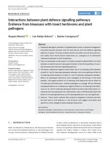
Results To study further the responses of the Xnr family of genes to signalling pathways known to be active in establishing and patterning the embryonic germ layers, and to explore systematically the possibility of combinatorial effects of such signalling pathways, we first examined the induction of the Xnr genes in blastula ectoderm (animal cap) explants cut from embryos that had been injected at the one-cell stage with mRNAs encoding Xwnt8, activin, or VegT, singly or in combination. The transcriptional responses of the six members of the nodal-related gene family, Xnr1 through Xnr6, were analysed by RT-PCR. All the experiments were repeated several times, with reproducible outcomes. The RT-PCR analysis was performed at stage 9 (late blastula, shortly after the initiation of zygotic transcription at the midblastula transition), and at stage 10 (blastopore formation, early gastrula). By stage 10 adequate time has elapsed for genes encoding possible secondary signalling proteins to have been transcribed and translated, and to have exerted their effects. Among these secondary signalling proteins are the multiple TGFβ family members that have been shown to be part of the network of zygotically-expressed endoderm-inducing and mesoderm-inducing factors initiated by the VegT transcription factor (Clements et al., 1999; Kofron et al., 1999; Agius et al., 2000; Xanthos et al., 2001). It is immediately apparent from a comparison of the analyses of Xnr gene induction at stages 9 and 10 (Fig. 1) that there are a number of clear differences between them, especially in the combinatorial effects (to be discussed in detail below) of the inducing factors at the two developmental stages. For example, Xnr6 expression shows strong synergy with all three inducing factors at stage 9, but almost no synergy at stage 10. The interactions are evidently dynamic, changing substantially and rapidly as development proceeds.
Multiple Interactions Control Xnr Genes
A
B
C
Fig. 1. Transcriptional responses of Xnr genes to Xwnt8, activinβB and VegT mRNAs singly or in combination. (A) One-cell Xenopus embryos were injected with three different amounts of mRNA (covering a four-fold range) for Xwnt8, activinβB and VegT. Animal caps were dissected and cultured to stage 9 or 10, at which point RNA was prepared for RT-PCR analysis. (B) RT-PCR analysis at stage 9. (C) RT-PCR analysis at stage 10. Only Xnr3 was strongly induced by Xwnt 8. Activin induced Xnr1,2 and 4. VegT induced Xnr1,2,4,5, and 6. Xwnt8, activin and VegT mRNAs were injected pairwise and as a mixture of all 3 mRNAs. In pairwise combinations the amount of each mRNA was the same as the lowest amount used singly. Positive synergy was observed for Xnr1,2 and 4 in response to Xwnt8 and activin. The synergy for Xnr4 was seen at stage9 but not at stage 10. Synergy was seen for Xnr4,5 and 6 for Xwnt8 and VegT at stage 10. When all 3 factors were combined at stage 9 Xnr6 displayed a strong synergistic induction. VegT showed a strong negative effect on the induction of Xnr3 by Xwnt8. Whole stage 9 or stage10 embryo cDNA was used for the linearity series.
219
Transcriptional Responses of Xnr Genes to XWnt8, Activin, or VegT, Singly Preliminary experiments (not shown) were performed to determine the minimal amounts of Xwnt8, activin, and VegT synthetic mRNAs that were required to give clear transcriptional activation of responding Xnr genes in animal cap explants. The amounts used subsequently were based on these preliminary experiments so as to give dosage-dependent responses of the caps to each of the three mRNAs, as made evident by graded induction of at least one responding Xnr gene (although simple dosedependence was not observed for all Xnr genes at each developmental stage; see below). In the experiments shown in Fig. 1, for each mRNA a set of three mRNA doubling amounts was injected, covering a fourfold range. For comparisons of the levels of induction achieved in animal caps with the amount of Xnr gene transcript found naturally in whole embryos, we note that the amount of animal cap RNA analysed was the same as the amount of RNA analysed from whole embryos in the ‘1.0 µl’ track of each linearity series shown in the figures. Figure 1B (analysis at stage 9), and Fig. 1C (analysis at stage 10) show that Xnr3 was strongly induced by Xwnt8 in animal cap explants, as expected from previous work (Smith et al., 1995; McKendry et al., 1997). None of the other five Xnr genes was induced in these caps. This absence of induction of Xnr1, 2, 4, 5, and 6, by Xwnt8 is consistent with the fact that Xwnt8 cannot induce mesoderm formation in early blastula animal caps (Sokol 1993), whereas each of these five Xnrs is capable of inducing mesoderm robustly in animal caps (Jones et al., 1995; Joseph and Melton, 1997; Takahashi et al., 2000). The non-responsiveness of the endogenous Xnr1 gene contrasts with our previous work showing that a 616 bp fragment of the Xnr1 promoter does indeed respond to wnt signalling in a transient assay, using a luciferase reporter, in which DNA was injected into early embryos (Hyde and Old, 2000). The Xnr1 promoter fragment used in that transient assay included a 14 bp sequence that is identical to a functional ‘wnt responsive’ element shown also to be present in the Xnr3 promoter (Hyde and Old, 2000; McKendry et al., 1997). This element does not in fact bind Lef1/Tcf (McKendry et al., 1997), but possibly mediates transcriptional activation of Xnr3 through the action of the homeodomain proteins siamois and twin, which are induced directly by the wnt/β-catenin pathway (Brannon and Kimelman, 1996; Carnac et al., 1996; Laurent et al., 1997). It therefore
220
M. Rex et al.
A
B
Fig. 2. The negative effect of VegT on Xnr3 induction. (A) The negative effect of VegT does not act through inhibition of siamois induction. Onecell Xenopus embryos were injected with Xwnt8 mRNA (50 pg), VegT mRNA (500 pg) or a mixture of the two mRNAs. Animal caps were harvested at stage 9 for RT-PCR analysis. Xwnt8 induced both Xnr3 and siamois expression. VegT inhibited the Xwnt8-induction of Xnr3 but not siamois. (B) VegT has a strong negative effect on the transcriptional activation, by Xwnt8, of the Xnr3 promoter. A 294 bp promoter fragment was linked to a luciferase reporter gene. The promoter-reporter DNA (30 pg) was injected into one-cell Xenopus embryos with or without Xwnt8 (1 ng) and VegT (2 ng) mRNA. Embryos were harvested at stage11 to assay luciferase activity. Samples of ten embryos were analysed and each bar represents the average of duplicate samples. The induction of Xnr3 by Xwnt8 and the negative effect of VegT were confirmed by multiple independent experiments, although the extent of induction/inhibition varied between experiments.
appears that the sensitive transient assay of the Xnr1 promoter fragment can reveal responses that are different from those of endogenous genes, either because the promoter fragment in question lacks negatively-acting DNA elements, or because there is a finite basal level of (‘leaky’) expression in sensitive transient assays, and this basal level is detectably stimulated by transcriptional activators that may not normally act alone (see below). Activin efficiently induced Xnr1, 2, and 4, but not 3, 5, or 6 (a slight induction of Xnr3 is apparent at stage 9). The dose-responses of the caps at the two developmental stages are complex. This may reflect the observation that the highest dose of activin had a profound effect upon the caps, visible as a pale coloration owing to the pigment redistribution that accompanies bottle cell formation (Kurth and Hausen, 2000). As expected from previous data, VegT induced Xnr1, 2, 4, 5, and 6 (Clements et al., 1999; Takahashi et al., 2000), but not Xnr3 (Hyde and Old, 2000).
Transcriptional Responses of Xnr Genes to Combinations of Factors: Positive and Negative Interactions Xwnt8, activin, and VegT mRNAs were injected in all pairwise combinations, and also as a mixture of all three mRNAs (Fig. 1). In the pairwise combinations, the amount of each component injected was the same as the lowest amount used in each set of singlemRNA injections, e.g. the ‘Xwnt8 plus VegT’ mixture contained 55 pg Xwnt8 mRNA plus 500 pg VegT mRNA. Clear positive synergy was observed for Xnr1, 2, and 4, in response to Xwnt8 plus activin. Xwnt8 alone did not induce these three Xnr genes, but Xwnt8 was capable of potentiating the responses to activin for each of them. In the case of the Xnr4 gene, the synergy was evident at stage 9 (Fig. 1B), but not stage 10 (Fig. 1C). The combination of Xwnt8 plus activin was not effective in inducing Xnrs 5 and 6. Xwnt8 and VegT were synergistic for induction of Xnr4, 5, and 6, even though Xwnt8 alone did not induce these genes. For Xnr5 and 6, strong synergy was apparent at stage 10, and this result is consistent with previous findings that β-catenin potentiates VegT in
inducing these two genes (Takahashi et al., 2000). When all three factors, Xwnt8, activin, and VegT were combined, at stage 9 Xnr6 displayed very strong synergistic induction above that the relatively weak induction produced by VegT alone, and even above the synergistic induction by VegT and Xwnt8 together, even though Xwnt8 and activin were incapable of inducing the gene either alone or when combined as a pair. In addition to these positive synergistic effects of VegT and Xwnt8, there was a simultaneous strong negative effect of VegT upon the ability of Xwnt8 to induce Xnr3. In the presence of VegT, the characteristically robust induction of Xnr3 by Xwnt8 was dramatically reduced, in caps analysed at stage 9 (Fig. 1B). This negative interaction was stronger at stage 9 than at stage 10, where a weaker but still substantial negative effect of VegT was evident (Fig. 1C). A possible mechanism for the inhibitory effect of VegT could be mediated through inhibition of siamois expression. The siamois gene is a direct target of wnt-signalling, and siamois induces Xnr3 (Brannon and Kimelman, 1996; Carnac et al., 1996; Kessler 1997), so an inhibition of siamois expression might cause a loss of Xnr3 induction. We therefore asked if VegT led to a reduction in siamois induction in animal caps coinjected with Xwnt8 and VegT mRNAs. The analysis of siamois expression (Fig. 2A), shows that the expected induction of siamois by wnt signalling is not reduced by expression of VegT in the animal caps. This indicates that the reduction of Xnr3 expression in these animal caps is not due to a failure of siamois expression.
VegT Inhibits the Ability of Xwnt8 to Activate Transcription from a 294 bp Region of the Xnr3 Promoter A general inhibition of wnt activity as a mechanism for the strong inhibition of Xnr3 induction by VegT is excluded because in the same animal caps where the negative interaction with respect to Xnr3 induction occurred, the combination of VegT and XWnt8 was positively synergistic for the induction of Xnrs 4, 5, and 6. To examine this further, we asked if the proximal promoter region of the Xnr3 gene mediated the negative interaction.
Multiple Interactions Control Xnr Genes The wnt-inducibility of the Xnr3 promoter has been studied previously. A 294 bp DNA region adjacent to the transcriptional start site of the gene was shown to direct wnt-inducible transcription, and wnt-responsive sequence elements were identified within this promoter region. Two of these elements bound LEF-1/TCF proteins (one element was a high affinity site, one a low affinity site). A third element did not bind these proteins, but contains a homeodomainbinding consensus sequence (McKendry et al., 1997). Since the homeodomain protein siamois is an inducer of Xnr3 (Kessler 1997), and siamois is itself a direct target of the wnt pathway (Carnac et al., 1996; Brannon and Kimelman 1996)), it is possible that siamois and/ or related proteins act through this third element. We cloned the 294 bp Xnr3 promoter fragment upstream of a luciferase reporter gene to create a Xnr3::luciferase promoterreporter plasmid construct. Plasmid DNA was injected into 1 cell embryos near the animal pole, coinjected together with Xwnt8 mRNA alone, or with VegT mRNA. Injected embryos were allowed to develop until stage 11 and harvested to assay luciferase activity (Fig. 2B). Embryos receiving the Xwnt8 mRNA expressed about 20-fold more luciferase than those without Xwnt8 mRNA, consistent with previous findings (McKendry et al., 1997). When VegT mRNA was coinjected into these embryos in addition to the plasmid DNA and Xwnt8 mRNA, the luciferase activity was similar to the uninduced level obtained with plasmid DNA alone, indicating an inhibition of wnt-induction. Thus the reporter assays confirmed the inhibitory effect of VegT upon wnt-induction of Xnr3, and showed that the effect is mediated by DNA sequences within the 294 bp proximal promoter region.
Effects of Disrupting Signalling Pathways upon Xnr Gene Expression in Whole Embryos The animal cap experiments described above displayed a number of interactions between signalling pathways that are known to be active in establishing and patterning the mesoderm and endoderm. In order systematically to examine the roles of the signalling pathways in inducing Xnr gene expression in whole embryos, we have exploited a dominant negative derivative (Molenaar et al., 1996) of Xtcf3, dnXTcf3, which disrupts the function of the β-catenin/Tcf signalling pathway. To disrupt activin signalling we have used the truncated ActRIIB receptor construct, tAR (Hemmati-Brivanlou and Melton, 1992). This dominant negative activin receptor has broad specificity, inhibiting signalling by a number of TGFβ family members including, activin, Vg1, and all the Xnrs excluding Xnr3 (Kessler and Melton, 1994; Schulte-Merker et al., 1994, Clements et al., 1999; Takahashi et al., 2000) The roles of maternal VegT action in inducing Xnr genes have been comprehensively examined by others in loss-of-function experiments using the oligonucleotide-directed mRNA depletion technique (Kofron et al., 1999; Xanthos et al., 2001), and have not been pursued by further loss-of-function experiments here. The effectiveness of each of the dominant negative constructs, dnXtcf and tAR, was first established by injecting their synthetic mRNAs into animal caps and assessing their ability to prevent the action of co-injected Xwnt8 mRNA, or activin mRNA (Fig. 3A). As expected dnXtcf almost completely abolished the induction of Xnr3 by Xwnt8, and tAR abolished or greatly reduced the induction of Xnr1, 2, and 4, by activin. The effects of the dominant negative constructs were then examined in whole embryos, at stage 9, following a vegetally-placed
221
microinjection of their synthetic mRNAs into embryos at the 1-cell stage. Effects of the tAR were remarkably limited, the most substantial being a partial reduction in Xnr1 and Xnr2 expression (Fig. 3A). Effects on the other Xnrs were minor. As expected from previous work there was no reduction in expression of Xnr5 or Xnr6 (Takahashi et al., 2000). In the case of Xnr6 this lack of effect of tAR initially seemed surprising in view of our demonstration of the large synergistic contribution of activin to Xnr6 induction, described above, that was observed when activin was combined with VegT and Xwnt8 in
A
B
Fig. 3. Effect of dominant negative XTcf3 and a truncated activin receptor, tAR on Xnr gene expression. (A) The effectiveness of the dominant negative constructs was established in animal cap assays. Dominant negative XTcf3 mRNA (500 pg) was coinjected with Xwnt8 mRNA (10 pg); and tAR mRNA (1 ng), with activin mRNA (13 pg). Animal caps were harvested at stage 9 for RT-PCR analysis. Dominant negative XTcf3 reduced the level of Xnr3 induction by Xwnt8. The induction of Xnr1, 2 and 4 by activin was suppressed by tAR. To test the effects in whole embryos, one-cell Xenopus embryos were injected vegetally with dominant negative XTcf3 mRNA (500 pg) or tAR mRNA (1 ng). Whole embryos were harvested at stage 9 for analysis by RT-PCR. In whole embryos, Xnr1 expression was reduced by tAR. Xnr2 and 4 expression showed minor reduction. Dominant negative XTcf3 substantially reduced Xnr3, 5, and 6 expression. (B) In animal caps, VegT induction of Xnr5 and Xnr6 is dependent on β-catenin activity in the cap. One cell stage Xenopus embryos were injected with VegT mRNA (1 ng), and half the embryos were subsequently injected with dominant negative Xtcf3 mRNA (1 ng). Animal caps were harvested at stage 9 for RTPCR analysis. Dominant negative XTcf3 substantially reduced the induction of Xnr5 and 6 by VegT.
222
M. Rex et al.
animal cap explants, analysed at stage 9. Thus, given (i) the presence of VegT protein throughout the vegetal region of blastula embryos (Stennard et al., 1999), and (ii) an active dorsally-localised β-catenin pathway within the vegetal region, where (iii) activin-like TGFβ family members are also thought to be present (Weeks and Melton, 1987 Fukui et al., 1994; Oda et al., 1995), it might be expected that tAR would eliminate any synergistic contribution of the activin-like signalling to Xnr6 expression. However, immunodetection of Smad2 phosphorylation (Lee et al., 2001), reveals that activin-like signalling is not detectable before stage 9. Therefore there is little or no temporal overlap between activin-like signalling and other pathways before stage 9, and hence little opportunity for synergy to occur at this stage. Consistent with expectations from previous work (Smith et al., 1995; McKendry et al., 1997), dnXtcf caused a loss of Xnr3 expression in whole embryos (Fig. 3A). The dnXtcf also caused an almost complete loss of Xnr5 and 6 expression, despite the fact that these two genes are not induced by Xwnt8 in animal cap explants. We concluded from the animal cap experiments that Xwnt8 can act synergistically with VegT in inducing Xnr5 and 6. The effect of dnXtcf in whole embryos leads us to conclude further that the β-catenin/Tcf pathway is absolutely necessary for expression of Xnr5 and 6 in whole embryos. Since Xwnt8 alone cannot induce Xnr5 or Xnr6, the role of Xwnt8 can be described as permissive for the induction of these two Xnrs. This conclusion that wnt activity is necessary prompts the question: How does the induction of Xnr5 and Xnr6 occur at all in animal caps injected singly with VegT mRNA? We reason that induction of these two genes in animal caps is indeed dependent upon β-catenin activity, and that activation of the β-catenin pathway is another consequence of the VegT expression in the caps. It is
known that VegT induces Xwnt8 (Zhang and King, 1996). To confirm the deduction that the induction of Xnr5 and Xnr6 by VegT in animal caps is dependent upon β-catenin activity, we examined the effect of dnXtcf upon the induction of the Xnrs by VegT in animal caps. As shown in Fig. 3B, our deduction is confirmed by the failure of VegT to induce the two Xnrs in the presence of the inhibitor of the β-catenin pathway. The conclusion that VegT activates β-catenin/Tcf pathway in animal cap explants raises a further question: Xwnt8 and activation of the β-catenin/Tcf pathway robustly induce Xnr3, so why does VegT not lead to the induction of Xnr3 in animal caps? This question is answered by the observation, described above for the pairwise combination of VegT and Xwnt8 mRNAs, that VegT has a striking negative effect upon the induction of Xnr3 by Xwnt8. So although the β-catenin/Tcf pathway is activated in the caps, this is not able to induce Xnr3 in the presence of VegT. Xnr Gene Expression in Embryos Depleted of β-Catenin As a further approach towards examining the roles of β-catenin signalling in whole embryos, we have exploited morpholino oligonucleotide methodology to inhibit translation of maternal β-catenin mRNA (Heasman et al., 2000). Vegetal injection at 1-cell stage of 23ng of antisense morpholino oligonucleotide directed against βcatenin mRNA resulted in embryos which developed with a characteristic lack of dorsal development (not shown). Analysis of Xnr gene expression in injected embryos at stage 9 revealed the expected absence of Xnr3 expression (Fig. 4). Expression of Xnr5 was also almost abolished, Xnr1 and Xnr6 transcripts were substantially reduced (to 12-25 % control values), Xnr2 partially reduced, and Xnr4 essentially unaltered. These effects are broadly
Fig. 4. (Left) Effect of a β-catenin antisense morpholino oligonucleotide on Xnr gene expression. Embryos were injected vegetally at the one-cell stage with a β-catenin morpholino antisense oligonucleotide, and then harvested at stage 9 for RT-PCR analysis. Xnr3 and Xnr5 expression was abolished; Xnr1 and 6 were substantially reduced and Xnr4 was unaffected by the morpholino. Fig. 5. (Right) Effects of inhibiting protein synthesis on expression of Xnr genes. Embryos were treated continuously with cycloheximide (CHX) from stage 7 onwards, and analysed by RT-PCR at stage 9, in parallel with control, untreated embryos. Mixer expression is dependent on protein synthesis from zygotic transcripts and was completely lost after CHX treatment. Xnr1, 2 and 3 gene expression was significantly reduced by CHX treatment, indicating that they require zygotic proteins. Xnr 4, 5 and 6 expression was unaffected.
Multiple Interactions Control Xnr Genes TABLE 1 INDUCING FACTORS AND THEIR INTERACTIONS, FOR INDIVIDUAL Xnr GENES IN THE ANIMAL CAP ASSAY Gene
Factors which induce the gene in animal cap explants
Xnr1 Xnr2 Xnr3 Xnr4 Xnr5 Xnr6
VegT; or activin (synergised by β-catenin) VegT (synergised by β-catenin); or activin (synergised by β-catenin) β-catenin in absence of VegT (VegT inhibits) VegT; or activin (synergised by β-catenin) VegT plus β-catenin (both essential) VegT plus β-catenin (both essential) (synergised by activin, stage 9)
similar to those observed with dnXtcf, but they are not identical. Xnr6 was less sensitive than Xnr5 to the morpholino, an effect not seen with dnXtcf for reasons that may reflect unknown different specificities of the two methods for interfering with β-catenin signalling. Taken together with the previous results, we conclude that β-catenin signalling is required for normal expression of Xnr5 and Xnr6, but not for expression of Xnr4.
Effects of Inhibiting Protein Synthesis upon Expression of Xnr Genes We have examined the effect of inhibiting the translation of zygotically expressed mRNAs by treating embryos with cycloheximide continuously from developmental stage 7, and analysing expression of the Xnr genes, at stage 9, by RT-PCR. The effectiveness of this treatment was demonstrated (Fig. 5) by the almost complete absence of Mixer transcripts, a gene whose expression is known to be dependent upon protein synthesis from zygotic transcripts and subsequent signalling by secreted factors, probably including TGFβ family members (Yasuo and Lemaire, 1999). In cycloheximide-treated embryos, the amount of expression of Xnrs 4, 5, and 6, was essentially unaffected, whereas expression of Xnr 1, 2, and 3 was substantially reduced by about 50%. The effects upon Xnr1 and 2 are consistent with previous suggestions that the early expression of these two genes is partly dependent upon (zygotic) expression of other Xnr genes such as Xnrs 4, 5, and 6 (Clements et al., 1999; Kofron et al., 1999; Agius et al., 2000; Takahashi et al., 2000). That expression of the three Xnr genes 4, 5, and 6 in late blastula embryos is almost totally insensitive to the cycloheximide treatment indicates that their expression is entirely or largely controlled by signalling proteins and/or transcription factors whose synthesis takes place before the onset of zygotic transcription.
Discussion Combinations of Factors: Conclusions From our analysis of interactions of maternal signalling pathways, as they affect Xnr gene expression, we have made a number of novel observations. The results are relevant to emerging models of the molecular circuitry of germ layer formation and patterning in early Xenopus embryos. Understanding of this molecular circuitry is currently at a very basic level with regard to the potential for, and probable importance of, interactions between signalling pathways. We have shown that β-catenin signalling is a requirement for the induction of Xnr 5 and 6 by VegT, and we have concluded that βcatenin signalling and activin together have the ability to make a very large synergistic contribution to the early induction of Xnr6 by VegT in animal cap explants. We have also concluded that VegT
223
prevents the induction of Xnr3 by wnt signalling, and that this effect is mediated by the proximal promoter region of Xnr3. Our conclusions about regulation of the individual Xnr genes by combinations of factors are summarised in Table 1. Xnr3 is induced by wnt signalling in the absence of VegT. The five mesoderm-inducing Xnr genes comprise two groups with broadly similar (but not identical) patterns of inducting factors. Xnrs 1, 2, and 4, form one group induced by VegT, or activin synergised by β-catenin, and the other group comprises Xnrs 5 and 6, induced by VegT in the presence of β-catenin signalling. While these results are fundamental to understanding Xnr gene induction in early embryos, there are many remaining questions. For example the differences in expression patterns of Xnrs 5 and 6 at gastrulation are unexplained. In interpreting the role of activin-like signalling in early embryos, it is important to note that it has recently been shown that Smad 2 activation first becomes detectable at stage 9. The activation is first apparent on the dorsal side and then spreads as a wave across the embryo towards the ventral region (Lee et al., 2001). It therefore seems likely that the earliest phase of transcription in the embryo, immediately following the mid-blastula transition, occurs in the absence of activin-like signalling which then soon, by the late blastula stage, becomes active dorsally.
Wnt/β-catenin Signalling is Permissive for Xnr5 and Xnr6 Induction by VegT In animal caps, we have shown that activation of the wnt signalling pathway occurs in response to VegT expression and that the induction of Xnrs 5 and 6 in caps is dependent upon both VegT and the induced activation of β-catenin. The results of experiments on whole embryos in which β-catenin mRNA translation or TCF function were perturbed have led us to conclude that wnt is required but not sufficient for Xnr5 and 6 induction by VegT, extending the previous suggestion that wnt signalling potentiates the induction by VegT (Takahashi et al., 2000). There is a growing list of examples of the permissive role for wnt signalling. Examples include even skipped and lethal of scute expression in the Drosophila mesoderm (Carmena et al., 1998). It has been suggested that the LEF1/Tcf effectors of wnt signalling play a role similar to other HMG boxcontaining proteins in establishing a chromatin structure that permits instructive transcription factors to function effectively and stably (reviewed by Sharpe et al., 2001). Immunolocalisation has shown that VegT protein is found in nuclei throughout the vegetal region in early to late blastula embryos (Stennard et al., 1999). The requirement for wnt signalling is the probable mechanism for limiting early expression of Xnr5 and 6 to the dorsal side of the vegetal region of the early embryo. VegT Inhibits wnt-Mediated Induction of Xnr3 We have found that VegT inhibits the ability of wnt signalling to induce Xnr3 in animal caps at stage 9, the effect being substantial but less strong at stage 10. It is likely that this effect explains the inability of VegT to induce Xnr3 in animal caps despite the concomitant activation of the wnt pathway in the caps. It is tempting to speculate that this negative effect of VegT has a natural role in refining the spatial pattern of expression of Xnr3. Consistent with this role for VegT, in situ hybridisation experiments have shown that at stage 9 Xnr3 transcripts are located in the dorsal marginal region, including deep cells of this region, but are absent from the vegetal region (Takahashi et al., 2000). By stage 10.25 Xnr3 expression extends
224
M. Rex et al.
into the dorsal vegetal region close to the vegetal pole (Darras et al., 1997), possibly partly as a result of relaxation of the inhibitory effect of VegT at this stage. What is the mechanism by which VegT effects and wnt-signalling converge upon Xnr3 transcription? A general effect of VegT upon wnt signalling is ruled out because of the positive synergistic interaction between them for induction of Xnr5 and 6, simultaneously with their negative interaction for Xnr3 induction. For Xnr3 induction it is known that LEF-1/Tcf effectors are involved (McKendry et al., 1997), and we have shown, by use of a dominant negative Tcf3 construct, that Tcf function is also required for Xnr5 and 6 induction. Hence the Xnrs 3, 5 and 6 genes are induced by the same arm of the wnt signalling pathways and we therefore can exclude a mechanism requiring a differential effect of VegT upon different arms of wnt signalling pathways. We have also excluded an effect upon siamois expression. Whatever the mechanism, the effect can be mediated at a 294 bp Xnr3 promoter region, which has been characterised in some detail previously (McKendry et al., 1997). It is possible that DNA elements that respond to a repressive signal are associated with the gene, in which case they must lie within this proximal promoter region. This is open to further investigation. A deduction from this negative interaction between VegT and wnt signalling is that Xnr3 expression should be increased in embryos lacking maternal VegT, in which mesoderm forms ectopically from the vegetal area (Zhang et al., 1998). Kofron et al. (1999) have already examined this and found that, contrary to our simple deduction, the amount of Xnr3 transcript was reduced to about 20% or 60% of normal control values, at stage 10, in VegT mRNA-depleted embryos. Intriguingly, however, the embryos injected with 6ng of antisense VegT oligonucleotide contained the higher amount of Xnr3 transcript (60% of normal), whereas embryos injected with 5ng of antisense oligonucleotide had the lower amount (20% of normal). It seems likely that the effects of depleting VegT so profoundly disrupt the embryo that it is difficult to interpret quantitative effects upon some downstream genes such as Xnr3.
Understanding Xnr Gene Induction at the DNA Sequence Level We would like to be able to relate the regulation of the six Xnr genes to their genomic DNA sequences. Limited functional analysis of regulatory elements has been performed previously for VegT regulation of Xnr1 (Kofron et al., 1999; Hyde and Old 2000), and for TGFβ family members controlling the asymmetric expression of Xnr1 in post-gastrula embryos (Osada et al., 2000). In the case of Xnr3 wnt-responsive elements have been identified by functional assays (McKendry et al., 1997). In order to take this further, we have determined sequences from the Xnr2, 4, 5, and 6 loci, and extended the known sequences for Xnr1 (Kofron et al., 1999, Hyde and Old 2000, Osada et al., 2000). These sequences have been submitted to Genbank (Accession numbers: Xnr1 AF410903, AF410904; Xnr2 AF410801; Xnr4 AF410800; Xnr5 AY050647; Xnr6 AY050648). Inspection of the sequences shows, perhaps surprisingly, that the obvious similarities between Xnr1 and Xnr3 in their proximal promoter regions (Hyde and Old 2000), do not extend simply to these other Xnr genes. Further analysis of these promoter regions in the light of conclusions reported here should be revealing. The Xnr Gene Family: Different Roles and Regulation The six members of the Xnr gene family have different expression patterns, and there are some well-documented differences in their roles. Despite this it is not clear why X. laevis should have 5 distinct mesoderm inducing Xnrs (ignoring pseudotetraploid, A and B cop-
ies), especially in view of the single nodal gene in the mouse. Experiments reported here using cycloheximide to inhibit translation support the emerging view that among the Xnr genes, Xnrs 4, 5, and 6 are ‘primary’ Xnr genes whose expression in mid to late blastula embryos is controlled solely by proteins present in the embryo before the mid-blastula transition. By contrast Xnrs 1 and 2 are substantially, but not completely, controlled by proteins synthesised after the midblastula transition. Among the proteins contributing to Xnr1 and Xnr2 induction are nodal-related proteins themselves and other TGFβ family members (Clements et al., 1999; Yasuo and Lemaire, 1999). Hence Xnr1 and Xnr2 can be termed ‘secondary’ Xnr genes.
Materials and Methods Embryos, Treatments and Injections Embryos were cultured and dissected by standard methods, and staged according to Nieuwkoop and Faber (1967). Synthetic mRNA injections were performed at the 1-cell stage using a Drummond microinjector to inject 13.8 nl. For animal cap explants, injection was into the animal pole. During injection, and for subsequent incubation, embryos were maintained in 6% Ficoll in 0.1x Barth’s saline and cultured at 13°C or 18°C. Animal caps were dissected at stage 8.5. Where indicated, embryos were treated continuously with cycloheximide at 10 µg/ml in from stage 7 onwards and analysed at stage 9. Plasmids and Transcriptions for Injection Capped mRNA was synthesised using the mMessage mMachine (Ambion) kits with templates prepared as described by suppliers. The VegT and activin cDNA plasmids were as described (Clements et al., 1999), and were the kind gift of Dr D. Clements. The Xwnt8 plasmid (Smith and Harland, 1991), and the dominant negative activin receptor ActRIIB (Hemmati-Brivanlou and Melton, 1992) were as described. The dominant negative Tcf3 construct, pc∆NXTcf3, was the gift of Dr. M. Molenaar, (Molenaar et al., 1997). RT-PCR Analysis Total RNA was prepared from embryos and explants as described previously (Hyde and Old, 2000). 0.5-1 µg of total RNA was reverse transcribed using Superscript RT (Gibco BRL). All PCRs were carried out with an annealing temperature of 55°C, except for Xnrs 3, 5 and 6, which were carried out at 58°C. PCR primers used with number of cycles used in brackets: EF1α F CAG ATT GGT GCT GGA TAT GC and R CAC TGC CTT GAT GAC TCC TA (25 cycles); Xnr1 F GAG AGG CTC AGG TAT GAG and R CTA CTA GCT TTC TCT ATG TC (31 cycles); Xnr2 F GAA AAG CAG CTA AGA TCC and R CGA TTG CCC ACT ACA ACA C (32 cycles); Xnr3 F GTG CAG TTC CAC AGA ATG AG and R CCA TGG ATC GGC ACA ACA GA (30 cycles); Xnr4 F ACT TGG CTG CTC TAC CTC and R CAG CAA GTT GAT GTT CTT CC (31 cycles); Xnr5 F TCA CAA TCC TTT CAC TAG GG and R GGA ACC TCT GAA AGG AAG C (32 cycles); Xnr6 F TCC AGT ATG ATC CAT CTG TTG C and R TTC TCT GTT CCT CTT GTG CCT T (32 cycles); siamois F ACC CCA CCA GGA TAA ATC TG and R GGT ACT GGT GGC TGG AGA A (35 cycles). Luciferase Reporter Gene Assay The 294 bp region of the Xnr3 promoter as described (McKendry et al., 1997) was obtained by PCR from genomic X. laevis DNA and cloned into the luciferase reporter vector, pGL3Basic (Promega). DNA sequence analysis showed the cloned fragment to be identical in sequence to that published previously. To test for promoter activity one-cell embryos were
Multiple Interactions Control Xnr Genes injected with 30 pg of Xnr3 promoter plasmid and 30 pg of pRL-TK (Renilla luciferase control promoter-reporter plasmid; Promega). Where indicated embryos were also injected with 1 ng of Xwnt8 mRNA as a source of Xwnt8 ligand, and with 2 ng of VegT mRNA. Embryos were harvested at stage 11 for luciferase assay as previously described (Hyde and Old, 2000). In each experiment approximately 10 embryos were used to make the cell extract and 3 embryo equivalents were used in each assay. The assays were carried out in duplicate. The amount of Renilla luciferase was used to normalise the luciferase values in each experiment.
Morpholino Oligonucleotide The antisense morpholino oligonucleotide, directed against β-catenin mRNA, was identical to that used previously (Heasman et al., 2000), having the sequence TTT CAA CCG TTT CCA AAG AAC CAG G, and was obtained from Gene Tools Inc. Acknowledgements This work was supported by a Biotechnology and Biological Sciences Research Council award to E.H., and by the Wellcome Trust. We are grateful to Hugh Woodland, Debbie Clements, and M. Molenaar for donating plasmids.
References AGIUS, E., OELGESCHLÄGER, M., WESSELY, O., KEMP, C., and DE ROBERTIS, E.M. (2000). Endodermal nodal-related signals and mesoderm induction in Xenopus. Development 127: 1173-1183. BRANNON, M., and KIMELMAN, D. (1996). Activation of Siamois by the Wnt pathway. Dev. Biol. 180: 344-347. CARMENA, A., GISSELBRECHT, S., HARRISON, J., JIMENEZ, F., and MICHELSON, A.M. (1998). Combinatorial signaling codes for the progressive determination of cell fates in the Drosophila embryonic mesoderm. Genes Dev. 12: 3910-3922. CARNAC, G., KODJABACHIAN, L., GURDON, J.B., and LEMAIRE, P. (1996). The homeobox gene Siamois is a target of the Wnt dorsalisation pathway and triggers organiser activity in the absence of mesoderm. Development 122: 3055-3065. CLEMENTS, D., FRIDAY, R. V., and WOODLAND, H.R. (1999). Mode of action of VegT in mesoderm and endoderm formation. Development 126: 4903-4911. COLLIGNON, J., VARLET, I., and ROBERTSON, E.J. (1996). Relationship between asymmetric nodal expression and the direction of embryonic turning. Nature 381: 155-158. CREASE, D.J., DYSON, S., and GURDON, J.B. (1998). Cooperation between the activin and Wnt pathways in the spatial control of organizer gene expression. Proc. Natl. Acad.Sci. USA 95: 4398-4403. CUI, Y., TIAN, Q., and CHRISTIAN, J.L. (1996). Synergistic effects of Vg1 and Wnt signals in the specification of dorsal mesoderm and endoderm. Dev. Biol. 180: 22-34. DARKEN, R.S., and WILSON, P.A. (2001). Axis induction by Wnt signaling: target promoter responsiveness regulates competence. Dev. Biol. 234: 42-54. DARRAS, S., MARIKAWA, Y., ELINSON, R.P., and LEMAIRE, P. (1997). Animal and vegetal pole cells of early Xenopus embryos respond differently to maternal dorsal determinants: implications for the patterning of the organiser. Development 124: 4275-4286. ECOCHARD, V., CAYROL, C., FOULQUIER, F., ZARAISKY, A., and DUPRAT, A.M. (1995). A novel TGF-β-like gene, fugacin, specifically expressed in the Spemann organizer of Xenopus. Dev. Biol. 172: 699-703. FUKUI, A., NAKAMURA, T., UCHIYAMA, H., SUGINO, K., and ASASHIMA, M. (1994). Identification of activins A, AB, and B and follistatin proteins in Xenopus embryos. Dev. Biol. 163: 279-281. GLINKA, A., DELIUS, H., BLUMENSTOCK, C., and NIEHRS, C., (1996). Combinatorial signalling by Xwnt-11 and Xnr-3 in the organizer epithelium. Mech. Dev. 60: 221-231. HEASMAN, J., CRAWFORD, A., GOLDSTONE, K., GARNER-HAMRICK, P., GUMBINER, B., MCCREA, P. KINTNER, C., YOSHIDA-NORO, C., and WYLIE, C. (1994). Overexpression of cadherins, and underexpression of β-catenin
225
inhibit dorsal mesoderm induction in early Xenopus embryos. Cell 79: 791-803. HAMILTON, F.S., WHEELER, G., and HOPPLER, S. (2001). Difference in Xtcf-3 dependency accounts for change in response to β-catenin-mediated Wnt signalling in Xenopus blastula. Development 128: 2063-2073. HANSEN, C.S, MARION, C.D., STEELE, K., GEORGE,S., and SMITH, W.C. (1997). Direct neural induction and selective inhibition of mesoderm and neural inducers by Xnr3. Development 124: 483-492. HEMMATI-BRIVANLOU, A., and MELTON, D.A. (1992). A truncated activin receptor inhibits mesoderm induction and formation of axial structures in Xenopus embryos. Nature 359: 609-614. HYDE, C.E. and OLD, R.W. (2000). Regulation of the early expression of the Xenopus nodal-related 1 gene, Xnr1. 127: 121-1229. JONES, C.M., KUEHN, M.R., HOGAN, B.L.M., SMITH, J.C., and WRIGHT, C.V.E. (1995). Nodal-related signals induce axial mesoderm and dorsalize mesoderm during gastrulation. Development 121: 3651-3662. JOSEPH, E.M., and MELTON, D.A. (1997). Xnr4: a Xenopus nodal-related gene expressed in the Spemann organizer. Dev. Biol. 184: 367-372. KESSLER, D.S. (1997). Siamois is required for formation of Spemann’s orgainizer. Proc. Natl. Acad. Sci. USA 94: 13017-13022. KESSLER, D.S., and MELTON, D. A. (1994). Vertebrate embryonic induction mesodermal and neural patterning. Science 266: 596-604. KOFRON, M., DEMEL, T., XANTHOS, J., LOHR, J., SUN, B., SIVE, H., OSADA, S.I., WRIGHT, C., WYLIE, C., and HEASMAN, J. (1999). Mesoderm induction in Xenopus is a zygotic event regulated by maternal VegT vis TGFβ growth factors. Development 126: 5759-5770. KURTH, T., and HAUSEN, P. (2000). Bottle cell formation in relation to mesodermal patterning in the Xenopus embryo. Mech Dev. 97: 117-31. LARABELL, C.A., TORRES, M., ROWNING, B.A., YOST, C., MILLER, J.R., WU, M., KIMELMAN, D., and MOON, R.T. (1997). Establishment of the dorso-ventral axis in Xenopus embryos is presaged by early asymmetries in beta-catenin that are modulated by the Wnt signalling pathway. J. Cell. Biol. 136: 1123-1136. LAURENT, M.N., BLITZ, I.L., HASHIMOTO, C., ROTHBACHER, U., and CHO, K.W. (1997). The Xenopus homeobox gene twin mediates Wnt induction of goosecoid in establishment of Spemann’s organizer. Development 124: 49054916. LEE, M. A., HEASMAN, J., and WHITMAN, M. (2001). Timing of endogenous activin-like signals and regional specification of the Xenopus embryo. Development 128: 2939-2952. LEVIN, M., and MERCOLA, M. (1998). Evolutionary conservation of mechanisms upstream of asymmetric Nodal expression: reconciling chick and Xenopus. Developmental Genetics 23: 185-193. LUSTIG, K.D., KROLL, K., SUN, E., RAMOS, R., ELMENDORF, H. and KIRSCHNER, M.W. (1996). A Xenopus nodal-related gene that acts in synergy with noggin to induce complete secondary axis and notochord formation. Development 122: 3275-3282. MCKENDRY, R., HSU, H.-S., HARLAND, R., and GROSSCHEDL, R. (1997). LEF1/TCF proteins mediate Wnt-inducible transcription from the Xenopus nodalrelated 3 promoter. Dev. Biol. 192: 420-431. MILLER, J.R., ROWNING, B.A., LARABELL, C.A., YANG-SNYDER, J.A., BATES, R. L. and MOON, R.T. (1999). Establishment of the dorsal-ventral axis in Xenopus embryos coincides with the dorsal enrichment of Dishevelled that is dependent on cortical rotation. J. Cell Biol. 146: 427-437. MOLENAAR, M., VAN DE WETERING, M., OOSTERWEGEL, M., PETERSONMADURO, J., GODSAVE, S., KORINEK, V., ROOSE, J., DESTRÉE, O., and CLEVERS, H. (1996). XTcf-3 transcription factor mediates β-catenin induced axis formation in Xenopus embryos. Cell 86: 391-399. NIEUWKOOP, P.D., and FABER, J. (1967). Normal Table of Xenopus laevis (Daudin). 2nd ed. Amsterdam: North-Holland Publishing Company. NISHITA, M., HASHIMOTO, M.K., OGATA, S., LAURENT, M.N., UENO, N., SHIBUYA, H., and CHO, K.W. (2000). Interaction between Wnt and TGF-beta signalling pathways during formation of Spemann’s organizer. Nature 403: 781785. ODA, S., NISHIMATSU, S., MURAKAMI, K., and UENO, N. (1995). Molecular cloning and functional analysis of a new activin beta subunit: a dorsal mesoderm-inducing activity in Xenopus. Biochem. Biophys. Res. Comm. 210: 581588.
226
M. Rex et al.
OSADA, S.-I., SAIJOH, Y., FRISCH, A., YEO, C-Y., ADACHI, H., WATANABE, M., WHITMAN, M., HAMADA, H., and WRIGHT, C.V.E. (2000). Activin/nodal responsiveness and asymmetric expression of a Xenopus nodal-related gene converge on a FAST-regulated module in intron 1. Development 127: 25032514. SAMPATH, K., CHENG, A.M.S., FRISCH, A., and WRIGHT, C.V.E. (1997). Functional differences among Xenopus nodal-related genes in left-right axis determination. Development 124: 3293-3302. SCHNEIDER, S., STEINBESSER, H., WARGA, R.M., and HAUSEN, P. (1996). Betacatenin translocation into nuclei demarcates the dorsalizing centers in frog and fish embryos. Mech. Dev. 57: 191-198. SCHULTE- MERKER, S., SMITH, J. C., and DALE, L. (1994). Effects of truncated activin and FGF receptors and of follistatin on the inducing activities of BVg1 and activin: does activin play a role in mesoderm induction? EMBO J. 13: 3533-3541. SHARPE, C., LAWRENCE, N., and MARTINEZ ARIAS, A. (2001). Wnt signalling: a theme with nuclear variations. BioEssays 23: 311-318. SMITH, W.C., and HARLAND, R.M. (1992). Injected Xwnt-8 RNA acts early in Xenopus embryos to promote formation of a vegetal dorsalizing center. Cell 67: 753-765. SMITH, W.C., MCKENDRY, R., RIBISI, S., and HARLAND, R.M. (1995). A nodalrelated gene defines a physical and functional domain within the Spemann organizer. Cell 82: 37-46. SOKOL, S.Y. (1993) Mesoderm formation in Xenopus ectodermal explants overexpressing Xwnt8: evidence for a cooperating signal reaching the animal pole by gastrulation. Development 118: 1335-1342. STENNARD, F., ZORN, A.M., RYAN, K., GARRETT, N., and GURDON, J.B. (1999). Differential expression of VegT and antipodean protein isoforms in Xenopus. Mech. Dev. 86: 87-98. SUN, B.I., BUSH, S.M., COLLINS-RACIE, L.A., LAVALLIE, E.R., DIBLASIO-SMITH, E.A., WOLFMAN, N.M., MCCOY, J.M., and SIVE, H.L. (1999). Derriere: a TGFβ family member required for posterior development in Xenopus. Development 126: 1467-1482.
TAKAHASHI, S., YAKOTA, C., TAKANO, K., TANEGASHIMA, K., ONUMA, Y., GOTO, J-I., and ASASHIMA, M. (2000). Two novel nodal-related genes initiate early events in Xenopus Nieuwkoop center. Development 127: 5319-5329. WATABE, T., KIM, S., CANDIA, A., ROTHBÄCHER, U., HASHIMOTO, C., INOUE, K., and CHO, K.W.Y. (1995). Molecular mechanisms of Spemann’s organizer formation: conserved growth factor synergy between Xenopus and mouse. Genes Dev. 9: 3038-3050. WEEKS, D.L., and MELTON, D.A. (1987). A maternal mRNA localized to the vegetal hemisphere in Xenopus eggs codes for a growth factor related to TGF-β. Cell 51: 861-867. XANTHOS, J.B., KOFRON, M., WYLIE,C., and HEASMAN. J. (2001). Maternal VegT is the initiator of a molecular network specifying endoderm in Xenopus laevis. Development 128: 167-180. YASUO, H., and LEMAIRE, P. (1999). A two-step model for the fate determination of presumptive endodermal blastomeres in Xenopus embryos. Current Biology 9: 869-879. YASUO, H., and LEMAIRE, P. (2001). Generation of the germ layers along the animalvegetal axis in Xenopus laevis. Int. J. Dev. Biol. 45: 229-235. ZHANG, J., and KING, M.L. (1996). Xenopus VegT mRNA is localized to the vegetal cortex during oogenesis and encodes a novel T-box transcription factor involved in mesodermal patterning. Development 122: 4119-4129. ZHANG, J., HOUSTON, D.W., KING, M.L., PAYNE, C., WYLIE, C., and HEASMAN, J. (1998). The role of maternal VegT in establishing the primary germ layers in Xenopus embryos. Cell 94: 515-524. ZORN, A.M., BUTLER, K., and GURDON, J.B. (1999). Anterior endomesoderm specification in Xenopus by Wnt/beta-catenin and TGF-beta signalling pathways. Dev. Biol. 209: 282-297.
Received: November 2001 Reviewed by Referees: January 2002 Modified by Authors and Accepted for Publication: February 2002