HENRI FRISCHER*t, RICHARD NELSONt, CLAUDIA NOYESt, PAUL E. CARSON*t, JAMES E. BOWMANt§, KARL H. RIECKMANN*t, AND FRANCO AJMARI.
Proc. Nat. Acad. Sci. USA Vol. 70, No. 8, pp. 2406-2410, August 1973
NAD(P) Glycohydrolase Deficiency in Human Erythrocytes and Alteration of Cytosol NADH-Methemoglobin Diaphorase by Membrane NAD-Glycohydrolase Activity (polyacrylamide-gel electrophoresis/glutathione reductase/dichlorophenolindophenol)
HENRI FRISCHER*t, RICHARD NELSONt, CLAUDIA NOYESt, PAUL E. CARSON*t, JAMES E. BOWMANt§, KARL H. RIECKMANN*t, AND FRANCO AJMARI * Department of Medicine, Rush University 60612; t Departments of Medicine and § Pathology, University of Chicago 60637; t University of Illinois, Chicago, Ill. 60637; and I University of Genoa, Italy
Communicated by Leon 0. Jacobson, February 26, 1973 ABSTRACT Erythrocytic NADH methemoglobin diaphorase acquires NADH-dichlorophenolindophenol diaphorase activity when enzyme-associated NAD is removed. This transformation is reversible and can be mediated by membrane NAD glycohydrolase (EC 3.2.2.5) in hemolysates as well as in intact cells exposed to hydrogen peroxide. It is abolished either in NADH methemoglobin diaphorase deficiency or in NAD(P) glycohydrolase (EC 3.2.2.6) deficiency which is common in Afro-American but not in European-American adults. Activities of erythrocytic NADP glycohydrolase and NAD glycohydrolase appear to depend on a single membrane enzyme.
X g (40) and the supernate was passed twice through 0.8-,um Millipore filters. In addition to hemolysates with unremoved membranes, reconstituted membrane-containing hemolysates were prepared by addition of one volume of purified membranes (5, 6) to one volume of previously destromatized hemolysates. Hemolysates containing or lacking membranes were incubated with or without additives for 1 hr at 370 or 450, in a covered water bath. At the end of the incubation and before electrophoresis, hemolysates lacking membranes were kept at 40, while the membranes from the other samples were removed by centrifugation and Millipore filtration. In some experiments, destromatized hemolysates were also dialyzed for 90 min against a 0.15 M Tris buffer (pH 7.4; Zeineh Dialyser, Chicago) or passed through a Sephadex G-25 (Pharmacia) column equilibrated and eluted with 1 M NaCl in 167 mM Tris buffer (pH 7.4); dialysis conditions were established initially by monitoring the disappearance of hemolysate glutathione and lactate. In studies with intact erythrocytes exposed to hydrogen peroxide (7), the cells, washed three times with 0.162 N NaCl (40), were suspended at hematocrits approximating 20 in isosmolar NaCl-phosphate buffer of pH 7.4 (147 mM NaCl2.1 mM KH2PO4-8.9 mM Na2HPO4); the suspensions were incubated for 3 hr at 370 with or without D-amino-acid oxidase (EC 1.4.3.3, Sigma or Worthington) and D-alanine in concentrations generating 11.2 ,umol of hydrogen peroxide in 2.5 hr at an average rate of 27 nmol min-' ml-'. The cells were then washed five times in buffer and hemolyzed by freezethawing; before electrophoresis, the samples were destromatized by centrifugation and Millipore filtration. Vertical acrylamide electrophoresis was performed in discontinuous Tris-borate buffer systems at 0-40 in 3-mm thick gel slabs (7% cyanogum-41 Fisher or Biorad polymerized by N,N,N',N'-tetramethylethylenediamine Biorad, with ammonium persulfate as catalyst). Two nonpreparative electrophoresis systems were used; in system A, the samples (40 ,ul/1-cm slot) were subjected to electrophoresis for 17 hr in the cold room (40) in Smithie's vertical starch gel apparatus (Buchler Co.), at a gradient of 10 V/cm of gel (280-300 V); gel buffer: 45 mM Tris HCl-38.4 mM borate (pH 8.3); anode (lower chamber) buffer: 180 mM Tris HCl-154 mM borate (pH 8.3); cathode buffer: 129 mM Tris HCl, 110 mM borate (pH 8.3). Subsequently and more conveniently (system B), electrophoresis was also performed in a vertical acrylamidegel apparatus (EC Corp., Philadelphia), fitted with a constant pulsed power supply (Ortec); gel buffer: 380 mM Tris HCl (pH 8.9); electrode buffers: 5 mM Tris- HCI-4.4 mM borate
This report characterizes a previously unsuspected modification of the NADH-methemoglobin diaphorase of human erythrocytes by membrane NAD glycohydrolase (NADGHase). This interaction was found during investigation of the catalytic properties of erythrocytic NAD(P)H-oxidized glutathione reductase (GSSG-R; EC 1.6.4.2.) (1,2), and of membrane-induced alterations of the electrophoretic isozyme pattern of this enzyme (3), when a sharply localized band of NADH-dichlorophenolindophenol diaphorase activity appeared in hemolysates that had been incubated with membranes before electrophoresis (4). The data indicate that a membrane enzyme whose physiological function is as yet unknown, and the deficiency of which is frequent in AfroAmericans, can interact both in hemolysates and in intact erythrocytes stressed by hydrogen peroxide with the main enzyme responsible for methemoglobin reduction. Furthermore, the usual methods for staining methemoglobin diaphorase actually demonstrate a form of this enzyme modified by NAD removal as well as other diaphorases. METHODS Blood samples obtained in acid-citrate-dextrose (USP Solution B) were centrifuged for 10 min at 3000 X g (40), and the plasma and buffy coat were aspirated; the cells, washed three times in 0.15 M Tris * HC1 buffer (pH 7.4), were hemolyzed by freeze-thawing in a dry ice-acetone mixture. The hemolysate was diluted with three volumes of buffer and an aliquot was kept at 40; the other portion was centrifuged for 1 hr at 45,000 Abbreviations: NAD-GHase, nicotinamide-adenine dinucleotide (NAD) nucleosidase (NAD glycohydrolase, EC 3.2.2.5); NADPGHase, nicotinamide-adenine dinucleotide phosphate (NADP) nucleosidase (NADP glycohydrolase, EC 3.2.2.6); NDD, NADH dichlorophenolindophenol diaphorase; GSSG, oxidized glutathione; GSSG-R, oxidized glutathione reductase; G6PD, glucose 6-phosphate dehydrogenase; 6PGD, phosphogluconate dehydrogenase.
2406
Proc. Nat. Acad. Sci. USA 70
(1973)
(pH 8.5). The gels were loaded with 25,ul/1-cm slot (4 parts of hemolysate to 1 part of 40% sucrose) cooled by circulating ice water; electrophoresis was performed for 2.5 hr at 400 V with 300 pulses per sec and capacitance of 1 1uF. After electrophoresis, gels were washed in distilled water and incubated for 15 min in the dark at 370 in a filtered, freshly prepared solution of 1 mM 3(4,5-dimethylthiazolyl2)-2,5-diphenyltetrazolium bromide and 40 uM dichlorophenolindophenol in 0.15 M Tris HCI buffer (pH 8.2). The gels were then stained for 1.75 hr in the dark at 370 after the addition of either: (a) 1.2 mM NADH (for NADH-dichlorophenolindophenol diaphorase); (b) 1.2 mM NADPH (for NADPH-dichlorophenolindophenol diaphorase); (c) 1.2 mM NADH and 2.3 mM GSSG (for NADH dichlorophenolindophenol diaphorase and NADH-GSSG-R); (d) 1.2mM NADPH and 2.3 mM GSSG (for NADPH-GSSG-R). Methemoglobin diaphorase was assayed with ferric hemoglobin as substrate according to Hegesh (8). Assays for glucose-6-phosphate dehydrogenase (G6PD, EC 1.1.1.49) and phosphogluconate dehydrogenase (6PGD, EC 1.1.1.44) were according to Glock and McLean (9), for GSSG-R according to Long and Carson (10) (modified to give a pH of 6.9). The activities of NADP-glycohydrolase (GHase) and of NAD-GHase were assayed in intact erythrocytes as follows; the cells, washed three times in 0.162 N NaCl (40), were suspended to a monitored hematocrit of about 20, in a total volume of 4 ml, in isosmolar NaCl-phosphate buffer of pH 7.4 containing either NADP (for NADP-GHase) or NAD (for NAD-GHase) at a final concentration of 0.7 mM. In initial studies, comparing the activities of intact erythrocytes with that of their isolated membranes (5), the suspensions were adjusted to contain 106 particles per mm' as monitored by a Coulter Counter B. The NAD(P) content of the cell-free supernate was assayed before and after 2 hr of incubation at 370 in neutralized protein-free filtrates [to avoid possible interference by liberated erythrocytic NAD(P)dehydrogenases and hemoglobin]. Protein was precipitated with one volume of cold 0.6 M perchloric acid per volume of original supernate. Samples were neutralized with one-third volume of 2.2 M K2HPO4 per volume of protein-free supernate. NADP in 1.0 ml of filtrate was determined in a total volume of 3.02 ml at 340 nm (eM :6200) with excess G6PD (10 units) and G6P (11.8 mM) in a Tris* HC1 (67 mM)-MgCl2 (10 mM) buffer of pH 8.0; NAD was determined with excess alcohol dehydrogenase (EC 1.1.1.1, 10 units) and ethanol (118 mM) in 10 mM sodium pyrophosphate-10 mM semicarbazide buffer (pH 8.8). Results are expressed in units of /Amol of NAD (P) consumed hr' liter-I of erythrocytes (370, pH 7.4). No measurable NAD(P) was liberated in cell suspensions incubated without added coenzyme and no NAD(P) was consumed in the absence of erythrocytes or membranes; NAD(P)-GHase activity increased linearly with erythrocyte concentrations up to hematocrits of 40 and after 1, 2, or 3 hr of incubation (370). Comparisons of erythrocytic NADP-GHase activities of freshly drawn blood samples with those of blood samples stored in acidcitrate-dextrose solution at 40, did not reveal significant differences when tested repeatedly over a period of 3 weeks in samples from five individuals. Precision, as exemplified by 15 replicate NADP-GHase assays during several months, in blood samples from one subject was 1450 d= 129 SD units. In all persons tested, NAD(P)-GHase activity of intact erythrocytes approximated closely those measured in their isolated membranes (P > 0.5; paired t-test; n = 10); in a given
Membrane NAD(P)ase and Methemoglobin Diaphorase
2407
person, NAD-GHase and NADP-GHase activities were highly correlated with a mean NADP-GHase/NAD-GHase ratio of 0.60/1; no isolated NAD-GHase or NADP-GHase deficiency was detected and all NADP-GHase-deficient (see below and Figs. 4 and 7) individuals also had NAD-GHase deficiency in the range previously observed in cord and in adult blood samples (11, 12, 6; unpublished observations). NADP-GHase activity in erythrocytes from 33 apparently healthy European-American men was 1186 i 175 SD units; in this group, the activities were distributed with a single mode centered between 1200 and 1300 units, and no person with an activity of less than 780 units was found (range 7801581). In contrast, erythrocytic NADP-GHase activity was significantly lower in a group of 52 apparently healthy AfroAmerican men (843 i 295 SD, range: 92-1549 units); in this group, at least two activity modes located at 850 and 350 units were discernible and there was no significant difference between the NAD (P)-GHase activities of the 11 G6PD;deficient (GdA-) and of the 41 nondeficient individuals; 15 of 52 Afro-Americans had activities less than the lowest found in European Americans, and in six of these 15 (5 GdA+ and 1 GdA-), designated as NAD(P)-GHase deficient, the NADPGHase activities were less than 450 units (280 4± 131 SD; range 92-433). The difference between the two groups remained significant (P < 0.05) even when the six NAD(P)GHase-deficient Afro-American subjects were excluded (916 ± 221 SD; range 536-1546; n = 46). RESULTS
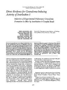
Fig. 1 shows that undialyzed hemolysates, which had been incubated before electrophoresis with their membranes (I-2), contain a sharply localized band of NADH-dichlorophenolindophenol diaphorase (NDD) activity undetectable in control hemolysates incubated without their membranes (1-1). This band, with Rf of 0.75 relative to hemoglobin A, appeared rapidly, reached maximal intensity within 2 hr of staining, was demonstrable by stains containing NADH (Figs. 1-I; 2-I and II), but not by stains containing NADPH (Figs. 1-II, 2-111 and IV), and was not affected by the presence or absence of GSSG (Fig. 2-I and II). In contrast, the slower electrophoretic bands with mean Rf of 0.33 with respect to Hb A (Figs. 1-I and II, 2, 5, 6-I, and 7), stained with NADPH as well as with NADH (Fig. 2) and were markedly intensified in the presence of GSSG (Fig. 1-I and II). Detection of rr
I
, @US*
*
GSSG-R
NDD
t 2
0 2
_4_ _HbA
% 2
2
FIG 1. Formation of NADH-dichlorophenolindophenol diaphorase activated by membranes. Nondialyzed hemolysates were incubated before acrylamide gel electrophoresis (system B) for 1 hr at 45° without (8sots 1) or with (slots 2) their membranes. A portion of the gel (I) was stained for NADH-dichlorophenolin-
dophenol diaphorase activity with dichlorophenolindophenol
and 3(4,5-dimethylthiazolyl-2)-2,5-diphenyltetrazolium bromide and without GSSG; another portion (II) was stained with NADPH and GSSG for NADPH-GSSG-R. HbA, hemoglobin A.
2408
Medical Sciences: Frischer et al.
mii =
JI
I
5
S
~~GSSG-R NDD HbA
_
2
2
1
Proc. Nat. Acadi. Sci. USA 70
2 1 2
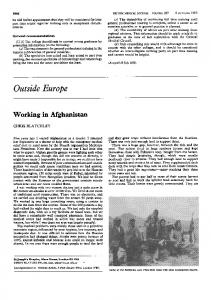
FIG 2. NDD induction in destromatized hemolysates by removal of NAD, suppression by addition of NAD, NADH specificity and differentiation from NAD(P)H GSSG-R. Destromnatized hemolysate was passed through a Sephadex 3-25 column equilibrated and eluted with 1 N NaCl in 157 mM Tris buffer (pH 7.4); slots I were loaded with aliquots of this hemolysate to which no NAD was added and slots 2 with aliquots to which 0.1 mM NAD was added after passage through the column. Acrylamide-grl electrophoresis, system B, was performed with borate in the electrode buffer raised to 5.7 mM. Gel portions I and II were stained for NADH diaphorase without (I) or with (II) GSSG; gel portions III and IV were stained with NADPH instead of NaDH either without (III) or with (IV) GSSG.
these slower bands in dialyzed hemolysates required GSSG in the stain (Figs. 2, 3, and 6-II). Further work with fractions eluted from gels, inhibitors, and activators of GSSG-R and with GSSG-R-deficient hemolysates, confirmed that the slower bands were isozymes of NAD(P)H-GSSG-R appearing as NAD(P)H diaphorases in undialyzed hemolysates. In contrast to these GSSG-R bands, the single faster (Rf 0.75) stromal-induced NDD was present in dialyzed and undialyzed hemolysates incubated with complete membranes as well as in
e~~~~~~~nc IM
3
NDD
_
bA ~~~~~H
W
^
w >_ 2
;lw it.
4
5
6 7
8
9
(1973)
hemolysates dialyzed after incubation with these membranes. Formation of NDD required incubation of hemolysates with their unremoved membranes (Figs. 1-I and 6-I) or with purified autologous or homologous membranes (Figs. 3, 4, and 6-Ij) in some blood samples, NDD could be induced by a membrane-hemolysate contact as short as 15 min at room temperature. Heating the membranes to 80° for 15 min abolished their effect; Tris buffer eluates or Triton X extracts of membranes did not contain NDD, nor could such membrane eluates or extracts induce NDD. Incubation of hemolysates containing membranes with either nicotinamide (Fig. 3) or NAD, but not with NADP or flavin adenine dinucleotide phosphate, prevented NDD formation. Membrane-induced NDD could be abolished by reincubation of destromatized hemolysates with NAD; in contrast, reincubation without additives or with NADP, nicotinamide (Fig. 3)j or flavin adenine dinucleotide phosphate did not remove NDD. NDD was also induced without membranes, by the following treatments designed to remove bound NAD from destromatized hemolysates: (a) incubation with Neurospora crassa NADase (Sigma D9880, 0.25 units ml-' hemolysate; 1 hr at 370; (Fig. 3-2)]; (b) exposure to acid-washed, activated charcoal (Norit: 20 mg ml-' hemolysate; 10 min at 250, Fig. 3-1); (c) dialysis against buffers of high ionic strength or passage through a Sephadex G-25 (Pharmacia) column equilibrated with and eluted by a high-ionic-strength buffer [167 mM Tris HCl, (pH 7.3) with 1 M NaCl; Fig. 2-I and II, slots 11; (d) electrophoresis in continuous Tris - borate buffer systems (Fig. 5-1). NDD induction by membranes or by high-ionic-strength dialysis can be reversed by addition of NAD before electrophoresis (Fig. 2-1 and II, slots 1), and the appearance of NDD after electrophoresis of destromatized hemolysates in Trisborate or other continuous buffer systems was also prevented by the addition of NAD (but not of NADP) to the gel and electrode buffers. Fig. 4 shows that membranes from erythrocytes deficient in NAD (P)-GHase fail to induce NDD in autologous or homologous normal hemolysates, whereas normal membranes can induce NDD in hemolysates from erythro-
10
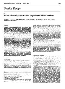
FIG. 3. Competitive inhibition of membrane-induced NDD by 0.1 M nicotinamide, failure of nicotinamide to reverse NDD, and formation of NDD in destromatized hemolysates by exposure to Neurospora NAD-CIHase and to Norit. Destromatized hemolysate, dialysis against 0.15 M Tris buffer (40, pH 7.4). Slots I and 2 were loaded with hemolysates treated with Norit and NeurTspora NAD-GHase, respectively (see text). All other hemolysates were incubated for 1 hr at 450; slot 3, with purified membranes alone; slot 4, without membranes; slot 6, without membranes but with nicotinamide; slots 6 and 7, initial incubation without membranes or nicotinamide then reintubation tion without membranes nor nicotinamide then reincubation (1 hr at 370) with or without nicotinamide, respectively; slots 8 and 9, initial incubation with membranes but without nicotinamide, followed by ghost removal and reincubation (1 hr at 37°) with or without nicotinamide, respectively; slot 10, incubation with membranes and nicotinamide. Acrylamide-gel electrophoresis (system A); NADH diaphorase stain. Destromatized hemolysates treated with Norit (1) or Neurospora NAD-GHase (2) develop NDD. All other hemolysates incubated without membranes lack NDD (4-7); rpembrane-induced NDD (3, 9) is absent if nicotinamide is present during membrane exposure (10); unlike NAD, nicotinamide does not reverse previously formed NDD (8).
e GSSG-R
..,
_P _
2 3 4
5
6 7 8
9
_
_
NDD
N~~bA
10 11 12
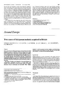
FIG. 4. Membranes from erythrocytes deficient in NAD(P)GHase fail to induce NDD in autologous or homologous hemolysates; normal membranes can induce NDD in
hemolysates from
NAD(P)-GHase-deficient erythrocytes. Nondialyzed, destromatized hemolysates and hemoglobin-free membranes were prepared from blood samples of two subjects with normal erythrocytic NADP-GHase (J:872 units; H:704 units), as well as from erythrocytes deficient in NADI-GHase (A:92 units; see Methods). Four aliquots of hemolysates from each individual were incubated (1 hr at 370) without membranes, with their own membranes, as well as with homologous membranes from each of the two other persons. Slots 1-4, 5-8, and 9-12 contain the hemolysates of J, H, and A, respectively. Acrylamide-gel electrophoresis (system B) was stained for NADH diaphorase without GSSG. No NDD was observed in hemolysates incubated without membranes (1, 5, 9) or incubated with membranes deficient in NAD(P)GHase (3, 7, 1i); all other systems developed NDD.
Proc. Nat. Acad. Sci. USA 70
(1973)
cytes deficient in NAD(P)-GHase. Membranes with normal NAD(P)-GHase activity from erythrocytes deficient in GSSG-R or G6PD, induced NDD in their hemolysates like those of normal erythrocytes. Fig. 6-1 demonstrates that a hemolysate with homozygous NADH methemoglobin diaphorase deficiency contains the slow GSSG-R bands and fails to develop NDD activity when incubated with their membranes; these membranes can induce NDD in normal hemolysates (Fig. 6-II). Fig. 7 indicates that NDD was induced in intact erythrocytes if the cells were exposed to H202, whereas no NDD band was detectable in control erythrocytes incubated in parallel without H202; the induction by H202 of NDD in intact erythrocytes cannot be demonstrated if the cells are NAD (P)-GHase deficient.
DISCUSSION Hemolysates incubated with erythrocytic memebranes acquire a new electrophoretic band with NADH-specific dichloro-
w
I
Membrane NAD(P)ase and Methemoglobin Diaphorase
2409
:II
IO o to HbA*
NDD
6
2 3 4 5
IHbA G)
7
8
9
FIG. 6. Hemolysate with homozygous NADH methemoglobin diaphorase deficiency fails to develop NDD when incubated with its membranes; these membranes can induce NDD in normal hemolysates. In I, nondialyzed hemolysates with normal NADH methemoglobin diaphorase activity (F.A.: 1.4 units; slots 1 and 2), with homozygous NADH methemoglobin diaphorase deficiency (A.C.: 0.1 units; slots 3 and 4), and with decreased NADH methemoglobin diaphorase (G.C., mother of
A.C.: 0.9 units; slots 6 and 6), were incubated (1 hr at 450) without (slots 1, 3, and 5) or with their membranes (slots 2, 4, and 6). Acrylamide-gel electrophoresis (system B with borate raised to 5.7 mM), stain for NADH-diaphorase in the presence of GSSG. NDD, present in the control normal hemolysate incubated with membranes (2), is faint in the membrane-treated, heterozygously deficient hemolysate (6), and absent in the membrane-treated, homozygous deficient blood (4) as in all hemolysates incubated without membranes (1, 3, 5). In II, erythrocytic membranes from A.C. were incubated with destromatized dialyzed hemolysate of normal NADH methemoglobin diaphorase activity (1.8 units, slot 7); this hemolysate was also incubated with (slot 8) and without its own membranes (slot 9). Acrylamide-gel electrophoresis (system B); NADH diaphorase stain without GSSG. Normal hemolysate exposed to membranes from the propositus with NADH methemoglobin diaphorase, develops NDD (slot 7).
phenolindophenol diaphorase activity (Figs 1 and 2). This activity is not eluted from the membranes and requires the
b
2
r' IC) rl LO
2
4
6
8
10 12 14
16 18 20 22 24 26 28 30 Eluted fractions
FIG. 5. NDD corresponds to NADH-methemoglobin diaphorase. Hemolysate proteins were first separated by electrophoresis: 6-mm preparative starch gel (Electrostarch 71 Connaught: 55 g/550 ml of buffer); 0.2 ml of destromatized hemolysate per 1-cm slot; 10-V gradient per 1 cm of gel for 17 hr at 40; continuous Tris-borate buffer system [gel and electrode buffer: 180 mM Tris *HCl-154 mM borate (pH 8.5)]. A strip of this gel, stained for NADH diaphorase, was used to locate the position of NDD (I, first peak of curve 1) and of hemoglobin A (I, second peak of curve 1). The rest of the gel was rotated 900 with respect to the direction of the first electrophoresis. Proteins were eluted by electrodialysis into 31 fractions [elution convection cell, EC, Philadelphia; electrodialysis: 4 hr at 4°; 20 V, 340 mA in 72 mM Tris -HCl-61 mM H3B03 (pH 8.5)]. The cleared gel was stained to document the disappearance of NDD and of hemoglobin (I, curve 2), and all eluted fractions were assayed for NADHmethemoglobin diaphorase (II, curve 1) and for their hemoglobin content, used as an additional position marker (II, curve 2). The only hemolysate fractions capable of reducing ferric hemoglobin with NADH, are located in the NDD area. I, gel scans: 1,-, before elution; 2, after elution. II, fraction assays: 1, NADH methemoglobin diaphorase; 2, hemoglobin.
interaction of a nondialyzable hemolysate component without NDD specificity, with a heat-labile constituent of autologous or homologous membranes. Nicotinamide prevents NDD formation if incubated with membranes and hemolysate; it does not abolish NDD when reincubated with membrane-free hemolysate in which this I
_a
II
-
6
_] &~~~~~SSG-R
NOD
HbA
2 3 4 S 6 7 8 12 3 4 5 6 7 8 FIG. 7. Induction of NDD in intact erythrocytes not deficient in NAD(P)-GHase but not in erythrocytes deficient in NAD(P)GHase exposed to hydrogen peroxide. Washed erythrocytes with normal membranes (I, NADP-GHase 1132 units) and with NAD(P)-GHase deficiency (II, NADP-GHase 21 units), were exposed to H202. Nondialyzed hemolysates were prepared from cells incubated without -aminoacid oxidase or D-alanine (slots 1), with n-alanine but without oxidase (slots 2), and with oxidase but without D-alanine (slots 3). Slots 4, 5, and 6, respectively, were loaded with hemolysates prepared from cells incubated with D-aminoacid oxidase and 1-, 1.5-, or 2-times the standard amount of n-alanine. Control destromatized (slots 7) and nondestromatized (slots 8) hemolysates from each person were incubated without H202 (3 hr at 37°) before electrophoresis. Acrylamide-gel electrophoresis (system B); NADK diaphorase stain.
2410
Medical Sciences: Frischer et al.
activity had been induced by prior exposure to membranes (Fig. 3). Since nicotinamide inhibits human erythrocytic membrane NAD(P)-GHase (6, 13, 14), these observations suggested that membranes acted through their NAD-GHase and/or their NADP-GHase activity. NAD, like nicotinamide, prevents NDD formation if incubated with membranes and hemolysates; unlike nicotinamide, however, reincubation of membrane-free hemolysates with NAD, can also reverse previously induced NDD; NADP neither inhibits nor reverses membrane-induced NDD. Thus, membranes seem active by virtue of their NAD-GHase activity. This suggestion was supported further by the activation of NDD in membranefree hemolysates by procedures designed to remove NAD and by the reversal of their effect with NAD but not with NADP (Figs. 3, 5, and Results). The requirement for membrane NAD-GHase was then established by the search for and detection of persons deficient in erythrocytic membrane NAD(P)-GHase activity (see Methods), and by finding that their membranes could not induce NDD in hemolysates in which this activity could be formed by normal membranes (Fig. 4). Furthermore, NDD can also be induced in intact normal but not in NAD (P)-GHase-deficient erythrocytes exposed to low amounts of H202 generation (Fig. 7). Since NADH methemoglobin diaphorase is often assayed as a dichlorophenolindophenol diaphorase (15), it seemed possible that this enzyme was the nondialyzable hemolysate component altered by NAD-GHase. This possibility was strengthened by finding that the only protein with NADH methemoglobin diaphorase activity (with ferric hemoglobin as substrate) elutable from preparative gels was located at the position of NDD (Fig. 5). This conclusion was then established when hemolysates with homozygous deficiency of NADH methemoglobin diaphorase, exposed to their membranes, failed to develop NDD, although these membranes could induce NDD in control hemolysates (Fig. 6). These studies thus demonstrate a reversible modification of NADH ferrihemoglobin diaphorase by NAD removal; this modification can be mediated in vitro by the NAD-GHase activity of human erythrocytic membranes, in intact cells stressed by exposure to hydrogen peroxide, as well as in hemolysates. The origin and mechanism of some previously reported NADH methemoglobin diaphorase "aging variants" (16-19) may now also be explained. Electrophoresis of NADH methemoglobin diaphorase reveals an NAD-stripped form of the native enzyme and requires control of exposure to membranes; furthermore interpretation of such studies for genetic analysis must also take into account that presently used methods for phenotyping NAD(P)H methemoglobin diaphorase can include NAD(P)H GSSG-R isozymes even when GSSG is omitted from the stain; GSSG-R appears then in undialyzed hemolysates as an NAD(P)H diaphorase, and its principal isozyme should not be misinterpreted as a slower NADH methemoglobin diaphorase band (and/or as a slow NADPH methemoglobin reductase band). Studies of NAD-GHase and NADP-GHase activities in membranes and in intact cells necessary to establish the nature of the membrane component interacting with methemoglobin diaphorase have confirmed the common occurrence of erythrocytic NAD-GHase deficiency in Afro-American adults
Proc. Nat. Acad. Sci. USA 70
(1973)
(12) and revealed that they are also deficient in erythrocytic NADP-GHase. Furthermore, erythrocytic NAD-GHase and NADP-GHase activities are highly correlated in a given individual, and isolated erythrocytic NAD-GHase or NADPGHase deficiency has not been detected. These findings strongly suggest, in contrast to previous reports (20, 21), that a single membrane enzyme determines the activities of both NADP-GHase and NAD-GHase. The physiological role of membrane NAD(P)-GHase and the reason for the high frequency of NAD(P)-GHase deficiency in Afro-Americans remain unknown. Further studies are thus necessary to determine whether erythrocytic NAD(P)-GHase deficiency can be associated, in vivo and under appropriate stress, with metabolic disturbances mediated by alterations of enzymes affected in vitro by NAD (P)-GHase activity (14, 6, 3). We thank Deatra Kinney, Edward C. Patterson, Sharon L. Ziegler, and Tanveer Ahmad for excellent technical assistance. This work was supported by U.S. Public Health Service Grant no. 7-RO 1 HE 14859, by U.S. Army Contract DADA-17-71-C1103, by U.S. Navy Contract NOOO 14-72-A-0288, and by a grant under the Mutual USA-Italy Educational and Cultural Exchange Act. 1. Frischer, H., Carson, P. E., Bowman, J. E. & Rieckmann, K. H. (1973) J. Lab. Clin. Med. 81, 603-612. 2. Frischer, H., Bowman, J. E., Carson, P. E., Rieckmann, K. H., Willerson, W. D. & Colwell, E. (1973) J. Lab. Clin. Med. 81,613-624. 3. Frischer, H., Noyes, C. & Nelson, R. (1971) J. Clin. Invest. 50, 34 abstr. 4. Frischer, H., Noyes, C., Nelson, R. & Kinney, D. (1971) Clin. Res. 19, 418. 5. Dodge, J. T., Mitchell, C. & Hanahan, D. J. (1963) Arch. Biochem. Biophys. 100, 119-130. 6. Ajmar, R., Scharrer, B., Hashimoto, F. & Carson, P. E. (1968) Proc. Nat. Acad. Sci. USA 59, 538-545. 7. Frischer, H., McNamara, J., Rieckmann, K. H., Stockert, T., Powell, R. & Carson, P. E. (1968) Clin. Res. 16, 303. 8. Hegesh, E., Calmanovia, N. & Avron, M. (1968) J. Lab. Clin. Med. 72, 339-344. 9. Glock, G. E. & McLean, P. (1953) Biochem. J. 55, 400-408. 10. Long, W. K. & Carson, P. E. (1961) Biochem. Biophys. Res. Commun. 5, 394-399. 11. Bergren, W. R., Ng, W. G. & Donnell, G. N. (1967) Pediatrics 40, 136-137. 12. Ng, W., Donnell, G. N. & Bergren, R. (1968) Nature 217, 64-65. 13. Webb, L. J. (1966) in Enzyme and Metabolic Inhibitors II (Academic Press New York), pp. 485-500. 14. Carson, P. E., Okita, G. T., Frischer, H., Hirasa, J., Long, W. K. & Brewer, G. J. (1963) in Proceedings of the Ninth Congress of European Societies of Hematology, Lisbon (S. Karger, Basel/New York), pp. 655-665. 15. Scott, E. M. & Griffith, I. V. (1959) Biochim. Biophys. Acta 34,584-586. 16. Brewer, G. J., Eaton, J. W., Knutsen, C. S. & Beck, C. C. (1967) Biochem. Biophys. Res. Commun. 29, 198-204. 17. Bloom, G. E. & Zarkowsky, H. S. (1969) N. Engl. J. Med. 281, 919-922. 18. Detter, J. C., Anderson, J. E. & Giblett, E. R. (1970) Amer. J. Human Genet. 22, 100-104. 19. Kaplan, J. C. (1971) in Red Cell Structure and Metabolism, ed. Ramot, B. (Academic Press, New York), pp. 125-133. 20. Gasiorowska, I. & Raczynska-Bojanowska, K. (1963) Bull. Acad. Pol. Sci. 11, 417-421. 21. Hofmann, E. & Noll, F. (1961) Acta Bid1. Med. Ger. 6, 1-6.