require the ability to distinguish live from dead nematodes. Vermiform-stage mobil- ity, spontaneous or induced, is an obvious indicator of viability. The lack of ...
Journal of Nematology 21(3):399-403. 1989. © T h e Society of Nematologists 1989.
Nematode Autofluorescence and Its Use as an Indicator of Viability T. A. FORGE AND A. E. M A C G U I D W I N 1 Abstract: Representatives of 15 nematode genera were viewed with 4 5 0 - 4 9 0 - n m epi-illumination and found to autofluoresce. T h e autofluorescence was limited to 1-5-#m-d globules in the intestinal cells of live nematodes. W h e n adult Pratylenchus penetrans or Caenorhabditis elegans were killed with formaldehyde, freezing, or heat, autofluorescence dispersed t h r o u g h o u t the body. Mixed stages of P. penetrans were killed by freezing at several different temperatures. Estimates of survival based on autofluorescence dispersal matched estimates based on mobility more closely than did estimates based on the vital stain, eosin-y. Key words: autofluorescence, lipofuscin, nematode viability, secondary lysosome.
Many areas of nematological research require the ability to distinguish live from dead nematodes. Vermiform-stage mobility, spontaneous or induced, is an obvious indicator of viability. T h e lack of mobility, however, does not indicate that a nematode is dead. This distinction is especially important in research on population dynamics and pesticide efficacy. In the first case, seasonal changes in nematode physiology such as extreme quiescence or diapause may drastically affect the ability to move. In the case of pesticides, especially those that are acetylcholinesterase inhibitors, sublethal doses may temporarily affect mobility (11). Stains have been used with variable success to distinguish live from dead nematodes (1,2,6,8,9,12-14). Stains work only if the event inducing death results in cuticle permeability (2,8,12). Consequently, the reliability of any particular stain is affected by the event inducing mortality, the harshness of that event, and the length of time between death and staining. Since it is dependent on cuticle permeability, the reliability of a stain can be expected to vary among species and among life stages within a species. Nematodes accumulate the fluorescent compound lipofuscin (5,10) and are therefore autofluorescent. Clokey and Jacobson (3) identified autofluorescent globules in Received for publication 8 November 1988. t Graduate student and Assistant Professor, Department of Plant Pathology, University of Wisconsin, Madison, W153706.
the intestinal cells of Caenorhabditis elegans as secondary lysosomes. We observed that the pattern of autofluorescence in dead nematodes appeared different from the pattern in live nematodes (7). Bird (1) stained live and dead nematodes with fluorescein diacetate and found fluorescence limited to globules in live nematodes and dispersed in dead nematodes. T h e fluorescence was attributed to hydrolysis of fluorescein diacetate by esterases, which dispersed upon nematode death. Davis et al. (4) treated Meloidogyne spp. juveniles with fluorochrome-lectin conjugates and found fluorescence in the lip region of live specimens and throughout the body of dead specimens. T h e purpose of this research was to determine if the dispersal of autofluorescence is a reliable indicator of death in nematodes. MATERIALS AND METHODS
Representatives of 15 genera (Aphelenchus, Caenorhabditis, Criconemella, Ditylenchus, Helicotylenchus, Heterodera, Hoplolaimus, Meloidogyne, Pratylenchus, Rotylenchus, Longidorus, Trichodorus, Tylenchus, Mononchus, and Rhabditis) were viewed with a c o m p o u n d m i c r o s c o p e (Zeiss S t a n d a r d GFL) equipped for epi-illumination. Only second-stage juveniles of Heterodera and Meloidogyne were observed. T h e light source and filter system consisted of a 50-watt mercury vapor bulb, band pass filter for 450-490-nm excitation, F T 5 1 0 chromatic beam splitter, and barrier filter for 520 nm 399
400 Journal of NematoIogy, Volume 21, No. 3, July 1989 and above. In addition to the 450-490-nm excitation, several specimens of C. elegans were viewed with a filter system that resulted in 365-nm peak excitation. Nematodes were photographed with black and white film (Kodak, ASA 400) and an exposure time of 5 seconds. To determine if autofluorescence dispersed when nematodes were killed, adult female Pratylenchus penetrans and adult hermaphrodites of C. elegans were hand picked into 0.5-ml microcentrifuge tubes containing 0.2 ml tap water. Ten replicate tubes containing 30 P. penetrans and six tubes containing 100 C. elegans were administered one of three lethal treatments: 1) 60 C for 75 seconds, 2) - 2 0 C for 12 hours, or 3) 0.2 ml of 10% formalin, resuiting in a 5% final concentration. T h e formalin was removed after 30 minutes by centrifuging, removing the supernatant, and resuspending in tap water. Controls were left at room temperature (ca. 24 C) for 12 hours. Twenty-four hours after the completion of each treatment, nematodes were viewed at 100x using epi-illumination. T h e percentage of nematodes with dispersed autofluorescence was recorded. Estimates of survival based on dispersed autofluorescence of mixed stages of P. penetrans frozen at three different temperatures were compared with estimates based on mobility and staining wit h eosin-y. In order to start the experiment with 100% live specimens, the nematodes were twice required to move through tissue on a Baermann funnel; survivors were then suspended to a density of approximately 250 individuals/ml tap water. One milliliter of the suspension was pipetted into each of 36 test tubes and the tubes were divided into four groups of nine. T h r e e groups were frozen at - 2 , - 4 , or - 8 C by placing each in a refrigerated ethylene glycol bath at one of the temperatures and adding a small ice crystal to each tube to initiate freezing. The fourth group served as unfrozen controls. After 24 hours at - 2 , - 4 , or - 8 C, the samples were placed at room temperature
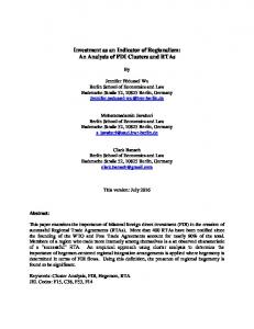
and 1.0 ml of 1.0% eosin-Y was added to three samples from each group. After an additional 24 hours, the stain was removed by twice adding 5.0 ml tap water, allowing the nematodes to settle, and then removing excess solution. Each sample was then viewed with a stereo microscope at 7 0 x , and the percentage of each stage taking up stain was determined. On the same day, three of the unstained samples from each group were viewed with a stereo microscope at 70 x, and the percentage of each stage not exhibiting any mobility was determined. This included not only spontaneous mobility, but any movement detected after the nematodes were tapped several times near the nerve ring with a fiberglass pick. T h e remaining three samples from each group were viewed with epi-illumination at 100 x, and the percentage of each stage exhibiting dispersed autofluorescence was recorded. The data for each stage were analyzed separately as a split-plot design, with freezing temperature as the main factor and assay type as the subfactor. Least significant differences were used for comparison of means. RESULTS All nematodes viewed with 450-490-nm epi-illumination e x h i b i t e d autofluorescence in the form of globules in the intestinal region (Fig. 1). Spicules and the lip region also fluoresced. T h e color, intensity, and quantity of autofluorescent globules differed slightly between genera as well as between individuals of a given species. T h e globules ranged in color from pale yellow to green. In C. elegans, the globules ranged in diameter from ca. 1 ~,m to 5 t~m and fluoresced blue when viewed at 365nm peak epi-illumination. Autofluorescent globules were faint in second-stage juveniles of Pratylenchus, Meloidogyne, and Het-
erodera. Regardless of how adult P. penetrans females or C. elegans hermaphrodites were killed, autofluorescence was dispersed in at least 97% of the individuals (Table 1). T h e
Nematode Autofluorescence: Forge, MacGuidwin 401
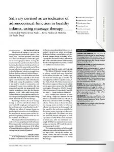
Fro. 1. Autofluorescence at junction of esophagus and intestine of an adult female Pratylenchus penetrans viewed with 450-490-nm epi-illumination. A) Alive. B) Immediately after freezing at - 2 0 C for 30 minutes and thawing.
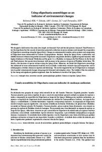
autofluorescence was dispersed in less than 2% of untreated controls. On the basis of lack of mobility, individuals with dispersed autofluorescence were classified as dead. Estimates of survival based on autofluorescence were significantly lower (P = 0.05) than estimates based on staining for every combination of stage and freezing temperature except third-stage juveniles frozen at - 2 C (Fig. 2). Estimates of survival based on autofluorescence were significantly higher than estimates based on mobility for second-stage juveniles frozen at all three temperatures, fourth-stage juveniles frozen at - 4 C, and females frozen at - 4 C. For all but the second juvenile stage, there was a significant (P < 0.01) interaction between the type of viability assay and temperature of freezing. For all three methods of assessing survival, the estimates for unfrozen controls were always 100%.
DISCUSSION
At 365-nm peak epi-illumination, the size, distribution, and color of autofluorescent globules in C. elegans match those described by Clokey and Jacobson as secondary lysosomes (3). T h e globules observed at 365-nm peak epi-illumination are the same size as those observed using 4 5 0 490-nm epi-illumination. That all nematodes viewed so far exhibit TABLE 1. Percentage of female Pratylenchus penetrans (Pp) and hermaphrodites of Caenorhabditis elegans (Ce) with dispersed autofluorescence after lethal treatments. Treatment
Pp
Ce
Freezing Formaldehyde Heat Controls
99.6 98.9 97.4 0,4
97.7 99.2 100.0 1.2
402 Journal of Nematology, Volume 21, No. 3, July 1989
I00. Second-stage
"6 "~
lOO:
Juveniles
80,
ao4
80.
8o.
40.
40-
20.
20-
AdultFemales
m •
100.
-i
-~
-i
°~'
-2
j
s°dj
80,
-g
-i
-a
AdultMales =
,°]
"5
~
-~
100 .
4o.1
40,
1
ao.l
2°.
//
0,
100,
:IV
g
.
--8
.
.
.
--6
H
~r,.or.
N
"==.=V
--4
--2
F o u r t h - s t a g e Juveniles
"6 .>
--8
--6
Temperature
--4
--2
(C)
80,
i:
eo.
W
40,
g~
j,~:
20. / .,./ r /
/
J
-
,-, ,=~g
/
H H Temperature
o~to~or.
mebmL 7
(C)
FIe. 2. Relationship between temperature of freezing and estimates of survival (+ SE) based on staining, autofluorescence, and mobility for each stage of Pratylenchuspenetrans.
autofluorescence is not surprising, since the a c c u m u l a t i o n o f lipofuscin is k n o w n to occur in m a n y cell types t h r o u g h o u t the m e t a z o a (3,5). Lipofuscin a c c u m u l a t i o n is related to age (3,5,10), which may explain why the juveniles do not fluoresce as intensely as o l d e r stages o f the same species. T h e association b e t w e e n d e a t h and dispersal o f autofluorescences suggests t h a t d e a t h may result in the b r e a k d o w n o f lysosomal m e m b r a n e s , allowing the lipofuscin to disperse. Death may also result in the dispersal or activation o f enzymes or
o t h e r molecules that cause biochemical reactions leading to autofluorescence in areas besides the secondary lysosomes. In addition to showing that a m e t h o d o f estimating survival works when the nematodes have b e e n killed by at least several different events, it is also informative to d e t e r m i n e how the m e t h o d c o m p a r e s with o t h e r m e t h o d s across several levels o f survival. Estimates o f survival based on autofluorescence dispersal paralleled estimates based on mobility m o r e closely than th~sse based on staining. / //
Nematode Autofluorescence: Forge, MacGuidwin 403 No mobile nematodes have been observed with dispersed autofluorescence. Since mobility is an absolute indicator of viability, autofluorescence does not err by categorizing live nematodes as dead. If and when mobility errs as a basis for estimating survival, it is because live nematodes are categorized as dead. It is likely that autofluorescence can distinguish between living nonmobile and dead nematodes. Nonmobile nematodes have been observed that do not exhibit dispersed autofluorescence. This may explain why estimates of survival based on autofluorescence were slightly higher than those based on mobility. Nonetheless, under the conditions of this experiment, mobility should provide highly accurate estimates of survival. Although low temperatures result in reduced metabolic rates and mobility (i.e., quiescence), We have found that mobility ofP. penetrans resumes within an hour of returning to room temperature (Forge, unpubl.). In other words, there is no evidence for a deep quiescence induced by short-term exposure to subzero temperatures. All other factors affecting the accuracy of estimates based on mobility should be reflected in the unfrozen controls, which always had values of 100% survival. Eosin-y was chosen for this comparison because it was previously reported to be effective in staining free living nematodes killed by freezing (2). If the accuracy of autofluorescence or mobility is accepted, then it is clear that eosin-y is not effective in staining P. penetrans killed by freezing. T h e discrepancy between survival estimates based on staining and estimates based on autofluorescence or mobility was least for second-stage juveniles and greatest for the adult stages. This phenomenon may be due to cuticle differences between stages, because staining efficacy is dependent on cuticle permeability. We have shown that a wide variety of nematodes are autofluorescent and that the autofluorescence is consistently dispersed in P. penetrans or C. elegans killed by formaldehyde, freezing, or heat. In addition, we have shown that estimates of survival based
on autofluorescence closely parallel estimates based on mobility over a range of survival levels. Although mobility is an indisputable indicator of viability, it must often be induced by prodding nematodes and can be time consuming. Dispersed autofluorescence is a reliable indicator of nematode mortality which can be quickly and easily determined. LITERATURE CITED 1. Bird, A. F. 1979. A method of distinguishing between living and dead nematodes by enzymatically induced fluorescence. Journal of Nematology 11:103105. 2. Chadhouri, N., R. I. Dick, R. S. Englebrecht, and J. H. Austin. 1966. Staining of free-living nematodes by eosin-y dye. Nematologica 12:337-342. 3. Clokey, G. V., and L. A. Jacobson. 1986. The autofluorescent "lipofuscin granules" in the intestinal cells ofCaenorhabditis elegans are secondary lysosomes. Mechanisms of Ageing and Development 35:79-94. 4. Davis, E. L., D. T. Kaplan, T. A. Permar, D. W. Dickson, and D.J. Mitchell. 1988. Characterization of carbohydrates on the surface of second-stage juveniles ofMeloidogyne spp. Journal of Nematology 20: 609-619. 5. Davis, B. O., Jr., G. L. Anderson, and D. B. Dusenbury. 1982. Total luminescence spectroscopy of fluorescence changes during aging in Caenorhabditis elegans. Biochemistry 21:4089-4095. 6. Fenner, L. M. 1962. Determination of nematode mortality. Plant Disease Reporter 46:383. 7. Forge, T. A., and A. E. MacGuidwin. 1986. Autofluorescence in nematodes and its use as an assay of viability. Journal of Nematology 18:637 (Abstr.). 8. Hollis, J. P. 1961. Nematode reactions to coal tar dyes. Nematologica 6:315-325. 9. Jatala, P. 1975. Efficiency of potassium permanganate in differentiating between live and dead nematodes. Annals of Applied Biology 80:109-113. 10. Klass, M. R. 1977. Aging in the nematode Caenorhabditis elegans: Major biological and environmental factors influencing life span. Mechanisms of Ageing and Development 6:413-429. 11. Marban-Mendoza, N., and D. R. Viglierchio. 1980. Behavioral effects of carbofuran and phenamiphos on Pratylenchus vulnus I. Motility and dispersion. Journal of Nematology 12:102-114. 12. Morlarty, F. 1961. The efficacy ofchrysoidin, new blue R and phloxine B for determining the viability of beet eelworm, Heterodera schachtii. Nematologica 10:644-646. 13. Ogiga, I. R., and R. H. Estey. 1974. The use of meldola blue and nile blue A for distinguishing dead from living nematodes. Nematologica 20:271276. 14. Shepard, A. M. 1962. New blue R, a stain that differentiates between living and dead nematodes. Nematologica 8:201-207.