bioRxiv preprint first posted online Aug. 26, 2018; doi: http://dx.doi.org/10.1101/394403. The copyright holder for this preprint (which was not peer-reviewed) is the author/funder, who has granted bioRxiv a license to display the preprint in perpetuity. It is made available under a CC-BY-NC 4.0 International license.
Huoviala et al., 26 August 2018 – BioRxiv preprint
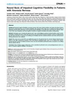
Neural circuit basis of aversive odour processing in Drosophila from sensory input to descending output Paavo Huoviala1,2 , Michael-John Dolan1,3,5, Fiona M. Love2,5, Shahar Frechter1, Ruairí J.V. Roberts2, Zane Mitrevica2, Philipp Schlegel2 , Alexander Shakeel Bates1, Yoshinori Aso3, Tiago Rodrigues1, Hannah Cornwall1, Marcus Stensmyr4, Davi Bock3, Gerald M. Rubin3, Marta Costa2, Gregory S.X.E. Jefferis1,2 1
Division of Neurobiology, MRC Laboratory of Molecular Biology, Cambridge CB2 0QH, UK Department of Zoology, University of Cambridge, CB2 3EJ, UK 3 Janelia Research Campus, Howard Hughes Medical Institute, United States 4 Department of Biology, Lund University, 22100 Lund, Sweden 5 Equal Contributions Contact:
[email protected] 2
https://doi.org/10.1101/394403 Abstract Evolution has tuned the nervous system of most animals to produce stereotyped behavioural responses to ethologically relevant stimuli. For example, female Drosophila avoid laying eggs in the presence of geosmin, an odorant produced by toxic moulds. Using this system, we now identify third order olfactory neurons that are essential for an innate aversive behaviour. Connectomics data place these neurons in the context of a complete synaptic circuit from sensory input to descending output. We find multiple levels of valence-specific convergence, including a novel form of axo-axonic input onto second order neurons conveying another danger signal, the pheromone of parasitoid wasps. However we also observe a massive divergence as geosmin-responsive second order olfactory neurons connect with a diverse array of ~75 cell types. Our data suggest a transition from a labelled line organisation in the periphery to one in which olfactory information is mapped onto many different higher order populations with distinct behavioural significance. Keywords: Neural circuits, Innate Behaviour, Olfaction, Valence, Connectomics Introduction Studying the neural circuit basis of innate behaviours in simple model organisms provides an ideal platform to discover how sensory signals are transformed into behavioural responses by the nervous system, arguably one of the fundamental goals of neuroscience. The olfactory system is a particularly shallow sensory modality where the sensory periphery is only two synapses away from higher brain areas important for organising behaviour and forming memories. In both insects and mammals, after processing in the first olfactory centre, divergent projections relay information to different higher olfactory centres, some of which appear to be specialised for innate behaviour and others for learning and memory [1–4]. The periphery of the Drosophila olfactory system consists of ~1300 sensory neurons (ORNs) of ~50 distinct types, on each side [5,6]. Each type, with a few exceptions, expresses only a single olfactory receptor that determines the neurons’ response profile and characteristics [6–8]. Many receptors are quite broadly tuned, and for this reason, odours are typically encoded in the combinatorial activity of this parallel array of odour channels. However in certain cases of odours of particular ethological relevance may be encoded in the activity of just one odour channel. This situation of functionally segregated pathways, is sometimes referred to as labelled line encoding [9–12]. The ORNs send their axons into the antennal lobe (AL) in the brain, where the ORNs expressing the same receptor converge onto just 1-10, presumably identical, second order projection neurons (PNs) and local neurons in ~50 distinct glomeruli [13,14]. From the AL the axons of the PNs then
go on to project to two higher olfactory processing centres: the mushroom body (MB) and the lateral horn (LH). The MB is crucial for associative learning, and key advances have been made in recent years regarding its circuit architecture and function, and the writing and retrieval of memories of different valence [15–18]. However, our understanding of the LH remains considerably more limited. The LH is thought to be important for innate odour responses [19], the PN axonal arbours in the LH have been shown to be hardwired and stereotyped across animals, and the general architecture of the brain area has been proposed to be organised by behavioural relevance of the afferent odour channels [20], thus demonstrating some kind of behaviourally defined topography. However, to date, no direct evidence for the role of lateral horn neurons (LHNs) in innate odourguided behaviours has been published. Furthermore, only the pheromonal cVA-processing circuit has been traced until third order neurons of the lateral horn [21–23]. The study of labelled line encoding potentially has significant experimental advantages for linking olfactory circuits to behaviour. Indeed an ideal approach would be to trace labelled line odour channels of varying and well understood behavioural significance to the LH in order to test if LHNs are required for innate odour-guided behaviours, and also sufficient to trigger them when activated. Such an approach might also reveal if the general organisation of the LH is indeed defined by behavioural relevance, so that odour channels containing information of similar meaning to the animal synapse onto the same LHNs, the activity of which would then trigger an appropriate behavioural response. A specific opportunity to achieve this is offered by studying
1
bioRxiv preprint first posted online Aug. 26, 2018; doi: http://dx.doi.org/10.1101/394403. The copyright holder for this preprint (which was not peer-reviewed) is the author/funder, who has granted bioRxiv a license to display the preprint in perpetuity. It is made available under a CC-BY-NC 4.0 International license.
Huoviala et al., 26 August 2018 – BioRxiv preprint
Figure 1: DA2 PNs labelled by R85E04-GAL4 are excitatory PNs that respond to geosmin and are required for geosmin avoidance behaviour. (A) Egg-laying two-choice preference indices (PI) to geosmin for wild type (Canton S) flies, anosmic mutants, and flies with silenced geosmin responsive sensory neurons (Or56a ORNs). The chemical structure for geosmin marks the stimulus side. Groups are compared by a Kruskal-Wallis rank sum test followed by planned comparisons of wild type group to the other groups by Wilcoxon rank sum tests with Holm-Bonferroni corrections. n=27 (left), n=22 (middle), and n=17 (right). (B) Eggs laid per wild type fly under geosmin or solvent exposure (Welch’s Two Sample t-test. n=12, n=11). (C) Expression pattern of R85E04, a GAL4 line that labels 5-6 DA2 PNs. A JFRC2 registered maximum intensity projection of a female brain, raw image data from Flylight database (http://flweb.janelia.org/cgi-bin/flew.cgi) [28]. Green channel is GFP and magenta, nc82 neuropil stain. (D) Raster plot of in vivo patch clamp recordings from DA2 PNs (R85E04-GAL4) to a panel of odorants [44]. Odour valve opening denoted by pale red bar, timebase ms. (E) Egg-laying two-choice preference indices (PI) to geosmin while silencing putative geosmin sensing PNs by expressing tetanus toxin light-chain (TNT) [30] via R85E04 (groups compared by a Kruskal-Wallis rank sum test followed by post-hoc comparisons of experimental group to each of the parental groups by Wilcoxon rank sum tests with Holm-Bonferroni corrections. n=38 for all groups). (F) Immunostainings against GFP, ChAT, and GABA in R85E04-GAL4, and the merge of the three. Single slices of confocal stacks showing the AL (magenta counterstain) and PN cell bodies (arrowheads). Significance values: * p10 synapses. Interestingly, among these 5
7
bioRxiv preprint first posted online Aug. 26, 2018; doi: http://dx.doi.org/10.1101/394403. The copyright holder for this preprint (which was not peer-reviewed) is the author/funder, who has granted bioRxiv a license to display the preprint in perpetuity. It is made available under a CC-BY-NC 4.0 International license.
Huoviala et al., 26 August 2018 – BioRxiv preprint
A
A’
A’’
B
B’
B’’
A’’‘’
A’’’ B’’’
B’’’’
C Normalized input strength
AD1a3 Aspis PV6a3 Caligari
ADLI Lazarus AV1a1 Duck
AV1a1 Pigeon
*
***
*
***
*
*
*
*
Figure 5: Some DA2 downstream targets receive input from multiple aversive PN channels. (A-A’’’’) Morphologies of five DA2 downstream targets of special interest. (A) Aspis (n synapses from DA2 PNs=108). (A’) Caligari (n=68). (A’’) Lazarus (n=31). (A’’’) Duck (n=19). (A’’’’) Pigeon (n=7). Cell bodies and primary neurites are coloured red, dendrites green, and axons blue, where such a division was possible. (B-B’’’’) Number of synaptic inputs onto Aspis (B) Caligari (B’), Lazarus (B’’), Duck (B’’’), and Pigeon (B’’’’) by PN type. (C) Heatmap representation of PN to selected LHN connectivity, normalised by the total PN inputs to each neuron, and colour-coded by putative behavioural relevance [12,33,35,36,51,52,54]. Odour channels implicated in aversive behaviour, specifically in an egg-laying context, are marked with asterisks [12,33,38,58].
8
bioRxiv preprint first posted online Aug. 26, 2018; doi: http://dx.doi.org/10.1101/394403. The copyright holder for this preprint (which was not peer-reviewed) is the author/funder, who has granted bioRxiv a license to display the preprint in perpetuity. It is made available under a CC-BY-NC 4.0 International license.
Huoviala et al., 26 August 2018 – BioRxiv preprint
there are several PN classes that are thought to carry aversive odour information: DC4 [52], DC2 [35,51], and DL4 (parasitic wasp pheromones [33]). Thus, this LHON appears to be an ideal candidate to perform valence-based integration, a computation we hypothesised might take place in the LH. Unfortunately, the neuron does not morphologically match any previously described neurons, and we were unable to find a driver line targeting it. Thirdly, a large, brain spanning neuron Lazarus receives strong bilateral synaptic input from DA2 PNs in the LH (right side=31, left side= 19, ~0.5% of DA2 synapses in the LH), and sends bilateral projections to the calyx of the MB, as well as PLP, SLP, AVLP, SCL, inferior clamp, and superior medial protocerebrum (fig 5A’’). Strikingly, DA2 PNs are its sole source of uniglomerular PN input in the LH (fig 5B’’&C). An NBLAST [32] search against the FlyCircuit database [31] found a good match for the neuron (fig S2F). Based on the morphology and the presence of neuropeptidergic dense core vesicles (fig S2G), Lazarus is likely to be the ADLI neuron described in [53], which expresses natalisin, a neuropeptide widely conserved in arthropods, potentially involved in reproductive behaviours. Finally, we identified two type AV1a1 [44] LHONs: Duck and Pigeon (fig 5A’’’&A’’’’). These LHONs receive strong (Duck) to moderate (Pigeon) input from both DA2 and DL4 PNs (fig 5B’’’&B’’’’). For Duck, we found 19 synaptic connections from DA2, and 51 from the single DL4 PN (fig 5B’’’), making it the second strongest downstream target of DL4 PNs. Pigeon received 7 synaptic connections from both DA2 and DL4 PNs (fig 5B’’’’). Combined, these neurons receive ~0.4% of the DA2 synapses in the LH. Although, as mentioned above, DA2 PNs also synapse onto DL4 PNs, putatively providing a somewhat higher level of input to these neurons than what is suggested by the number of synapses from DA2 PNs alone. However, AV1a1 neurons receive a relatively broad set of PN inputs (fig 5B’’’-B’’’’ & fig 5C) including the supposedly aversive (DL5, DC2, D [35,51]), food [51,54] and even pheromonal inputs (VA1v [55]) (although notably, activating the VA1v pathway in isolation was recently shown to be strongly aversive in an egg-laying context [38]). Interestingly, many of the strongly connected food related PNs (VC5,VL2a, VL2p, VM1) receive sensory information via the family of ionotropic IR receptors [56]. These mainly respond to acids and polyamines [57]. The latter have been shown to be attractive in a T-maze but aversive in an egg-laying context (although this may involve the gustatory system) [58]. While the AV1a1 neurons were not the strongest targets of DA2 PNs, nor as selective to it as some of the other LHNs, there are nevertheless a few factors that make them particularly interesting. Firstly, the same neuron type (AV1a1) came up in our sampling twice (Duck and Pigeon). Secondly, morphologically similar neurons have been suggested to be important for aversive odour processing based on functional imaging experiments performed on a relatively broad driver line [59]. Thirdly, the dendrites of the neurons arborise in the ventralposterior LH where most aversive PNs send their axonal projections. A recent paper, using a novel olfactogenetic approach suggests that this area of the LH may mediate negative egg-laying decisions similar to the avoidance of geosmin [38]. A statistical cross-comparison of the AV1a1 inputs and the odour channels recently reported to be most aversive in an egg-laying context [38] shows that both neurons receive more inputs from aversive channels than would be expected by chance: Duck receives strong (>10 synapses) input from 3/5 of the most aversive PNs (VA1v, DL4, DL5 but not VC2, VA3), and only 3/19 from the rest (Chi-squared test p=0.04); For Pigeon the numbers are 2/5 (VA1v, DL5,) and 1/19, respectively (Chi-squared test p=0.04). Thus, in summary, despite the considerable divergence of DA2 onto many LHN targets, we find neurons that receive inputs from multiple aversive PN channels, and could in principle act as valence based
integrators for behavioural aversion either in an egg-laying context or more generally (see fig 5C for overview). Light-level in silico anatomical screen for DA2 downstream targets finds cell type-specific driver lines For our parallel light-level screening for DA2 downstream neurons and driver lines, we used registered confocal stacks of R85E04 converted into a binary mask showing the axonal projections pattern of the PNs in the LH (fig 6A). This mask was then co-visualised together with Janelia FlyLight GAL4 lines [28] that had been annotated to have expression in the LH (a total of 351 GMR lines annotated by Frechter et al. [44]) (see fig 6B-C for an example). The GMR lines were then manually scored for amount of overlap (reflecting the a priori probability of synaptic connectivity), and sparseness of expression pattern (allowing better identification of LHN cell type and more specific targeting of LHN types). For most of the best candidate LHN types we were also able to identify a Split-GAL4 line from a collection being made as a part of an ongoing effort to create a comprehensive cell type-specific driver line library for LHNs (Dolan et al., in preparation). Overall, the in silico screen narrowed down the number of putative DA2 downstream targets to 18 LHN types, 12 of which could be accessed relatively specifically through either GAL4 or Split-GAL4 lines (details will be described in Dolan et al., in preparation). AV1a1 LHNs found in LH728 and LH1983 trigger aversion and are necessary for geosmin avoidance in egg-laying To look for behaviourally relevant LHNs potentially downstream of DA2 PNs, we next carried out a behavioural optogenetic activation screen on the hits of the in silico anatomical screen for aversion triggering LHNs. As the Split-GAL4 lines in most cases had stronger expression levels as well as less off-target expression, they were used instead of the GMR GAL4 lines where possible. A total of 27 driver lines (for 12 LHN types) were tested (the full details of the screen in Dolan et al., in preparation). The behavioural experiments were carried out similarly to [17], using a four-field arena (fig 6D), and only female flies were used for the screen. However, only two of the tested driver lines that had LHNs putatively downstream of DA2 PNs triggered aversion in this assay (in comparison to the mildly phototactic genetic control Empty-Split GAL4) (fig 6E, S3A-C): LH728 and LH1983 (fig 6F-G). Both Split-GAL4 lines shared a parent line (R22D02, fig 6B) and appear to label a small group of neurons with their somas ventral and medial to the ventrolateral protocerebrum (AVLP). Both lines appear to contain the same two strongly labelled neurons sending dendrites to the (ventral) LH and axons to the AVLP, and no other LHNs. These LHNs belong to the neuron type AV1a1 according to a recent LHN annotations [44], are cholinergic (Dolan et al., in preparation), and appear morphologically very similar to the AV1a1 neurons Duck and Pigeon found downstream of DA2 PNs in the EM sample (fig 6H). To quantitatively match the neurons labelled by LH728 and LH1983 to the EM tracings we generated light-level tracings of the neurons in LH1983. LH1983 was used for this as the signal to noise ratio of the line was deemed better (see fig 6F-G). As the processes of the two LHNs were in many places too close to resolve, the tracing resulted in a single hybrid skeleton of the two AV1a1 neurons found in the line. However, overlaying the light-level tracing (black) with Duck and Pigeon (green, magenta) reveals remarkably similar morphology (fig 6I). Moreover, a quantitative comparison of morphology between the light-level tracing and all the 33 neurons taking the AV1 tract using the NBLAST algorithm [32] shows that the top matches are Duck (NBLAST similarity score=0.69) and Pigeon (NBLAST similarity
9
bioRxiv preprint first posted online Aug. 26, 2018; doi: http://dx.doi.org/10.1101/394403. The copyright holder for this preprint (which was not peer-reviewed) is the author/funder, who has granted bioRxiv a license to display the preprint in perpetuity. It is made available under a CC-BY-NC 4.0 International license.
Huoviala et al., 26 August 2018 – BioRxiv preprint
A
B
C
In silico anatomy screen of 351 GMR lines:
50 um
DA2 PN binary mask
binary mask outline
50 um
50 um
R22D02
D
E
****
1.0
***
PI (to light)
Optogenetic behavioural screening of 27 GMR and Split-GAL4 lines (for 12 LHN types):
0.0
-1.0
Gr
66
G
F
a
LH728
19
83
pty
Sp
lit
LH1983
H
Matching lines to EM data:
Loss of function behavioural experiments and functional imaging:
I
AV1a1 Duck AV1a1 Pigeon Light-level tracing (LH1983)
GFP (LH1983) AV1a1 Duck AV1a1 Pigeon
J
K *
8
50 um
50 um
1.0
72
csChrimson::mVenus nc82
csChrimson::mVenus nc82
M
L
**
*
Em
LH
LH
1.15
n.s.
*
3
1.0
0.0
∫ΔF/F (a.u.)
0.0
ΔF/F
PI (to yeast)
PI
1.10
1.05
2
1
1.00 -1.0
-1.0
0
LH
72
8/
LH
LH
72
TN
8/
T-a c
72
+
tiv
10
e
8/
TN
T-a c
tiv T-i e / + na cti ve
TN
LH
8/
0.0
Em
72
pty
TN
tiv
e
4.0
6.0
time (s)
Sp
lit
T-a c
2.0
Geosmin
/T
NT -ac
tiv
H2O solvent
e
Vinegar
8.0
Geosmin
H2O solvent
Figure 6: Identifying Split-GAL4 lines labeling DA2 PN downstream targets. (A) An LH close-up of the binary mask generated from registered confocal images of DA2 PNs (R85E04) axonal projections (JFRC2 template brain). (B) An example of a good hit (R22D02) in the in silico screen. A maximum intensity projection of a confocal stack (JFRC2 template brain). (C) An example of the binary mask (as an ROI, in yellow) together with a GAL4 driver line (R22D02, expression pattern in green). A partial projection (JFRC2 template brain). (D) A schematic representation of the optogenetic four quadrant assay [17] used for behavioural screening. 20 female flies explore a circular arena for 30 seconds before two of the quadrants are illuminated with red light for 30 seconds, after which the protocol is repeated by illuminating the remaining two quadrants. (E) PI (to the light quadrants) for the last 5 seconds of the stimulation epochs. Two split-GAL4 lines (LH728 and LH1983) are compared to the parental control via twosample t-tests with Holm-Bonferroni corrections for multiple comparisons. Gr66a-GAL4 shown as a positive aversive control. (F-G) Expression patterns of LH728 and LH1983 (JFRC2013 registered maximum intensity projections of female brains). The arrowheads mark the cell bodies (white), dendrites (light grey), and axons (dark grey) of the type AV1a1 LHNs. (H) A 3D rendering of EM reconstructions of Duck and Pigeon (black) overlaid with the LH1983 expression pattern (green). Bridging registrations were used to transform the EM skeletons into the JFRC2013 reference brain. (I) A light-level tracing of the AV1a1 LHONs from LH1983 (black) overlaid with EM-level tracings of Duck and Pigeon (green and magenta, respectively) in the JFRC2013 reference brain. (J) Egg-laying two-choice preference indices (PI) to geosmin while silencing putative DA2 downstream targets by expressing tetanus toxin light-chain (TNT) [30] via LH728 (groups compared by a Kruskal-Wallis rank sum test followed by post-hoc comparisons of experimental group to each of the parental groups by Wilcoxon rank sum tests with Holm-Bonferroni corrections. n=38 for all groups). (K) Egg-laying two-choice preference indices (PI) to yeast odour (groups compared by a Wilcoxon rank sum test with continuity correction). (L) Odour-evoked in vivo two-photon imaged calcium responses (GCaMP3.0) from LH dendrites of AV1a1 neurons labelled by LH728 (n=5). (M) Area under curve for the stimulation epoch for geosmin and its solvent control (green). Data same as L. (Paired samples t-test, n=5). Significance values: * p