Neural processing of imminent collision in humans Article
Billington, J., Wilkie, R. M., Field, D. T. and Wann, J. P. (2010) Neural processing of imminent collision in humans. Proceedings of the Royal Society B-Biological Sciences. ISSN 0962-8452 (In Press) Available at http://centaur.reading.ac.uk/15523/ Accepted Version
It is advisable to refer to the publisher’s version if you intend to cite from the work. Published version at: http://dx.doi.org/10.1098/rspb.2010.1895
To link to this article DOI: http://dx.doi.org/10.1098/rspb.2010.1895 Publisher: The Royal Society
All outputs in CentAUR are protected by Intellectual Property Rights law, including copyright law. Copyright and IPR is retained by the creators or other copyright holders. Terms and conditions for use of this material are defined in the End User Agreement.
www.reading.ac.uk/centaur CentAUR Central Archive at the University of Reading Reading’s research outputs online
Submitted to Proceedings of the Royal Society B
Neural processing of imminent collision in humans
r Fo Journal:
Manuscript ID: Article Type:
Complete List of Authors:
Draft
Research n/a
Re
Date Submitted by the Author:
Proceedings B
Subject: Keywords: Proceedings B category:
ew
vi
Billington, Jaclyn; Royal Holloway, University of London, Psychology Wilkie, Richard; University of Leeds, Institute of Psychological Sciences Field, David; Univeristy of Reading, Centre for Integrative Neuroscience & Neurodynamics Wann, John; Royal Holloway, University of London, Psychology Neuroscience < BIOLOGY
tectopulvinar, motor preparation, looming, fMRI, collision Neuroscience
ly
On http://mc.manuscriptcentral.com/prsb
Page 1 of 21
Submitted to Proceedings of the Royal Society B
Neural processing of imminent collision in humans Jac Billington1, Richard M. Wilkie2, David T. Field3, and John P. Wann1
r Fo ew
vi
Re On
1 Department of Psychology, Royal Holloway, University of London, Egham, Surrey,
ly
TW20 0EX, UK; 2 Institute of Psychological Sciences, University of Leeds, Leeds LS2 9JT, UK; and 3 Centre for Integrative Neuroscience & Neurodynamics, School of Psychology and CLS, University of Reading, Reading, RG6 6AL, UK.
Corresponding Author: John Wann; Department of Psychology, Royal Holloway, University of London, Egham, Surrey, TW20 0EX, UK; Tel: 01784 414368; Email:
[email protected]. 1
http://mc.manuscriptcentral.com/prsb
Submitted to Proceedings of the Royal Society B
Page 2 of 21
Abstract Detecting a looming object and its imminent collision is imperative to survival. For most humans it is a fundamental aspect of daily activities such as driving, road crossing, and participating in sport, yet little is known about how the brain both detects and responds to such stimuli. Here we use fMRI to assess neural response to looming stimuli in comparison to receding stimuli and motion controlled static stimuli. We demonstrate for the first time that, in the human, the SC and the pulvinar nucleus of the thalamus respond to looming in addition to cortical regions associated with motor preparation. We also implicate the anterior insula in making timing computations for collision events.
r Fo
Key Words: Tectopulvinar; motor preparation; looming; collision; fMRI
ew
vi
Re ly
On 2
http://mc.manuscriptcentral.com/prsb
Page 3 of 21
Submitted to Proceedings of the Royal Society B
Introduction
The detection of looming and estimation of time to collision (TTC) are fundamental for survival in the environment. These facilities are readily observable in most locomotor animals, and influence drivers’, cyclists’ or pedestrians’ critical decisions on a daily basis. For an object moving at a constant speed, TTC can be computed using instantaneous distance and velocity. In most environments, however, such parameters are not directly available to the observer, and require estimation from 3D scene information. As an object approaches the observer the optical size of the object on the retina (θ) increases exponentially with time, as
r Fo
does the rate of expansion of the object, or looming (Gibson, 1958). It has been proposed that the optical variable tau, based on the relative rate of image dilation, can be used as an estimation of TTC (Lee, 1976). Consistent with this interpretation, fear or defence responses to symmetrical expansion of a closed contour object in the visual field have been elicited in
al., 1977).
vi
Re
human infants and a of variety animal species (Schiff, Caviness, & Gibson, 1962; Yonas et
Despite a wealth of research on behavioural aspects of looming detection and TTC
ew
computation in humans, little has been done to establish how processes are represented in the human CNS. In the search to understand the neural basis of looming detection a large proportion of research has been carried out using invasive methods on insect and avian visual
On
systems. In the locust, the Lobula Giant Movement Detector (LGMD) neuron and the Descending Contralateral Movement Detector (DCMD) have been found to respond to looming stimuli (Peron & Gabbiani, 2009). In the pigeon the tectofugal pathway in particular
ly
has been implicated in the detection of looming. This pathway consists of the optic tectum, nucleus rotundus and telencephalic entopallium. Three classes of neurons (tau, rho and eta cells) have been located in both layer 13 (the stratum griseum centrale) of the optic tectum (Wu, Niu, Yang, & Wang, 2005) and the nucleus rotundus (Sun & Frost, 1998), which are sensitive to time to collision, angular velocity and the moment when an object reaches a specific optical size. The tectofugal pathway is the avian homologue of the superior colliculus (SC) and pulvinar nucleus (Pu) in mammalian species (Wu et al., 2005) with layer 13 of the optic tectum thought to be the equivalent of the intermediate stratum of SC (Stein, 1984). The SC has also been found to be involved in defensive, escape or cringe behaviour in avian species (Wang & Frost, 1992) and rodents and it is a subpopulation of cells in deep and 3
http://mc.manuscriptcentral.com/prsb
Submitted to Proceedings of the Royal Society B
Page 4 of 21
intermediate grey regions of the SC which are more closely associated with these behaviours (Northmore, Levine, & Schneider, 1988; Brandao, Cardoso, Melo, Motta, & Coimbra, 1994). Such behaviours would be consistent with a potential threat, such as a looming predator in central or peripheral vision. Furthermore, the SC has connections to multiple regions of the pulvinar nucleus in primates (Grieve, Acuna, & Cudeiro, 2000), which in turn has reciprocal connections with a distributed network of cortical regions involved in vision, attention and multisensory processing. Thus, both these subcortical structures operate within a corticothalamic network and could allow for the modulation of motor and visual processing that is necessary for rapid response to threatening stimuli.
r Fo
More recently, two studies have used human fMRI in order to determine the neural correlates of looming detection and TTC decision making. Within our own lab comparative judgments on pairs of looming stimuli were found to cause increases in BOLD response in superior
Re
parietal and motor cortex (Field & Wann, 2005). Complementary to this, the supplementary motor area was found to vary with the likelihood of collision in an egocentric TTC judgement
vi
task (Coull, Vidal, Goulon, Nazarian, & Craig, 2008). This sensorimotor response suggests the forward engagement of motor preparation in response to an approaching object even
ew
though execution is not intended, underlining the direct and impelling nature of looming events. The methodology employed in these studies, however, did not look at subcortical activations associated with looming, particularly those in the superior colliculus and thus
On
could not confirm any equivalence to avian models. Furthermore, neither of these two studies employed a task which necessitated accurate TTC judgments, but rather used binary choice judgments.
ly
In the present study we aim to look at the neural correlates, in humans, of estimating TTC for the approach of looming objects. Looming is associated with an increase in the relative rate of image dilation on the retina. Measuring the neural response to looming stimulus provides a challenge, as it is also inherently linked with changes in both local and global luminance and motion properties, which themselves can be represented neurally. In order to account for low level visual effects we used point dot stimuli that minimised the overall change in luminance across the time-course of an event and allowed us to present stimuli with similar local, but not global motion properties. A second challenge is to establish whether neural systems are responding to potential collision or just a change in a low level motion property that might be 4
http://mc.manuscriptcentral.com/prsb
Page 5 of 21
Submitted to Proceedings of the Royal Society B
dissociated from a collision stimulus. Gibson (1958) noted that “magnification reaches an explosive rate in the last few moments before contact...This accelerated expansion specifies imminent collision.” (p. 188). In this respect a system that is sensitive to accelerated expansion (looming) is sufficient to act as a collision warning system, although this will not provide a precise estimate of when the collision will occur.
In this first foray into
establishing human neural processing of collisions we focus specifically on the response to accelerated expansion and contrast this with accelerated contraction, but and also compare it with stimuli that have the same average local motion speed, and contain elements that change direction (accelerate), but do not display accelerated expansion.
r Fo ew
vi
Re ly
On 5
http://mc.manuscriptcentral.com/prsb
Submitted to Proceedings of the Royal Society B
Page 6 of 21
Methods Participants. 10 neurotypical, paid volunteers aged between 20 and 40 (7 males and 3 females) took part in this study. The study was approved by a local ethical committee; all participants were screened according to standard fMRI scanning guidelines and gave their written consent to take part in the study. Stimulus Presentation. Stimuli were projected on to a screen at the end of the scanner bore via a projector in the scanner control room. Participants viewed the screen whilst lying in the bore of the scanner via a mirror positioned ~15 cm from their eyes. The screen refresh rate
r Fo
was 60 Hz and the resolution was 1024*768 pixels. The horizontal and vertical extent of the screen was 34º and 30º respectively. Stimuli were presented monocularly to the left eye, with the right eye covered by a patch for the duration of the experiment. 30 repetitions of each condition were presented in total using an n-back sequence so that each stimulus was
Re
preceded by each other stimulus an equal number of times. Furthermore, the interstimulus interval was varied between 7.5s and 9s. Both these procedures seek to reduce bias in estimating event related % signal change because of activity related to previous trials.
vi
ew
Stimuli were generated using Vizard (Worldviz) software that uses OpenSceneGraph libraries to present perspective correct 3D stimuli. This allows all stimuli to undergo the transformations that would occur with a solid object moving in depth. For the moving
On
stimulus we used a ball composed of point lights, placed at 500 vertices around a sphere. These points expanded during flight as would texture elements on a real sphere, but the size of each element did not increase. In addition we added a rotation of 45°/s, to each sphere
ly
during flight (1s), randomized across three axes (pitch, roll, and yaw) for each trial, which gave a compelling impression of a structured object moving in depth.
For all stimuli two vertical lines where set behind the main stimuli at 3.87m and their colour (green, yellow, or blue) indicated what trial type was about to occur. These lines were also used as part of the TTC task for the receding condition. The experimental sequence always commenced with the ball fading into the scene over 1.5s. This then underwent motion for 1s and then disappeared some time before a TTC button response was required. The looming ball (Figure 1a) was 24cm in diameter and travelled at 3.1m/s, it disappeared 0.25s before reaching the observer (0.77m from observer and 17.7o optical size). The receding ball 6
http://mc.manuscriptcentral.com/prsb
Page 7 of 21
Submitted to Proceedings of the Royal Society B
(Figure 1b) created exactly the same series of images as the looming ball, but starting with the largest size (17.7o), and contracting down to the smallest. The observer pressed a button when they judged the ball would reach the size that would pass through two yellow vertical lines 3.87m away (although this size was never actually reached on any trial). This condition provided matched stimuli for the looming ball in terms of representing a structured 3D object in motion which required a TTC judgment and also had an equivalent distribution of local motion, but it presented accelerated contraction, rather than accelerated expansion. The random condition faded in at the final size of the looming ball (17.7o), but the dot elements then underwent random motion at a speed equivalent to the mean rate of dot motion in the
r Fo
looming ball approach. The observer pressed a button when a blue square appeared approximately 0.25s after the dot motion ceased. This presented a control for the manual response in the other conditions, but in this case the response was not based on anticipated arrival. This condition also acted as a low level visual control. The dot elements had
Re
equivalent local motion properties averaged across the 1s event and the elements changed direction of motion (directional acceleration), but importantly there was no isotropic
vi
acceleration, which is a key feature of motion in depth.
The comparison of Random,
Looming, and Receding stimuli allow us to identify areas that respond to motion patterns
ew
signalling motion in depth and then whether this activation is due to approach (collision) or just any motion in depth. All velocities and temporal parameters were randomized by +/10% across trials so as to avoid habituation when making a TTC motor response.
ly
On
FIGURE 1
fMRI data acquisition and pre-processing. fMRI data was collected using a Siemens Trio 3 Tesla scanner with an 8 channel head array coil. Functional images were collected using 38 slices covering the whole brain (slice thickness 3 mm, inter-slice distance 0 mm, in-plane resolution 3mm×3mm) with an echo planar imaging sequence (TR = 3 s, TE = 29 ms, flip angle = 90 degree). All experiments in this study employed an event related design and data was collected over the runs of 141 each, with the first four volumes of all runs being discarded to allow for T1 equilibration effects. fMRI data pre-processing and data analysis were carried out using Brain Voyager QX. Prior to analysis, all images were corrected for slice timing using the middle slice as a reference slice. Images were realigned to the first image in the first session and resultant realignment parameters were used as regressors in 1st 7
http://mc.manuscriptcentral.com/prsb
Submitted to Proceedings of the Royal Society B
Page 8 of 21
level GLM analysis. All images were transformed to Talairach space. ROI analysis at the individual level was carried out on unsmoothed data; however, whole brain contrasts were carried out on smoothed (5mm) images.
1st and 2nd level GLM analysis. Beta values were estimated using the general linear model (GLM) in order to convolve the haemodynamic response (HRF) with the time series of events. We removed low frequency noise with a high pass (GLM-Fourier) filter. An event in this case was classed as the onset of the stimulus fade in to the offset of the stimulus some time before TTC (2500ms). The period between offset and TTC was not included in the event
r Fo
time in order to avoid directly modelling early motor responses. A correction for serial correlations was employed using a first order autoregressive model (AR-1). Six regressors were added to each model in order to model potentially confounding rotational and translational minor head movements in x, y and z coordinates; this was considered
Re
particularly important in the event that looming stimuli was associated with minor ‘avoidance’ head movements. At the group level a p < 0.001 (uncorrected (unc.) threshold)
vi
was used alongside a 10 voxel (vx.) cluster level extent threshold for general comparisons.
ew
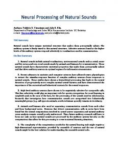
Region of interest (ROI) event related analysis. The SC is a particularly difficult structure to image using fMRI due to its small size and its proximity to major blood vessels in the brain stem, making it subject to cardiac noise (DuBois & Cohen, 2000). Wall, Walker, & Smith,
On
(2009) recently presented data which questioned the validity of using a standard (6 second) canonical haemodynamic response function (HRF) citing that a 4/5 second HRF was actually optimum for modelling activity in the SC. They noted a significant improvement in detected
ly
activation in the SC without the need for complex scanning procedures or analysis. Thus, in order to maximise statistical inference we did an additional ROI analysis on anatomically defined regions of the left and right SC. Each ROI comprised of a 27mm3 cube drawn over (and within) the SC using landmarks derived from a standard brain atlas (Mai, Paxinos, & Voss, 2007), see figure 2. In order to allow us to assess BOLD responses which may not be precisely modelled by a 6 second HRF raw event related time courses were extracted from each individual SC ROI. These % signal change time courses were averaged across all participants for left and right separately, adjusted by subtracting the mean of the 0 & 1 second time points and then normalised by dividing by the maximum data point in the data set.
8
http://mc.manuscriptcentral.com/prsb
Page 9 of 21
Submitted to Proceedings of the Royal Society B
FIGURE 2
Bell, Meredith, Van Opstal, and Munoz (2006) found that a high intensity stimulus which prompts a saccade resulted in earlier neural activation (~50ms) in the primate SC than a lower intensity stimulus, suggesting that neural responses in the SC can be mediated by light intensity. However, Bell and colleagues (2006) used a stimulus that was 160 times brighter than its comparator, whereas the brightness of our stimuli in the fovea only varied by a factor of 5 for a distant ball (point lights compressed) vs. a near ball (point lights dispersed). As our looming and receding stimuli have perfectly matched but reciprocal intensity ramps, the raw
r Fo
time courses also allowed us to determine whether there were temporal differences in the BOLD peak response due to specific clusters of frames. The looming event presented a dense patch of dots in the centre of the screen at onset and a larger sparser patch of dots at completion; however the converse were true for the receding stimuli. Examining time course
Re
values within the SC ROIs allowed us to assess this influence. If the BOLD response is consistently time locked to the start of the event (regardless of sequence order) we would
vi
expect a temporally consistent response peak for each condition regardless of the strength of that response; however, if the BOLD response is time locked to frames with a high level of
conditions.
ew
luminance in central visual field we would expect a consistent one second offset across
ly
On 9
http://mc.manuscriptcentral.com/prsb
Submitted to Proceedings of the Royal Society B
Page 10 of 21
Results Behavioural Results. Compared to a veridical time estimate (TTC = 0) participants tended to respond only fractionally before TTC for the receding balls (mean = -0.043s, SD = 0.229: t = -0.59, p = 0.57) and significantly after TTC for the looming balls (mean = 0.233s, SD = 0.205: t = 3.60, p < 0.01). Late responses to looming balls may have been related to where participants considered their face to be in relation to the screen when asked to press the button “when they thought the ball would hit them in the face”. Although participants reported finding the receding condition harder the more accurate responses may relate to being able to visibly see the point of contact. Given this pattern of results, the receding
r Fo
condition was considered a good cognitive as well as low level visual control for the looming stimulus. Motor responses to the square cue in the random condition were compatible with a reaction time to the onset of a stimulus (mean = 0.537s after, SD = 0.107: t =15.74, p random, p < 0.001 unc., vx > 10) activated middle frontal gyrus (MFG), superior frontal gyrus (SFG), inferior
ew
parietal lobe (IPL), and post central gyrus (PCG), see Table 1. In contrast, making a TTC computation specific to looming (looming > random, p < 0.001 unc., vx > 10) stimuli activated MFG, cingulate gyrus and anterior insula. Viewing looming balls in comparison to
On
a high level TTC control (looming > receding, p < 0.001 unc., vx >10) resulted in activation in inferior frontal gyrus, anterior and mid insula as well as basal ganglia and thalamic medial
ly
Pu.
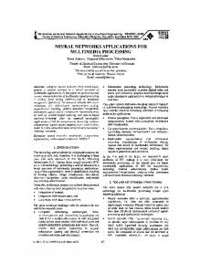
Following the onset of stimulus fade in each condition a six second peak BOLD response in left and right SC for the looming ball (see figure 3). ROI t-tests showed that there were significantly increased responses in left and right SC when viewing looming stimuli compared to both receding stimuli (L: t =1.634, p random; this activation was located on the anterior end of the calcarine fissure (x=7,y= -72, z= 9) and
r Fo
occipital gyrus (x=-24, -85, -12), anatomically corresponding to V1v and V2d respectively. The looming condition resulted in no additional visual activation in comparison to receding or random stimuli; thus strengthening our claim that SC response to a looming object is not simply a result of low level visual features.
vi
Re ew
FIGURE 3 TABLE 1
ly
On 11
http://mc.manuscriptcentral.com/prsb
Submitted to Proceedings of the Royal Society B
Page 12 of 21
Discussion
This study has shown that an extensive network of sub-cortical and cortical regions respond preferentially to visually looming stimuli compared to salient stimuli with no accelerated expansion properties. At the sub-cortical level a response to looming in human SC and mPu is in line with research findings in both the pigeon and the locust (Sun & Frost, 1998; Wu et al., 2005; Peron & Gabbiani, 2009). This finding also fits well with the role of the SC in detecting visually salient stimuli and orientating or defensive movements of the head and eye
r Fo
to salient stimuli (Brandao et al., 1994). Activation associated with looming stimuli in the basal ganglia is also consistent with previous research. Inhibitory mechanisms in the basal ganglia network have been inferred to be involved in ceasing ongoing motor behaviour when a looming, potentially dangerous, object is detected (Redgrave, Prescott, & Gurney, 1999).
Re
The basal ganglia output nucleus has efferent projections to the thalamus and pre-motor areas of the brain stem, including SC (Comoli et al., 2003) and may play a role in mediating motor
ew
vi
responses to looming stimuli entering personal space.
The mPu receives inputs from intermediate layer of the SC and as well as having projections to and from striate and extrastriate regions. It is also connected to a range of higher cortical regions such as; parietal, temporal, orbital frontal and cingulate cortex as well as the
On
amygdala (Grieve et al., 2000), and could thus play an important role in motor preparation with regard to looming stimulus.
A recent review by Kaas and Lyon (2007) which
ly
predominantly focused on primate literature implicates a subset of medial nuclei in the pulvinar as the subcortical component of the dorsal visual stream, with the majority of pulvinar projections to middle temporal visual area (MT) emanating from medial regions. A neural network which circumvents V1 in this way may provide a more direct pathway for the purpose of responding to approaching stimulus. Given that the pulvinar nucleus of the thalamus plays a role in attentional modulation and orientating (Kastner & Pinsk, 2004), looming stimuli may simply be more attentionally engaging, thus resulting in tectopulvinar activation. However, we argue that a looming detection mechanism which works on such a principle would still be an effective looming detection mechanism for rapid response to approaching object. 12
http://mc.manuscriptcentral.com/prsb
Page 13 of 21
Submitted to Proceedings of the Royal Society B
Activations across frontal, parietal and cingulate cortex were present when making a TTC judgement to both looming and receding stimuli in comparison to random motion control. These activations are
consistent with previous research and reflect the high cognitive
demand of the task (Field & Wann, 2005). Additional activation present in frontal and parietal cortical regions when making a TTC judgement to receding stimuli are consistent with the cognitive difficulty associated with making a computational TTC judgement in comparison to a low level control (Livesey et al., 2007) and encoding spatially salient aspects of the external environment (Culham & Valyear, 2006). Previously, comparative judgements
r Fo
made between two looming stimuli have been found to selectively increase the BOLD response in superior parietal and motor cortex (Field & Wann, 2005); in our study these regions were only activated for receding stimuli. The receding condition was the only condition which required the use of multiple objects in the simulated environment as the
Re
participants had to judge time to collision to a distant set of lines rather than to the face. This suggests that there is either a specific or additional role for parietal or motor cortex in making
judgements with a single object.
ew
vi
relative TTC judgements involving multiple objects in visual space as opposed to absolute
The anterior insula was additionally active when making a TTC judgement to looming stimulus in comparison to both the control and receding stimuli. This may bear some
On
connection to this region’s involvement in making duration judgments (Livesey et al., 2007), error detection (Klein et al., 2007) and interoceptive awareness on a variety of tasks (Craig, 2009). Our current findings, taken together this research suggests that this cortical region may
ly
be particularly attuned to making timing judgements with respect to objects in the environment which are approaching personal space.
Future research needs to focus on elucidating the role of attentional orientating in looming detection and associated tectopulvinar response. The two may be closely linked as looming stimuli tends to be alerting. Furthermore, disambiguating the contribution of different visual cues which are inherently linked to a looming object is a challenging next step. In this experiment we controlled for global changes in luminance using point light stimuli, but manipulating object properties such as visible texture can bias the percept of looming (Jacobs & Diaz, 2010). The progression from point light stimuli, towards more natural textures that 13
http://mc.manuscriptcentral.com/prsb
Submitted to Proceedings of the Royal Society B
Page 14 of 21
will produce different luminance ramps, is a balance that needs to be struck between ecological validity and experimental control in MRI.
We conclude that an extensive network of regions is involved in both low level detection of looming, sensorimotor response and higher level TTC estimation. The network contains the tectopulvinar early warning system, a motor preparatory system and possibly a more sophisticated computation system involving the anterior insula. These preliminary findings do have implications for humans’ performance during every day activities, such as driving or sports. If errors occur in collision detection, these may be due to failure at a sub-cortical level
r Fo
rather than in higher level cognitive processing. The dedicated nature of these early detection systems may also shed light on the “footballer’s dilemma” where we are able to react very rapidly to objects on a collision course, but after the event are unable to elucidate what we perceived or what we did to intercept or avoid an object.
ew
vi
Re ly
On 14
http://mc.manuscriptcentral.com/prsb
Page 15 of 21
Submitted to Proceedings of the Royal Society B
Funding This work was supported by an award from the UK Engineering and Physical Sciences Research Council [EP/D055342/1] to JPW, DTF & RMW.
r Fo ew
vi
Re ly
On 15
http://mc.manuscriptcentral.com/prsb
Submitted to Proceedings of the Royal Society B
Page 16 of 21
References
r Fo
Bell, A. H., Meredith, M. A., Van Opstal, A. J., & Munoz, D. P. (2006). Stimulus intensity modifies saccadic reaction time and visual response latency in the superior colliculus. Experimental Brain Research, 174(1), 53-59. Brandao, M. L., Cardoso, S. H., Melo, L. L., Motta, V., & Coimbra, N. C. (1994). Neural substrate of defensive behaviour in the midbrain tectum. Neuroscience and Biobehavioral Reviews, 18(3), 339-346. Comoli, E., Coizet, V., Boyes, J., Bolam, J. P., Canteras, N. S., Quirk, R. H., et al. (2003). A direct projection from superior colliculus to substantia nigra for detecting salient visual events. Nature Neuroscience, 6(9), 974-980. Coull, J. T., Vidal, F., Goulon, C., Nazarian, B., & Craig, C. (2008). Using time-to-contact information to assess potential collision modulates both visual and temporal prediction networks. Frontiers in Human Neuroscience, 2, 10. Craig, A. D. (2009). How do you feel - now? The anterior insula and human awareness. Nature Reviews Neuroscience, 10(1), 59-70. Culham, J. C., & Valyear, K. F. (2006). Human parietal cortex in action. Current Opinion in Neurobiology, 16(2), 205-212. DuBois, R. M., & Cohen, M. S. (2000). Spatiotopic organization in human superior colliculus observed with fMRI. Neuroimage, 12(1), 63-70. Field, D. T., & Wann, J. P. (2005). Perceiving time to collision activates the sensorimotor cortex. Current Biology, 15(5), 453-458. Gibson, J. J. (1958). Visually controlled locomotion and visual orientation in animals. British Journal of Psychology, 49(3), 182-194. Grieve, K. L., Acuna, C., & Cudeiro, J. (2000). The primate pulvinar nuclei: vision and action. Trends in Neurosciences, 23(1), 35-39. Jacobs, D.M., & Diaz, A. (2010). Judgements of time to contact are affected by the rate of appearance of visible texture. The Quarterly Journal of Experimental Psychology 63(6), 1041-1048. Kaas, J. H., & Lyon, D. C. (2007). Pulvinar contributions to the dorsal and ventral streams of visual processing in primates. Brain Research Reviews, 55(2), 285-296. Kastner, S., & Pinsk, M. A. (2004). Visual attnetion as a multilevel selection process. Cognitive, Affective and Behavioral Neuroscience. 4(4), 483-500. Klein, T. A., Endrass, T., Kathmann, N., Neumann, J., von Cramon, D. Y., & Ullsperger, M. (2007). Neural correlates of error awareness. Neuroimage, 34(4), 1774-1781. Lee, D. N. (1976) A theory of visual control of braking based on information about time to collision. Perception. 5, 437-459. Livesey, A. C., Wall, M. B., & Smith, A. T. (2007). Time perception: Manipulation of task difficulty dissociates clock functions from other cognitive demands. Neuropsychologia, 45(2), 321-331. Mai, J.K., Paxinos, G., & Voss, T. (2007) Atlas of the Human Brain (3rd Edition). New York: Academic Press. Northmore, D. P. M., Levine, E. S., & Schneider, G. E. (1988). Behavior Evoked by Electrical-Stimulation of the Hamster Superior Colliculus. Experimental Brain Research, 73(3), 595-605. Peron, S., & Gabbiani, F. (2009). Spike frequency adaptation mediates looming stimulus selectivity in a collision-detecting neuron. Nature Neuroscience, 12(3), 318-326. Redgrave, P., Prescott, T. J., & Gurney, K. (1999). The basal ganglia: A vertebrate solution to the selection problem? Neuroscience, 89(4), 1009-1023.
ew
vi
Re
ly
On
16
http://mc.manuscriptcentral.com/prsb
Page 17 of 21
Submitted to Proceedings of the Royal Society B
r Fo
Schiff, W., Caviness, J. A., & Gibson, J. J. (1962). Persistent fear responses in rhesus monkeys to optical stimulus of looming. Science, 136(3520), 982-983. Stein, B. E. (1984). Multimodal representation in the superior colliculus and optic tectum. In H. Vanegas (Ed.), Comparative Neurology of the Optic Tectum (pp. 819–841). New York and London: Plenum. Sun, H. J., & Frost, B. J. (1998). Computation of different optical variables of looming objects in pigeon nucleus rotundus neurons. Nature Neuroscience, 1(4), 296-303. Wall, M. B., Walker, R., & Smith, A. T. (2009). Functional imaging of the human superior colliculus: An optimised approach. Neuroimage, 47(4), 1620-1627. Wang, Y. C., & Frost, B. J. (1992). Time to collision is signalled by neurons in the nucleus rotundus of pigeons. Nature, 356(6366), 236-238. Wu, L. Q., Niu, Y. Q., Yang, J., & Wang, S. R. (2005). Tectal neurons signal impending collision of looming objects in the pigeon. European Journal of Neuroscience, 22(9), 2325-2331. Yonas, A., Bechtold, A. G., Frankel, D., Gordon, F. R., McRoberts, G., Norcia, A., et al. (1977). Development of sensitivity to information for impending collision. Perception & Psychophysics, 21(2), 97-104.
ew
vi
Re ly
On 17
http://mc.manuscriptcentral.com/prsb
Submitted to Proceedings of the Royal Society B
Page 18 of 21
Table 1: Activated regions for timing computation and looming. The whole brain contrasts receding ball > random motion and looming ball> random motion were run in order to identify brain regions which were involved in making a time to collision judgement to a moving stimulus in comparison to a low level visual and motor control stimulus. The contrast looming > receding was run to identify activation which was specific to perceiving an approaching object in comparison to a control matched for making a TTC judgement. All activations presented were at the p < 0.001 uncorrected level with a voxel extent threshold =10.
r Fo
Table 1
Peak Y
Voxel N
64 10 40 64
6.29 6.67 5.20 5.49
67 120 26 33
45 3 -36
29 11 19
22 44 4
5.21 6.34 5.43
24 68 14
-9 -9 9 48 39 36
2 -1 -31 26 22 -7
4 -5 -2 13 7 -11
5.58 5.45 6.03 5.78 6.41 7.01
25 23 16 11 20 29
ly
Looming > Receding Head of Caudate Nucleus External Globus Pallidus Medial Pulvinar nucleus of the Thalamus Inferior Frontal Gyrus Anterior Insula Mid Insula
t
-1 56 -46 -49
On
Looming > Random Middle Frontal Gyrus Cingulate Gyrus Anterior Insula
Peak Z
-6 39 51 -21
ew
vi
Receding > Random Superior Frontal Gyrus Middle Frontal Gyrus Inferior Parietal Lobule Postcentral Gyrus
Re
Condition
Peak X
http://mc.manuscriptcentral.com/prsb
Page 19 of 21
Submitted to Proceedings of the Royal Society B
r Fo ew
vi
Re On
ly
Figure 1: Sequence of events in a looming trial (a.) and receding trial (b.). 1. Lines change colour to cue stimulus type. 2. Ball appears by slowly fading in for 1.5 seconds. 3. Ball looms towards/ recedes from viewer for 1 second. 4. Ball disappears and participant has to make a TTC judgement after a random interval. 73x68mm (600 x 600 DPI)
http://mc.manuscriptcentral.com/prsb
Submitted to Proceedings of the Royal Society B
r Fo vi
Re ew
Figure 2. 27mm3 ROI of left and right SC (inset) and the location of SC within the brain (main image). 224x148mm (72 x 72 DPI)
ly
On http://mc.manuscriptcentral.com/prsb
Page 20 of 21
Page 21 of 21
Submitted to Proceedings of the Royal Society B
r Fo
Figure 3: Event related time course % signal change values for bilateral SC. The event start and end points are marked by the dashed grey line; looming onset is marked by the solid grey line. 59x22mm (600 x 600 DPI)
ew
vi
Re ly
On http://mc.manuscriptcentral.com/prsb