Auch gilt mein Dank dem Kuratorium der Gustav-Adolf-Lienert. Stiftung ...... Lienert-Foundation to CK and from the EU (Neuro-IT.net, IST-2001-35498) to AKE. 47 ...... 4 âT' bezieht sich auf das Wort âtarget', dem englischen Begriff für Zielreiz. 97 ...
Neural Correlates of Target Detection in the Attentional Blink
Dissertation
zur Erlangung des akademischen Grades
doctor rerum naturalium (Dr. rer. nat.)
genehmigt durch die Fakultät für Naturwissenschaften der Otto-von-Guericke Universtät Magdeburg
von
Dipl.-Psych. Cornelia Kranczioch
geb. am
09.09.1976
Gutachter:
Prof. Dr. A.K. Engel
in
Dresden
Prof. Dr. H. Hinrichs PD Dr. A. Keil
Eingereicht am
23.04.2004
Verteidigt am
09.09.2004
‚But then, how is it we are able to continuously perceive our environment?’ To which I (…) respond, ‘But how do you know for sure that we do?’ (Shapiro, 2001b, Preface)
Danksagung Viele Menschen haben direkt oder indirekt zum Entstehen dieser Arbeit beigetragen. Ihnen möchte ich an dieser Stelle meinen Dank aussprechen. An erster Stelle gilt mein Dank Prof. Andreas Engel, der den Anstoß für die vorliegende Arbeit gab, und ihr Entstehen durch seine Ideen und durch fruchtbare Diskussionen stets hilfreich begleitete. Bei Prof. Christoph Herrmann möchte ich mich für die geduldige Einführung in den Bereich der Wavelet-Analyse und für die inhaltliche Betreuung in diesem Bereich bedanken. Prof. Rainer Goebel und seiner Arbeitsgruppe danke ich für die große Gastfreundschaft, mit der ich in Maastricht aufgenommen wurde, und für die Unterstützung beim Erlernen der fMRT. Auch gilt mein Dank dem Kuratorium der Gustav-Adolf-Lienert Stiftung, welches mir durch ein Stipendium den Aufenthalt in Maastricht möglich machte. Von der eher technischen Seite her möchte ich mich für den Software-Support und die hilfreichen Erläuterungen zur Wavelet-Analyse durch Maren Grigutsch bedanken. Für seine tatkräftige Unterstützung bei der Aufnahme der EEG-Daten und seine große Hilfsbereitschaft danke ich Oliver Haumann. Bei der Aufnahme der MR-Daten wurde ich großartig unterstützt durch Paul Gallman. Auch sei denjenigen Mitarbeitern des Forschungszentrums Jülich und des Universitätsklinikums Eppendorf gedankt, welche dazu beigetragen haben, dass der Umzug von Jülich nach Hamburg so reibungslos von statten ging. Von unschätzbarem Wert war für mich die wissenschaftliche und persönliche Unterstützung durch Stefan Debener. Stefan danke ich neben unzähligen anderen Dingen vor allem für sein unerschütterliches Vertrauen in die Vollendung dieser Arbeit und dafür, dass er mich immer wieder aufgebaut hat, wenn etwas nicht so ganz glatt lief. Danke auch dafür, meine Launen in den letzten Wochen so geduldig abgefangen zu haben. Letztlich möchte ich meiner Familie danken. Meinen Eltern danke ich besonders dafür, dass sie es mir ermöglichten zu studieren. Meinen Geschwistern danke ich für die Selbstverständlichkeit, mit der sie mich bei dem was ich tue unterstützen.
Neural Correlates of Target Detection in the Attentional Blink
Contents CHAPTER 1 - Introduction .............................................................................................. 1 1.1 The Attentional Blink ......................................................................................... 2 1.2 Models of the Attentional Blink ......................................................................... 4 1.3 Electrophysiology of the Attentional Blink ........................................................ 9 1.4 fMRI Studies of the Attentional Blink.............................................................. 17 1.5 Objectives of the Thesis.................................................................................... 20 CHAPTER 2 -Methods ................................................................................................... 22 2.1 The Electroencephalogram ............................................................................... 22 2.2 Functional Magnetic Resonance Imaging......................................................... 26 CHAPTER 3 - Event-Related Potential Correlates of the Attentional Blink.................. 29 Abstract................................................................................................................... 29 3.1 Introduction....................................................................................................... 29 3.2 Materials and Methods...................................................................................... 32 3.3 Results............................................................................................................... 36 3.4 Discussion......................................................................................................... 42 CHAPTER 4 - Neural Correlates of Conscious Perception in the Attentional Blink ..... 47 Abstract................................................................................................................... 47 4.1 Introduction....................................................................................................... 47 4.2 Materials and Methods...................................................................................... 50 4.3 Results............................................................................................................... 54 4.4 Discussion......................................................................................................... 62 CHAPTER 5 - Investigating the Early Evoked Gamma Response in the Attentional Blink ................................................................................................................................ 70 5.1 Introduction....................................................................................................... 70
I
Contents 5.2 Materials and Methods...................................................................................... 71 5.3 Data Analysis and Results ................................................................................ 72 5.4 Discussion......................................................................................................... 75 CHAPTER 6 - EEG Gamma-Band Activity in Rapid Serial Visual Presentation.......... 77 Abstract................................................................................................................... 77 6.1 Introduction....................................................................................................... 78 6.2 Methods ............................................................................................................ 79 6.3 Results............................................................................................................... 83 6.4 Discussion......................................................................................................... 86 CHAPTER 7 - Summary and Conclusions ..................................................................... 89 7.1 Summary of the Findings.................................................................................. 89 7.2 Conclusions....................................................................................................... 90 Deutsche Zusammenfassung ........................................................................................... 97 References ..................................................................................................................... 103 Curriculum Vitae........................................................................................................... 115 Publikationsliste ............................................................................................................ 117
II
Neural Correlates of Target Detection in the Attentional Blink
CHAPTER 1
Introduction Research on the temporal aspects of dual-task interference revealed that directing attention to a target prevents accurate report of a second target presented within approximately 500 ms of the first (Broadbent & Broadbent, 1987). This impairment has been termed the ‘attentional blink’ (Raymond, Shapiro, & Arnell, 1992). So far, the attentional blink paradigm has been primarily in the focus of investigations of the time course of attention. The time course of attention refers “to the temporal availability of whatever property (or properties) of the brain is or are responsible for enhancing perception” (Shapiro, 2001a, p. 1). Since its first description numerous behavioral studies have been conducted investigating the attentional blink phenomenon, and thus, the time course of attention (for review see Shapiro, 2001b; Shapiro, Arnell, & Raymond, 1997). In consequence, some insight has been gained about what is required for an attentional blink to occur, about how the magnitude of the attentional blink can be affected, and under which circumstances no attentional blink is obtained. Furthermore, concurrent models aimed to explain behavioral findings were tested, and modified. Yet what exactly is the fate of the second target? Where in the processing stream is it lost? What might distinguish a target that is correctly reported from a similar target that cannot be reported? How is the time course of the attentional blink accomplished? Questions like these are intimately related to assessing the neural events associated with the attentional blink. Methods investigating neural activity can provide a more direct correlate of cognitive processes than do behavioral measures. For instance, functional magnetic resonance imaging (fMRI) allows to identify brain areas involved in a certain cognitive process, and eventrelated potentials (ERPs) enable to follow the temporal structure of information processing. Thus, methods investigating the activity of the brain can help to distinguish more thoroughly whether impaired performance on the second target is associated with processes of perception, identification, or consolidation of information, or even with response selection. Studying the neural correlates of the attentional blink thus significantly contributes to a better understanding of the characteristics of the time course of human information processing. Moreover, because models of the attentional blink allow different predictions with respect to
1
Introduction its neural correlates, it does also contribute to the discussion which model offers the best explanation of the phenomenon, or whether models should be modified. Because with the attentional blink a paradigm is available that allows comparing brain activation to identical stimuli once explicitly perceived and once missed, the paradigm is of particular interest for yet another branch of cognition research, namely the study of the neural correlates of consciousness (NCC). The search for the NCC concentrates on identifying and characterizing neural activity patterns that co-vary with conscious experience. (Crick & Koch, 1990; Engel & Singer, 2001; Rees, Kreiman, & Koch, 2002). A factor of consciousness seen as accessible for experimental quantification and theoretical explanation is sensory awareness (Crick & Koch, 1990; Engel & Singer, 2001). The attentional blink paradigm allows quantifying sensory or visual awareness as explicit report of the presence of a target stimulus. Thus, by comparing brain activation to physically identical events that have different behavioral outcomes further insight might also be gained into the correlates of visual awareness. Thereby conclusions might be drawn regarding the NCC. Investigating neural correlates of the attentional blink might contribute to the generation of models regarding the attentional blink. It may further provide insight into neural events that can account for the time course of attention as reflected in the attentional blink. And lastly, specifically the neural correlates of target detection in the attentional blink should add to the knowledge about neural correlates of sensory awareness. In the remainder of this chapter the attentional blink paradigm will be described in detail. Models of the attentional blink will be outlined, and previous studies investigating neural correlates of the attentional blink will be introduced and discussed in the context of the models. Furthermore, a recent ‘electrophysiological’ model of the attentional blink’s time course is introduced in detail and put into a broader context. Against this background, the introductory chapter closes with an outline of the studies presented in this dissertation, their aims and questions.
1.1 The Attentional Blink Other than the term might imply the attentional blink is no physical eye blink, but rather something that can be described as a blink of the mind. The attentional blink is characterized by a transient reduction of attention which occurs if more than one target has to be processed in a series of stimuli that rapidly succeed one another. Typically, the attentional blink is investigated in rapid serial visual presentation (RSVP) tasks. Here, series of 15-20 stimuli such as letters, digits, words, or keyboard symbols are presented at a single location with a frequency of about 10 per second. In such a series of distractor stimuli two target stimuli are 2
Neural Correlates of Target Detection in the Attentional Blink embedded. Target stimuli can for instance be defined by their color, identity, or category, e.g., ‘the blue digit’ or ‘the black X’. Critical for the attentional blink is the temporal distance between the targets. It is either expressed as stimulus onset asynchrony (SOA) or, more frequently, as the serial position of target two (T2) relative to target one (T1). In the latter case, the relative serial position of T2 is referred to as lag, that is, if T2 comes next after T1 this corresponds to lag 1 and so forth. Figure 1.1.a shows the illustration of a trial in which the non-targets or distractors are black letters. T1 is the only grey letter and T2, presented at lag 2, is the black X. If both T1 and T2 are task-relevant and are presented in close temporal proximity, task performance on T2 decreases as a function of distance between the two targets. At the example of the trial shown in Figure 1.1.a, such a dual-task condition would be to firstly identify whether the grey letter (T1) was a vowel, and then to decide whether an X (T2) was present. The decrease in T2 performance is most pronounced for an SOA of between 200 and 400 ms (Figure 1.1.b), task performance is better for longer SOAs. A reduction or elimination of the impairment in task performance is observed in standard attentional blink experiments when T2 appears at lag 1, or, at an SOA of about 100 ms. This effect is also known as ‘lag 1 sparing’ (Chun & Potter, 1995; Potter, Staub, & O'Connor, 2002). Thus, the term ‘blink’ refers to this u-shaped pattern in performance, because a comparable course of performance would be expected if the first target actually triggered an eye blink (Raymond et al., 1992). Initially described was the attentional blink phenomenon in the ninety-eighties (Broadbent & Broadbent, 1987; Reeves & Sperling, 1986; Weichselgartner & Sperling, 1987).
Figure 1.1. (a) Illustration of a typical attentional blink trial. For the two targets (T1, T2) in the stimulus stream un-speeded responses are required. (b) Detection accuracy of the second target (T2) as a function of distance (stimulus onset asynchrony, SOA) between the first target (T1) and T2 (adapted from Kranczioch, Debener, & Engel, 2003). In the single target condition only T2 had to be detected, whereas in the dual target condition tasks were to identify T1 and to detect T2.
3
Introduction
1.2 Models of the Attentional Blink Most frequently discussed in literature are the two-stage model of the attentional blink by Chun and Potter (1995) and the interference model of the attentional blink by Shapiro and colleagues (Isaak, Shapiro, & Martin, 1999; Shapiro, Raymond, & Arnell, 1994). In the twostage model as originally proposed by Chun & Potter (1995) it is assumed that for being reportable, targets have to be processed in two stages. During the first stage all stimuli are processed to the point of conceptual representation. In the second stage the representation is then consolidated into a durable and reportable form. For the second processing stage attention is needed, the attentional resources are thought to be limited in capacity however. Hence, as long as T1 occupies the second stage attentional resources are not sufficient for consolidation of T2. While waiting for Stage 2, T2 is prone to be lost and thus not to be reported. This two-stage model is illustrated in Figure 1.2. In a recent extension and modification of the model (two-stage competition model, Potter et al., 2002) it is also proposed that in the first stage all stimuli are initially analyzed. If during this stage object properties are detected that make the stimulus a likely target, attentional resources are allocated. The model assumes further that attention at Stage 1 is labile. Hence, if two potential targets are detected in temporal proximity, the second target will attract attention away from the first target. That is, in attentional blink paradigms it is assumed that there is ongoing competition between potential targets for limited processing resources in Stage 1. The target on which sufficient resources are allocated is identified and then enters and monopolizes the second stage. Here the target is consolidated in short-term memory and can thus be reported. The other potential target that cannot immediately enter Stage 2 is vulnerable to interference or forgetting during the wait for Stage 2. Thus, in contrast to earlier versions of the two-stage model it is now assumed that either T1 or T2 can first enter and monopolize Stage 2. The likeliness for T2 to enter Stage 2 first is assumed to depend on the T1-T2 SOA. At SOAs of around 100 ms T2 can attract resources away from T1, and therefore chances are increased that T2 is identified first. At longer SOAs however, T1 is identified first and occupies Stage 2, and T2 is likely to be lost. Two other models follow the two-stage approach by Chun and Potter (1995). In the object substitution model (Brehaut, Enns, & Di Lollo, 1999) it is specifically focused on the role of the item immediately following T1 and T2, that is, their masks. It is suggested that the role of T1 masking is to introduce a delay in processing T2. This role, however, could also be
4
Neural Correlates of Target Detection in the Attentional Blink
Figure 1.2. Illustration of the two-stage model by Chun and Potter (1995). All items of the RSVP including distractors (D) are identified in Stage 1. The first target (T1) is selected for consolidation in Stage 2 and can thus be reported. As long as T1 occupies Stage 2, this processing stage cannot be entered by the second target (T2). Due to interference with subsequent stimuli or decay in Stage 1, T2 is prone to get lost. T2 is thus likely not to be transferred to Stage 2 even after T1 consolidation. What can be reported besides T1, however, depends on the information that can be transferred from Stage 1 to Stage 2 after T1 has been consolidated. If T2 has been lost, other items might be reported instead of T2.
played by other ways of introducing a delay in T2 processing. Because during the delay T2 is not attended, it is assumed to be vulnerable of being substituted by its mask while still in Stage 1 (Enns, Visser, Kawahara, & Di Lollo, 2001). It has been postulated that for obtaining the diminished T2 accuracy typical for the attentional blink, interruption masking of T2 is essential. However, recently this assumption has been modified (Enns et al., 2001; Kawahara, Zuvic, Enns, & Di Lollo, 2003). That is, interruption masking of T2 seems to be necessary whenever the T2 stimulus belongs to a class of overlearned stimuli such as letters or digits. If this is not the case, an attentional switch between targets seems to be sufficient to produce the attentional blink. Attentional switching is required when the two targets or tasks differ. It is suggested that due to the switch, the visual system has to be reconfigured. Reconfiguration results in the failure to encode T2, that is, in this case it does not even enter Stage 1. On the other hand, Jolicoeur, Dell’Acqua and colleagues (Dell'Acqua, Jolicoeur, Pesciarelli, Job, & Palomba, 2003; Jolicoer, Dell'Acqua, & Crebolder, 2001; Jolicoeur & Dell'Acqua, 1998) introduced the idea of a central processing bottleneck as Stage 2 to the two-stage model. That is, it is postulated that short-term consolidation of T2 is not only postponed by concurrent consolidation of T1, but also by interference with other processes such as response selection. In other words, it is argued that part of the processing required to generate a reportable representation of a target is subject to central capacity limitations. Figure 1.3 summarizes the two-stage approach underlying the central bottleneck model, but also the original and 5
Introduction modified two-stage model (Chun & Potter, 1995; Potter et al., 2002), and the object substitution model (Brehaut et al., 1999).
Figure 1.3. Model of the structure of information processing to explain dual-target performance in attentional blink, task switching, and short-term memory consolidation paradigms. Information from the sensory modalities converges to a central bottleneck of information processing. The central processor (Jolicoeur, 1998, 1999; Pashler, 1994) is assumed to be limited in capacity, and interference at this central stage of processing leads to a decrease in performance. Adapted from Chun and Potter (2001, p. 28, Figure 2.7)
Beside the two-stage model, the interference model of the attentional blink (Isaak et al., 1999; Raymond, Shapiro, & Arnell, 1995; Shapiro et al., 1994) is frequently discussed. It comprises likewise two stages of target processing. In the model it is postulated that first each RSVP item is given a perceptual description matched for similarity to templates of the target. Based on similarity with the templates, and based on temporal features, weights are assigned to the items. That is, T1 and T2 are likely to get high weights. Yet because of their temporal proximity to the targets the same applies for T1 mask and T2 mask. Items that receive a high weight are passed on to the second stage or visual short-term memory (VSTM). Capacity of and total weighting in VSTM is assumed to be limited. The first item that enters VSTM will have a high weight, but due to the capacity limitations weighting for items entering later is diminished. From VSTM items are then retrieved for report based on their relative weights. Because T1 usually has the highest weight it can easily be reported. Weights of T2, T1 mask, and T2 mask on the other hand are reduced. Therefore these items interfere, and the wrong item will frequently be retrieved from VSTM as T2. At longer SOAs T2 is assumed to have a 6
Neural Correlates of Target Detection in the Attentional Blink higher chance of being reported, because the weight of T1 in VSTM decays, or because T1 is erased from VSTM when it is passed to a response stage. Figure 1.4 shows an illustration of the interference model.
Figure 1.4. Illustration of the interference model. All items including distractors (D) and target masks (M1, M2) are identified in a first processing stage. Because of similarity with a target template, T1 and T2 are assigned high weights in this stage. M1 and M2 also receive higher weights than other distractors because of their proximity to the targets. T1, M1, T2, and M1 are transferred to visual short-term memory (VSTM). Because it arrives first, T1 receives the highest weight within VSTM and can thus easily be retrieved from VSTM and reported. As total weighting within VSTM is limited, the weights of remaining items are greatly attenuated as compared to T1. Therefore, in retrieval from VSTM, the masks and T2 interfere. In consequence, the wrong item is frequently selected for report.
Interference and two-stage models have been brought together in a hybrid model of the attentional blink (Vogel, Luck, & Shapiro, 1998). In the hybrid model it is assumed that after full identification all items in the RSVP stream are initially stored in a conceptual short-term memory (CSTM) buffer. The items in this buffer are not yet available for report at this stage and inclined to decay and to replacement by other stimuli. The selection of items for transfer from CSTM into visual working memory (VWM) is based on the similarity between the representation of the item in CSTM and a template of the target. For the transfer into VWM attention is needed, but attentional resources are assumed to be limited. Thus, as long as T1 is transferred to VWM, T2 cannot be consolidated in a more durable and reportable form. As a result errors are made in the report of T2. These errors are not random though, but depend on the content of CSTM after the consolidation of T1. That is, in order to report T2, the system will attempt to transfer any remaining information from CSTM to VWM. Within CSTM however, the representations of items interfere and decay, so that not T2 but the wrong item will frequently be transferred into VWM. This is illustrated in Figure 1.5. A related proposal to bring together the diverse models of the attentional blink has been made by Shapiro and colleagues (1997).
7
Introduction
Figure 1.5. Illustration of the hybrid model (Vogel et al., 1998). All items reach a level of conceptual representation and are initially stored in a conceptual short-term memory (CSTM) buffer. Items are selected for transfer from CSTM to visual working memory (VWM) based on the degree of match between their representation in VSTM and a target template. As long as attentional processes are engaged in transferring T1 into VWM they are unavailable for T2. As soon as attentional resources are available again, information remaining in CSTM will be transferred into VWM. Due to decay and interference in CSTM frequently this will not be T2. In consequence, T1 can be reported, whereas in reporting T2 errors occur.
A further alternative account of the time course of attention that can neither be assigned to the two-stage nor the interference approach is the attentional dwell time model (Duncan, Ward, & Shapiro, 1994; Ward & Duncan, 1996). In this model it is suggested that the multiple attributes of an object are processed in parallel. Separate objects, on the other hand, are assumed to be processed serially. The amount of time required to complete the parallel processes are referred to as dwell time (Brehaut et al., 1999). Parallel processing is limited in capacity and if more than one relevant object appears within several hundred milliseconds, the majority of resources is engaged in processing the first object. Only gradually do these resources become available for other objects (Ward & Duncan, 1996). That is, according to this account attention dwells for several hundred milliseconds on T1. During this epoch T2 is not encoded and thus lost. In sum, all of the models introduced here assume that due to T1 processing a capacity limited process cannot, or not sufficiently, be applied to T2. The result is that T2 is lost to awareness. Yet the models differ with regard to when T2 is assumed to get lost. According to the dwell time model (Duncan et al., 1994; Ward & Duncan, 1996) T2 is not processed until after processing of T1 is complete. Similarly, recent modifications of the object substitution model (Enns et al., 2001; Kawahara et al., 2003) predict that with a significant task switch between T1 and T2 no representation of T2 is formed. By contrast, the models related to the 8
Neural Correlates of Target Detection in the Attentional Blink two-stage model (Brehaut et al., 1999; Chun & Potter, 1995; Jolicoeur & Dell'Acqua, 1998) and the hybrid models by Vogel et al. (1998) and Shapiro et al. (1997) postulate that T2 is fully identified. The representation of T2 however is lost prior to working memory. Finally, in the interference model (Isaak et al., 1999; Shapiro et al., 1994) it is assumed that T2 firstly enters working memory, but is lost due to interference or competition in working memory. Based on these assumptions, different experimental outcomes would be predicted for studies on the neural correlates of the attentional blink. That is, either correlates of stimulus perception or correlates of target consolidation would be expected to be affected by the attentional blink. In this context, in the following results from event-related potential studies and a model of the attentional blink based on electrophysiological variables are presented and discussed. Thereafter, fMRI studies on the attentional blink are summarized.
1.3 Electrophysiology of the Attentional Blink ERP Studies of the Attentional Blink ERPs are derived by averaging over many repetitions the electrical activity of the brain in relation to an event. The positive and negative deflections of the ERP are seen as a stimuluslocked measure of information processing (see Figure 1.6 for an illustration), which, for instance, provides information about the depth of or variations in processing in dependence of experimental variations (for a review see Rugg & Coles, 1995). Hence ERPs may help to answer the question which process related to the processing of T2 is disturbed in the attentional blink. Previous ERP studies of the attentional blink primarily focused on four ERP components: P1, N1, P3, and N400.
Figure 1.6. Example event-related potential (ERP) waveform. Time 0 represents the onset of the stimulus elicting the ERP. Note that negative is plotted upward.
9
Introduction The P1 and N1 components are a positive and a negative wave elicited early in the time course of stimulus processing, that is, within 200 ms following stimulus presentation. It has been shown that both are sensitive to the physical characteristics of the eliciting stimulus (Kaskey, Salzman, Klorman, & Pass, 1980) and that they are enhanced for stimuli presented at attended locations (Heinze, Luck, Mangun, & Hillyard, 1990; Luck & Hillyard, 1994). P1 and N1 have been interpreted as reflecting a ‘gain control’ mechanism over sensory/perceptual processing (Mangun & Hillyard, 1995). This joint interpretation was challenged by Luck and colleagues (Luck, Heinze, Mangun, & Hillyard, 1990) who assume that only the P1 reflects enhanced stimulus-evoked activity while in the N1 the engagement or orienting of attention to a task-relevant location is represented. The P3, P3b or target-P3, is elicited by rare, task-relevant stimuli (for reviews see Kok, 2001; Verleger, 1997). It is largely assumed that the P3 is a correlate of working memory processes. However, notions differ regarding the functional interpretation of P3. For Donchin (Donchin & Coles, 1988) the P3 is a correlate of the update of a model of the environment hold in working memory. An alternative view is that the P3 reflects context closure or the termination of a perceptual epoch (Verleger, 1988). That is, it is assumed that the P3 is elicited if stimulus evaluation results in a match with the context-based expectation. The N400 is a negative deflection in the ERP peaking approximately 400 ms post-stimulus onset. The most prevalent view on the N400 is that it reflects integration in a semantic context (Kutas & Federmeier, 2000). Accordingly, it would be expected that a word that matches a previously established semantic context elicits a small N400, whereas a mismatch would elicit a large N400. This is also known as semantic priming effect.
McArthur, Budd, and Michie (1999) focused in their study on the P3 ERP evoked by T1. They argue that both the attentional blink and the P3 have a similar time course, and that both have been linked to inhibitory processes. Thus, the study was aimed to investigate whether there is an association between the amplitude of the P3 elicited by T1 and the size of the attentional blink. Therefore the attentional blink waveform based on eight lags (SOA 90 ms) was shifted forward by 235 ms with reference to T1 presentation, and correlated with the P3 ERP. For both experiments conducted, McArthur and co-workers found that under these conditions, P3 amplitude and the percentage of detected T2 followed a similar course. In addition, reduction of task difficulty had similar effects on P3 and attentional blink magnitude at the group level, that is, both were attenuated. However, contrary to the expectations, the magnitude of the attentional blink and the amplitude of the P3 were found to be negatively
10
Neural Correlates of Target Detection in the Attentional Blink correlated in one of the two experiments. This, however, could not be replicated in the other experiment, where the two measures did not correlate. The results were interpreted as indicating a moderate association between the attentional blink and the P3 elicited by T1, suggesting that there is an impairment in visual processing coinciding with the course of P3 (McArthur et al., 1999). The study however leaves unresolved which stage of T2 processing is assumed to be impaired by the P3, as the motivation for the 235 ms shift of the attentional blink wave form is not easy to follow. On the one hand it is argued that this shift should account for the propagation delay between T2 onset and the arrival of the signal at the cortex. For this explanation, however, the shift is rather large, as the first major component of the visual evoked cortical potential, the C1 component, has an onset latency between 40-70 ms and a peak latency between 60-100 ms (for review see Di Russo, Martinez, Sereno, Pitzalis, & Hillyard, 2002). On the other hand, it is mentioned that the shift is an estimate of the latency of neural processing underlying T2 discrimination, and corresponds to the latency of the N2 ERP. However, no evidence is provided that the T2-evoked N2 and hence the processes reflected in this component are affected by T1 processing or the T1-evoked P3. Thus, even though in the study of McArthur et al. (1999) an important issue is addressed, namely processing of T1 and its relation to T2 processing, it does not allow conclusions with respect to models of the attentional blink.
In contrast to McArthur et al. (1999), other studies focused on the electrophysiological correlates of T2 processing. In an elegant series of experiments, Vogel and colleagues (1998) utilized ERPs to determine the stage at which T2 processing is impaired. T1 and T2 were embedded in an RSVP stimulus stream, and T2 was the first, third, or seventh item after T1. In a first experiment Vogel et al. tested the hypothesis that the attentional blink reflects a suppression of sensory processing. They applied an irrelevant-probe technique, where in half of the trials a solid white square was presented behind T2. The P1-N1 components elicited by irrelevant probe stimuli were not reduced during the attentional blink period, while behavioral data confirmed the typical attentional blink pattern, that is, a significant reduction in performance for T2 items presented at lag 3, and nearly unimpaired detection accuracy for lags 1 and 7. Furthermore, the N400 was investigated to determine whether the attentional blink impairs word identification (see also Luck, Vogel, & Shapiro, 1996). A context word that was either related or unrelated to the T2 target word was presented before the RSVP stream. T1 was a number that needed to be identified. The N400 effect, that is, the N400 for 11
Introduction related as compared to unrelated T2, was of similar size for all three T2 lags. In contrast to the P1-N1 and N400 findings were the results of the experiment aimed at investigating the P3 component as an index that a stimulus has reached working memory. The T2 task was to detect the white letter E among the otherwise black RSVP items. In 85% of the trials T2 was a white letter other than E. In contrast to lags 1 and 7, the P3 was virtually absent in the ERP time-locked to T2 items presented at lag 3, that is, during the attentional blink. Vogel et al. (1998) concluded that the impairment in T2 processing associated with the attentional blink arises relatively late. They suggested that the impairment occurs after stimulus processing and after stimulus identification has been completed as evident by the unimpaired P1-N1 and N400 effect during the attentional blink period. Because P3 was found to be suppressed during this period, the impairment was assumed to most likely arise at the stage of working memory, that is, before or during a stable representation of the stimulus is formed in working memory. Dell’Acqua and colleagues (2003) carried out a study very similar to Vogel et al.’s (1998) P3 experiment. The T2 task was to indicate whether one of the letters following T1 was an E. T2 was presented at lags 1, 3, or 9. In this study a progressive P3 suppression with decreasing T2 lag was observed, that is, not only P3 at lag 3, but also P3 at lag 1 was suppressed. This is in contrast to Vogel et al. (1998) who found the P3 at lag 1 to be unimpaired. The authors suggest that this discrepancy might be due to differences in participants’ performance in the T2 task. Whereas in Vogel et al.’s study performance at lag 1 was nearly as good as at lag 7, performance in Dell’Acqua et al.’s study was clearly worse at lag 1 than at lag 9. In a second experiment Dell’Acqua and colleagues (2003) combined a speeded tone discrimination task (T1) with an un-speeded letter detection task (T2). Again, the T2-evoked P3 was reduced with decreasing lag. Because P3 was similarly altered in both the ‘classical’ un-speeded dual target condition and the speeded T1 task condition the results were interpreted as in line with accounts of the attentional blink that focus on a central processing bottleneck. That is, as consolidation and response selection interfere similarly with a temporally overlapping task as indicated by the P3 attenuation, both operations seem to require central resources that cannot be shared easily across tasks. The object substitution model (Brehaut et al., 1999) was tested in an ERP study by Vogel and Luck (2002). In the model it is proposed that T2 cannot be consolidated in working memory until after T1 consolidation. T2 is therefore in danger of being replaced by subsequent stimuli. In support of the object substitution model no attentional blink is observed if T2 is the last stimulus, because there are no following items that might substitute T2
12
Neural Correlates of Target Detection in the Attentional Blink (Giesbrecht & Di Lollo, 1998). Vogel and Luck (2002) found that in accordance with the model, P3 was not eliminated but only delayed if T2 was the last item of the RSVP sequence. If, on the other hand, T2 was followed by a masking item, the P3 was completely suppressed. Rolke, Heil, Streb, and Hennighausen (2001) investigated the role of automatic spread of activation in the generation of the semantic N400 effect by means of unattended semantic priming. T2 words that served as prime were presented at lag 3. Subsequently, probe words occurred that either had a strong, a weak, or no semantic association to T2. For data analysis trials in which the T2 word was identified and trials in which T2 was not identified were separated. Interestingly, the N400 priming effect of the probe was unimpaired by the detection of the prime (that is, T2). By contrast, the T2-evoked P3 was found only when T2 was correctly identified. In summary, the P1-N1 and N400 findings of these ERP studies provide evidence that T2 is lost after being fully identified (Luck et al., 1996; Rolke et al., 2001; Vogel et al., 1998). Thus, models assuming that T2 is not processed during the attentional blink (Duncan et al., 1994) are not supported. On the other hand, processes reflected in the P3 component seem to be impaired during the attentional blink. That is, there is evidence that working memory processes can either not be applied to T2 or are delayed (Dell'Acqua et al., 2003; Vogel & Luck, 2002; Vogel et al., 1998). The impairment of P3 is in support of all models that assume that T2 is identified, but for some reason cannot be consolidated in working memory. With exception of the interference model (Shapiro et al., 1994) this assumption is inherent in all the remaining models of the attentional blink. The interference model would predict that P3 is not suppressed, as T1 and T2 are thought to reach working memory (Vogel et al., 1998). Yet all ERP studies focusing on the P3 (Dell'Acqua et al., 2003; Vogel & Luck, 2002; Vogel et al., 1998) leave open whether the observed suppression of the P3 is related to T2 presentation lag (that is, T2 presented during the attentional blink do not evoke a P3), or might (also) depend on task performance (that is, undetected T2 do not evoke a P3, and during the attentional blink the number of undetected T2 is increased). In explanation, in these studies trials in which T2 was detected and in which it was not detected were averaged for ERP analysis. A possible consequence of this strategy of analysis can be illustrated at the example of the P3 experiment of Vogel and colleagues (1998). Here, mean discrimination accuracy for T2 was about 15% at lag 3, but about 85% or more at lags 1 and 7 (Vogel et al., 1998, Figure 8, p. 1667). Hence it is likely that the lag 3 ERP consisted mainly of trials in which T2 had not been detected, whereas for lags 1 and 7 T2-detected trials should clearly prevail in the ERP. For lag 3 the outcome would be a suppression of the P3 in the averaged data. Indeed, there is
13
Introduction first evidence that separate analysis of trials in which T2 is detected and in which it is not detected reveals a P3 for detected T2 (Rolke et al., 2001). This might indicate that the impairment assumed to operate at or before the stage of working memory may not be absolute, and the possibility that detected T2 items are actually detected and not only correct guesses cannot be ruled out. An Electrophysiological Model of the Attentional Blink Recent theoretical work (Fell, Klaver, Elger, & Fernandez, 2002) focuses similar to McArthur and colleagues (1999) on the interrelation between T1 and T2 processing. In detail, it is postulated that suppression of the T2-related early evoked gamma activity by the T1-evoked P3 ERP might cause the attentional blink. The current interest in oscillatory brain activity in the gamma frequency band (30-80 Hz) goes back to the late 1980s. At that time it was found that neurons in cat visual cortex synchronize their activity within the gamma frequency band when they are presented with coherently moving bars as compared to independently moving patterns (Eckhorn et al., 1988; Gray, König, Engel, & Singer, 1989). Importantly, this was in support of the binding hypothesis proposed by von der Malsburg and Schneider (1986). Central to the model of von der Malsburg is the neuroscientific problem of how the different aspects of a visual object that are processed in different modules of the visual system, are bound together to become one coherent representation of the object. In the model it is assumed that this so-called binding problem might be solved by a neuronal mechanism that relies on time-based coding. The idea is that neurons that represent one object are bound together by synchronization of their action potentials. They thereby form a functional neuronal assembly that can be distinguished from different assemblies on the basis of the relative timing of their discharges. The temporal binding hypothesis found further support in numerous animal studies (for review see Engel, Roelfsema, Fries, Brecht, & Singer, 1997; Gray, 1999) as well as in human scalp EEG recordings. When coherently moving bars were shown to participants, it was found that power in the gamma-band was enhanced as compared to bars moving into different directions (Lutzenberger, Pulvermüller, Elbert, & Birbaumer, 1995; Müller et al., 1996). For more complex stimuli such as schematic face stimuli it has been observed that when the orientation of these stimuli allowed perception of a face, gamma activity increased (Keil, Müller, Ray, Gruber, & Elbert, 1999; Rodriguez et al., 1999). TallonBaudry and colleagues (Tallon-Baudry, Bertrand, Delpuech, & Permier, 1997) presented their subjects pictures of black blobs that were either meaningless, or where the blobs were arranged in a way that a Dalmatian was hidden. When subjects perceived the dog, an enhancement in gamma activity was found. Gamma activity has also been shown to be 14
Neural Correlates of Target Detection in the Attentional Blink involved in multistable perception (Basar-Eroglu, Strüber, Schürmann, Stadler, & Basar, 1996; Strüber, Basar-Eroglu, Hoff, & Stadler, 2000). During states of perceptual switching gamma-band activity significantly increased. The enhancement was found to be larger in subjects with a high rate of perceptual switches. Besides its role in feature binding, synchronous neural activity in the gamma frequency band has been suggested to be the neuronal mechanism of attentional selection (Fell, Fernandez, Klaver, Elger, & Fries, 2003; Niebur, Hsiao, & Johnson, 2002). Selection is assumed to be implemented by enhancing the synchrony between neurons that represent the specific sensory information (Niebur et al., 2002). In the electrophysiological model of the attentional blink by Fell et al. (2002) the idea of attentional selection by synchronous gamma activity is also inherent. Specifically, it is proposed that due to a suppression of the early evoked gamma response (EEGR), attentional selection of T2 is impaired. The evoked gamma response is the fraction of total gamma activity strictly phase-locked to stimulus onset (Tallon-Baudry & Bertrand, 1999). Tiitinen et al. (1993) demonstrated that the auditory EEGR is enhanced for tones presented to the attended ear. In the visual modality the EEGR has been reported to be enhanced for an attended motion stimulus as compared to a condition where the same motion stimulus could be ignored (Sokolov et al., 1999). Furthermore, in studies in which participants had to discriminate target and non-target stimuli, the EEGR was found to be increased for targets (Debener, Herrmann, Kranczioch, Gembris, & Engel, 2003; Herrmann & Mecklinger, 2000, 2001). In line with these studies are findings from intracortical microelectrode recordings in monkeys (Fries, Reynolds, Rorie, & Desimone, 2001). Fries and colleagues presented simultaneously two stimuli on a screen, one inside and the other nearby the receptive fields of the recorded neurons in V4. Attention was directed to one of the two stimuli. If attention was shifted to the stimulus that was inside the receptive fields an increase in the evoked gamma frequency synchronization was observed. In sum, numerous animal and human studies provide evidence that the EEGR is sensitive to attention and that it may be of relevance for the selection and identification of target stimuli.
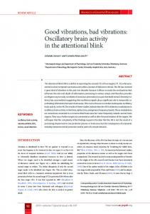
In their model of the attentional blink based on electrophysiological variables Fell and coworkers (2002) suggest that a process Pr1 triggered by T1 and indexed by the P3 component impairs a process Pr2 triggered by T2 and indexed by the EEGR. This is assumed to cause the attentional blink. The idea of the model, illustrated in Figure 1.7, is based on the observation
15
Introduction
Figure 1.7. Illustration of the model of the attentional blink as proposed by Fell et al. (2002). The upper panel depicts the time course of the attentional blink underlying the model, with the largest impairment in T2 detection accuracy at a T1-T2 stimulus onset asynchrony (SOA) of 300 ms. In the middle panel the P3 triggered by T1 and indicator of Process 1 (Pr1) is shown. The lower panel illustrates the early evoked gamma response (EEGR) triggered by T2 and indicator of Process 2 (Pr2) for different T1-T2 SOAs. When the P3 is maximal at 400 ms, the EEGR to T2 presented with 300 ms SOA is maximally suppressed.
that detection accuracy for T2 reaches its minimum about 300 ms after T1 presentation (Figure 1.7, upper panel). Accordingly, it is suggested that whatever impairs T2 processing must have at least a latency of about 300 ms. Due to its peak latency of about 400 ms in attentional blink experiments (McArthur et al., 1999; Vogel et al., 1998) the P3 is seen as a likely candidate for Pr1 (Figure 1.7, middle panel). Furthermore, because Pr1 has a latency of about 400 ms, the impaired Pr2 must have a latency of about 100 ms in order to account for the minimum in detection accuracy at about 300 ms. Fell et al. reason that despite their latency, the P1 and N1 components of the time-domain ERP are unlikely to be impaired by Pr1, because Vogel et al. (1998) did not observe an attenuation of these components during
16
Neural Correlates of Target Detection in the Attentional Blink the attentional blink. They also discard the possibility that the N2 might be an indicator of the impaired process Pr2 (McArthur et al., 1999), because the N2 does not fulfill the postulated requirement of a 300 ms time lag to Pr1. Instead they argue that the EEGR might be a reflection of the impaired Pr2, which in the visual modality has its peak around 100 ms (Figure 1.7, lower panel). In other words, if the latency of the EEGR to T2 coincides with the P3 to T1, the EEGR to T2 is assumed to be suppressed. This is specifically the case for T2 presented with an SOA of about 300 ms. By contrast, at an SOA of about 100 ms Pr1 has not yet evolved, and the EEGR and task performance as well are relatively unimpaired. Direct evidence in support of the assumptions by Fell and co-workers (2002) has not yet been provided. A verification of the model would suggest an early locus of the impairment causing the decrement in task performance. With respect to other models of the attentional blink this would favor those that postulate that initially T2 is not selected for processing in a stage necessary for consolidation of information. These are the models based on the two-stage approach and the hybrid-models of the attentional blink.
1.4 fMRI Studies of the Attentional Blink While the electrophysiological measures discussed above allow to follow the time course of stimulus processing thoroughly, fMRI enables a detailed localization of neural structures involved in cognitive processes. To isolate neural correlates of the attentional blink, Marois, Chun, and Gore (2000) conducted a series of experiments in which the degree of T1 masking was manipulated. They argued that because the attentional blink has been shown to depend on the perceptual interference generated by the T1 mask (Chun & Potter, 1995; Raymond et al., 1992), the identification of the neural correlates of the attentional blink requires isolation of T1, and not T2 processing. Respectively two different degrees of perceptual interference were implemented in three different RSVP experiments (Marois et al., 2000): The target letter (T1) was either followed by a blank or a letter, was embedded either in a stream of keyboard symbols or digits, or was flanked on either side with distractor letters either separated by a gap or immediately adjacent. Increased interference on T1 always led to an attenuation of T2 detection accuracy, as was confirmed prior to the fMRI experiments in behavioral studies. In the fMRI experiments participants performed only the T1 detection task. Comparison of high versus low interference conditions showed bilaterally increased activation in the intraparietal sulcus, in a lateral frontal area with the center of mass at the intersection between the middle, inferior frontal and precentral gyri, and in anterior cingulate areas. Most consistent was the activation in the right intraparietal sulcus. This area did also show no increase in activation in 17
Introduction an experiment designed to control for effects of general effort or task difficulty. Based on these findings Marois et al. (2000) suggested a parietofrontal network as the neural correlate of the attentional blink with a key component being the right intraparietal sulcus. A recent study by Marois, Yi, and Chun (2004) differs from their earlier work described above in that now T2 processing is addressed. In detail, Marois and colleagues investigated how activation differs between detected and not detected T2 stimuli. In the study T1 was a picture of a face and T2 was a picture of scene, as distractor stimuli served scrambled scenes. Compared was the hemodynamic response to identified, not detected, and correctly rejected T2. The latter condition comprised trials in that no scene was present, which had been correctly detected by the participant. Marois et al. expected that reported as well as unreported T2 should engage high-level visual areas. In accordance with this expectation the parahippocampal place area (PPA) was significantly activated in either case. On the other hand, it was hypothesized that differences between identified and not detected T2 should be specifically evident in the parietofrontal network observed previously (Marois et al., 2000). Only one region of the network was found in which activation followed this hypothesis: In lateral frontal cortex was the hemodynamic response enhanced for detected, correctly identified T2 as compared to not detected T2, and as compared to correct rejections of T2. By contrast, activation in anterior cingulate and parietal regions was not significantly different between detected and missed T2. This result was interpreted to reflect the predominant role of frontal cortex in reporting the consciously reported world. Marcantoni, Lepage, Beaudoin, Bourgoin, and Richer (2003) were interested in neural activation associated with different degrees of task interference in the attentional blink. In their study T1 and T2 letters were embedded in an RSVP stream of black distractor letters. T2 was presented either at lag 3 or lag 7. Behavioral experiments performed prior to the fMRI experiments confirmed that task performance was attenuated in the lag 3 condition. During the fMRI experiment only sub-vocal responses were required by the participants. Comparison of lag 3 and lag 7 conditions revealed that activation was increased for lag 3 in bilateral inferotemporal, lateral frontal and occipital cortex, and the cerebellum. In addition, a region in the left posterior parietal cortex was also more activated. Marcantoni and colleagues (2003) suggest that these regions are part of a ventral fronto-temporal network that together with parietal and cerebellar regions is involved in resolving the dual-task interference in the attentional blink. Marois et al. (2000) argue for a parietofrontal network as the locus of capacity-limited processing in the attentional blink. The design of their experiments allowed mainly 18
Neural Correlates of Target Detection in the Attentional Blink investigating neural correlates of global target-mask interference though. Masking of T1, however, is not necessary for obtaining an attentional blink (Brehaut et al., 1999; Jolicoeur & Dell'Acqua, 1998). Thus, by investigating neural correlates of perceptual interference between target and mask, the neural correlates of the attentional blink are not necessarily captured as well. Furthermore, as the attentional blink phenomenon itself was not investigated, the experiments do not contribute to the debate of which model might be most appropriate to describe the attentional blink bottleneck (Marois et al., 2000). Marcantoni and colleagues (2003) also focused on interference in the attentional blink, but in contrast to Marois et al. (2000) emphasis was on interference between T1 and T2. If T2 was presented close to T1, an increase in activation was observed in various brain regions. Interestingly, differential activation was observed in association areas, indicating that interference during the attentional blink is not restricted to visual areas. Marcantoni et al. concluded that a ventral frontotemporal network together with parietal and cerebellar regions might be involved in resolving dual-task interference in the attentional blink. With respect to models of the attentional blink two conclusions can be drawn from Marcantoni et al.’s results. First, a percept of T2 seems to be generated even at short T1-T2 SOA, because without a percept of T2 no dual-task interference should be expected. This is against models that assume that T2 is not encoded during the attentional blink (Duncan et al., 1994). Second, interference between T1 and T2 seems to be larger at short T1-T2 SOA. Yet this assumption is inherent in all models of the attentional blink, and therefore the finding does not contribute to any model specifically. On the other hand, in the study of Marcantoni and co-workers (2003) neither task performance was measured, nor were trials in which T2 was detected and in which it was missed analyzed separately. Thus, it cannot be distinguished whether increased activation in association areas was due to a generally increased interference between T1 and T2 at short lags, or whether increased activation was restricted to trials in which T2 was detected. Therefore, the study of Marcantoni et al. does not allow further conclusions concerning the processing stage critical for explicit perception of a target. Different the study of Marois et al. (2004), in which it was found that detected and not detected T2 activated a high-level visual area, whereas only detected T2 were associated with increased activation in frontal cortex. Thus, this study provides first fMRI evidence in support of two-stage models of the attentional blink that assume that stimuli are identified at an early stage of visual processing, but that for explicit report of a stimulus consolidation in working memory is necessary (Marois et al., 2004). Thereto belong the two-stage approaches (Brehaut et al., 1999; Chun & Potter, 1995; Jolicoeur & Dell'Acqua, 1998) and the hybrid model (Vogel et al., 1998).
19
Introduction
1.5 Objectives of the Thesis In the previous sections, models of the attentional blink have been introduced, and current studies of ERP and fMRI correlates of the attentional blink have been discussed. This evidence, however, is by no means unequivocal, neither with respect to the models of the attentional blink, nor regarding neural events associated with the time course of the attentional blink. Here ties in this thesis, which will specifically deal with two main issues. On the one hand, neural correlates of target detection in the attentional blink will be investigated. As outlined in the beginning, this directly relates to discussions about models of the attentional blink. Specifically, knowledge about the neural correlates of target detection can contribute to describing the stage at which T2 processing might be impaired more thoroughly. Moreover, by dealing with differences between the neural correlates of detected and not detected T2, the thesis also addresses an issue of the attentional blink that has been rarely taken into account so far, nor is regarded in current models of the attentional blink. This issue concerns the circumstances under which physically identical stimulation results either in detecting or in missing a stimulus. The investigation of differences in brain activation to detected and not detected stimuli furthermore contributes to the study of neural correlates of visual awareness. On the other hand, the thesis deals with a neural event, the EEGR, whose suppression has been postulated to account for the time course of the attentional blink. In the following, objectives of the experiments will be outlined. Specific questions and hypotheses are given in the respective chapters. Chapter 3 presents a study in which it has been focused on the P3 ERP in the attentional blink. The P3 is associated with working memory processes and has been found to be impaired during the attentional blink. Thus, there is evidence that that working memory processes cannot be applied successfully to T2. Yet first findings suggest that the T2-evoked P3 might not be suppressed if participants correctly identify T2. Aim of the EEG experiment presented in Chapter 3 was to replicate this finding, and to investigate its dependence on T2 lag. Therefore, an attentional blink experiment was conducted using letters as targets and distractors. T2 was presented at lags 1, 2, and 7, and ERPs were analyzed with regard to T2 detection. To further explore the neural correlates of target detection in the attentional blink, we then conducted an event-related fMRI study, which is described in Chapter 4. The experiment was designed similar to the EEG study. However, data analysis focused not only on the comparison between detected and missed T2, but also on the differences in brain activation between instances in which T2 was not detected and in which T2 was not present. Thus, by 20
Neural Correlates of Target Detection in the Attentional Blink this study it was aimed to describe neural correlates of target detection and to make inference about the neural fate of undetected targets. Chapter 5 is focused on the investigation of the EEGR in the attentional blink. Suppression of this response by the P3 ERP has been hypothesized to account for the time course of the attentional blink. This assumption was explored based on the data recorded in the EEG experiment described in Chapter 3. Therefore, P3 ERP and EEGR to targets were investigated. We expected that task-relevant, correctly identified T1 should evoke a P3, but also an EEGR. With respect to T2 it was expected that, specifically for not detected T2, the EEGR is suppressed. Chapter 6 presents a follow-up study of the study described in Chapter 5. In the followup study it was investigated whether increasing trial number and changing stimulus size affect the EEGR. To this end the original experiment was greatly simplified. That is, rare colored target letters were embedded in a continuous RSVP stream of black standard letters. Participants’ task was to silently count the target letters. It was expected that targets as compared to standards would be followed by event-related responses in the gamma-band, and that specifically the EEGR is enhanced for larger stimuli. Finally, in Chapter 7, the empirical findings of Chapters 3 to 6 will be summarized. Furthermore, the relevance of the presented research for models of the attentional blink, the time course of attention, and its implications for the search of neural correlates of visual awareness will be discussed. Yet before the studies of this thesis are presented and discussed, in Chapter 2 the brain imaging methods applied will be outlined, and an overview will be given of the origin of the measured signals, and of the analysis procedures chosen.
21
Methods
CHAPTER 2
Methods EEG and fMRI methods differ in their degree of temporal and spatial resolution (cf. Figure 2.1), and therefore can complement one another. Whereas the EEG allows determining precisely the time course of information processing, the strength of fMRI lies in localizing the brain areas involved. This section introduces EEG and fMRI with a focus on the event-related procedures of data collection and analysis used in the present work.
Figure 2.1. Temporal and spatial resolution of several methods to image functions of the brain. EEG – electroencephalogram, MEG – magnetoencephalogram, PET – positron emission tomography, fMRT – functional magnetic resonance imaging
2.1 The Electroencephalogram The EEG comprises the portion of bioelectric activity of the brain that can be measured at the surface of the head as voltage. Voltage fluctuations are measured in the EEG with millisecond precision. Because of this excellent temporal resolution the EEG is a valuable instrument in the non-invasive investigation of cognitive processes.
22
Neural Correlates of Target Detection in the Attentional Blink It is assumed that the electrical activity registered in the EEG is mainly a result of the summed postsynaptic potentials (PSP) of neocortical pyramidal cells. The PSP cause a displacement of electrically charged particles, or, in other words, an electrical current in the extra-cellular space. The resulting voltage difference can be measured as cortical field potential of one or multiple neurons in the neocortex. The extra-cellular space, but also meninges, cranial bone and scalp offer resistance to the movement of the particles. Thus, in order to be able to measure the electrical activity of the pyramidal cells on the scalp, cortical field potentials of sufficient strength have to be generated. This is the case when many neighboring neurons are active synchronously and when the electrical currents generated by these neurons are oriented in such a way, that their effects at the scalp cumulate. Moreover, electrical activity of the brain that contributes to the EEG has to have a dipolar open field structure (Fabiani, Gratton, & Coles, 1999; Zschocke, 2002). Ongoing voltage fluctuations in the EEG are partitioned into five frequency bands, which slightly differ between authors. For frequencies up to 30 Hz Zschocke (2002) suggests the following classification: delta-band 0.5-3.5 Hz, theta-band 3.5-7.5 Hz, alpha-band 7.512.5 Hz and beta-band 12.5-30 Hz. Frequencies above 30 Hz are referred to as gamma-band. With regard to the upper limit of the gamma-band, specifications range between approximately 70 to 100 Hz (e.g., Bertrand & Tallon-Baudry, 2000; Herrmann & Knight, 2001; Tallon-Baudry, Bertrand, & Pernier, 1999). Like most other EEG research, the present experiments were not focused on the ongoing activity of the brain, but on event-related changes in the EEG in differing experimental conditions. In detail, conditions were compared concerning the amplitude of the averaged event-related potential and concerning differences in the oscillatory activity of the EEG. Both approaches are described in more detail in the following sections. Event-Related Potentials ERPs are derived by averaging the brain’s electrical activity in response to a repeated event, like for instance the presentation of a sound. Averaging is necessary because in most cases the event-related voltage fluctuations are much smaller than the ongoing activity of the brain. Due to averaging, potentials that occur event-related, repeated, and with a similar temporal characteristic remain, while potentials without these characteristics are eliminated (Picton et al., 2000). ERPs allow to investigate the cortical correlates of information processing with high temporal precision (Picton et al., 2000). That is, the comparison of latency, amplitude, or 23
Methods topography of a certain peak or trough of the ERP in different conditions allows conclusions regarding the cognitive processes reflected in that portion of the ERP. If the functional significance of a potential or deflection has been sufficiently investigated, it is referred to as ‘component’. Deflections of the ERP are commonly labeled according to polarity (P-positive, N-negative) and latency in milliseconds (e.g., P300) or temporal order (e.g., P3, the third positive deflection of the ERP) (van Boxtel, 1998). It is assumed that the ERP mainly reflects neuronal activity evoked by the processing of an event or stimulus. The ongoing activity of the brain, which is also contained in the measured signal, is seen as noise or background activity. This noise is random in relation to the stimulus and is eliminated by the averaging procedure (Fabiani et al., 1999). Alternatively, the ERP is seen as resulting from changes in the dynamics of ongoing brain activity (e.g., Sayers, Beagley, & Henshall, 1974). In accordance with this assumption non-target ERPs have been shown to be mainly generated by a partial stimulus-induced phase resetting of multiple processes of the ongoing activity (Makeig et al., 2002). These two approaches for the explanation of the generation of ERPs are not necessarily exclusive, however (Makeig, Debener, Onton, & Delorme, in press). For instance, the early portion of the ERP might partly be a result of a stimulus-induced phase-resetting of ongoing brain activity, while the late portion might reflect activity evoked by information processing. Event-Related Oscillatory Activity Similar to the ERP, event-related oscillatory activity in different frequency bands has been found to correlate with cognitive processes (for review see Basar, Basar-Eroglu, Karakas, & Schürmann, 2001). From this perspective, ongoing activity is not seen as background noise. Prerequisite of utilizing event-related oscillations (ERO) for the investigation of cognitive processes is to extract the frequency information from the original signal. Frequently used methods therefore are Fourier transform, filtering, and wavelet analysis (Herrmann, 2003). Because of its good resolution in the time as well as in the frequency domain (Samar, 1999), wavelet analysis was chosen in the present work for analyzing ERO in the gamma-band. The wavelet analysis is a convolution of a signal with a wavelet (small wave). Convolving the signal, for instance the ERP, with the wavelet results in a new signal, in which each time point is a complex number. The absolute values of these complex numbers give amplitude and temporal characteristics of those frequencies the wavelet consists of. That is, the absolute values indicate whether and to which degree oscillations of a certain frequency are in the original signal, and they also give the time course of these oscillations. In addition,
24
Neural Correlates of Target Detection in the Attentional Blink wavelet transform with complex wavelets also gives phase information for each time point of the new signal. Phase values range between +π and -π, and phase statistics can be computed to indicate the degree of synchrony in brain activation between trials ore between recording sites. Mathematical details of the wavelet analysis with Morlet wavelets applied here have been published by Herrmann and colleagues (Herrmann, 2003; Herrmann & Mecklinger, 2000; Herrmann, Mecklinger, & Pfeifer, 1999). The wavelet is the basis function of the wavelet analysis, and it is relatively localized in frequency and time (Samar, 1999). That means that is a wavelet stretched or shrunk, its frequency spectrum is shifted, but also is its localization in time and frequency altered. Stretching a wavelet shifts its spectrum to lower frequencies, and the wavelet concentrates more over a smaller bandwidth. However, the wavelet is also more spread out, which reduces its localization in time. Shrinking a wavelet, on the other hand, makes it more localized in time. In this case the spectrum is shifted to higher frequencies, and the wavelet spreads out over a larger bandwith. That is, the wavelet is less localized in frequency. This principle of a trade-off between time and frequency localization obeyed by wavelets is known as the Heisenberg Uncertainty Principle (Samar, 1999), and is illustrated in Figure 2.2.
Figure 2.2. Trade-off between time and frequency resolution in wavelet analysis. A high frequency wavelet W1 has a relatively large bandwith (low frequency resolution), but a high resolution in time. Wavelets of lower frequencies, like W2, have a low time resolution, yet a high resolution in the frequency domain.
25
Methods ERO are categorized into evoked and induced oscillations. Evoked activity is, by definition, strictly phase-locked to stimulus onset. If the latency of the oscillation jitters from trial to trial, and phase-locking is not given, the ERO is termed induced. This difference between evoked and induced activity is illustrated in Figure 2.3.a. Furthermore, whereas evoked oscillations can be analyzed by applying the wavelet transform to the time domain average of the signal, the phase jitter of induced activity leads to a suppression of this part of the activity in averaged signal measures. As a result, investigation of induced activity demands for a strategy of analysis not based on the averaged signal. In Figure 2.3.b is schematically depicted how evoked and induced activity were analyzed in the present work. The figure shows that evoked activity is calculated by a wavelet analysis of the ERP, whereas computations of induced activity are based on single-trial wavelet analyses. Single-trial absolute values of the signal resulting from the wavelet transform are then averaged, resulting in a combined measure of evoked and induced activity. From this total activity the induced ERO are derived by subtracting the evoked signal, that is, WTinduced = WTtotal - WTevoked.
Figure 2.3. (a) Event-related oscillations phase-locked (left) and not phase-locked to stimulus-onset (right). (b) Scheme of analysis steps followed in the present study for the calculation of evoked and induced activity. WT – wavelet transform.
2.2 Functional Magnetic Resonance Imaging The fundamental signal for magnetic resonance imaging (MRI) comes from the hydrogen atoms (protons), which are abundant in the water molecules of the brain (Heeger & Ress, 2002; Schild, 1997). The protons rotate (they have a spin), and as a result each proton has its own magnetic field. In the presence of an external magnetic field, like for instance the magnetic field of the MR scanner, protons align parallel or antiparallel to this field. At the 26
Neural Correlates of Target Detection in the Attentional Blink same time they move like a spinner, which is called precession. The precession frequency, that is, the speed of the movement, depends on the strength of the external magnetic field. If a radio-wave of the same frequency is applied, energy is absorbed by the protons. After applying the radio-frequency excitation the protons emit the absorbed energy until they return to their equilibrium state. This is also called relaxation. The energy emitted by the protons is measured as frequency-signal, and this frequency signal is in turn the basis for obtaining MR images. The strength of the frequency-signal depends on the homogeneity of the magnetic field (Heeger & Ress, 2002). Inhomogeneity causes a slightly different magnetic field strength for each proton. As a result the emitted radio-frequencies cancel one another out after a short time and the image intensity is reduced. Blood oxygen level dependent (BOLD) functional magnetic resonance imaging (fMRI) techniques are designed to measure primarily changes in the inhomogeneity of the magnetic field that result from changes in blood oxygenation. Deoxyhaemoglobin, that is, haemoglobin that has emitted its oxygen, introduces an inhomogeneity into the magnetic field of the surrounding tissue. Oxyhaemoglobin, on the other hand, has little effect. An increase in deoxyhaemoglobin would therefore cause a decrease in image intensity and vice versa. The BOLD fMRI response is seen as reflecting the result of a transient increase in neuronal activity (Heeger & Ress, 2002). Due to increased neuronal activity cell metabolism and hence the fraction of deoxyhaemoglobin rise. In the BOLD response this is often reflected by a short decrease in image intensity, the so-called initial dip. This is followed by a large increase in oxygenated blood. Because much more oxygen is supplied than was consumed, the fraction of deoxyhaemoglobin declines, resulting in an increased BOLD signal. The oversupply of oxygenated blood slowly diminishes and the blood volume returns to baseline. During this phase the BOLD response initially goes back to below baseline and then returns to baseline as well. Amplitude and duration of the BOLD response depend on the duration of the stimulus (Donaldson & Buckner, 2001), but also on the cortical region (Handwerker, Ollinger, & D'Esposito, 2004; Miezin, Maccotta, Ollinger, Petersen, & Buckner, 2000). Event-Related fMRI Two main approaches in the design of fMRI studies can be distinguished (Donaldson & Buckner, 2001). In the most commonly used block design a series of trials in one condition is presented during a discrete period. The signal acquired during this block is integrated over time and compared to blocks involving different task conditions. The second approach is the
27
Methods event-related design, where the signal of individual trials is measured rather than the temporally integrated response. The event-related design offers several advantages over the block design (Josephs & Henson, 1999). It allows to randomize order of trials of different conditions, and to realize fMRI studies of experimental paradigms where events cannot be presented blocked, or where events occur unpredictably (e.g., ‘oddball’ paradigms). Further, event-related designs are more directly comparable with other trial-based methods such as ERP. And most importantly in the context of the present work, event-related fMRI (efMRI) enables to categorize trials post hoc according to subject’s performance (e.g., was a target stimulus detected or was it missed). EfMRI can be analyzed by calculating the mean and variance of the individual signals. This averaging procedure leaves signal changes that are systematic and invariant across trials and time-locked to the experimental event, and eliminates signal changes that are random (Donaldson & Buckner, 2001). Furthermore, efMRI data can also be analyzed within the general linear model (GLM). For this approach it is necessary to generate an explicit model of the hemodynamic response, which is derived from the layout of the experiment and contains predictors for the different trial events, as well as for possible confounding effects. For each voxel in a data set multiple regression analysis calculates estimates for the regression weights of the predictors such that the time course predicted from the model is as close as possible to the measured time course. Furthermore, the multiple correlation coefficients give the correlation between the predicted time course and the actually measured time course of the signal.
28
Neural Correlates of Target Detection in the Attentional Blink
CHAPTER 3 Event-Related Potential Correlates of the Attentional Blink1
Abstract The attentional blink phenomenon results from a transitory impairment of attention that can occur during rapid serial stimulus presentation. A previous study on the physiological correlates of the attentional blink employing event-related potentials (ERPs) suggested that the P3 ERP component for target items presented during this impairment is completely suppressed. This has been taken to indicate that the target-related information does not reach working memory. To reevaluate this hypothesis, we compared ERPs evoked by detected and missed targets in the attentional blink paradigm. Eighteen subjects performed a rapid serial visual presentation (RSVP) task in which either one target (control condition) or two targets had to be detected. ERPs elicited by the second target were analyzed separately for trials in which the target had been detected and missed, respectively. As predicted, detected targets did elicit a P3 during and after the attentional blink period. No clear P3 was found for detected targets presented before the attentional blink, that is, at lag 1. In contrast, missed targets generally did not evoke a P3. Our results provide evidence that targets presented during the attentional blink period can reach working memory. Thus, these findings contribute to evaluating theories of the attentional blink phenomenon.
3.1 Introduction The attentional blink is a phenomenon that can be described as a transitory impairment of attention that occurs if multiple targets have to be processed in close temporal proximity.
1
The chapter is based on an article by Kranczioch, C., Debener, S., & Engel, A. K.
(2003). Event-related potential correlates of the attentional blink phenomenon. Brain Research: Cognitive Brain Research, 17 (1), 177-187. We acknowledge the help of O. Haumann in data acquisition, and thank M. Grigutsch (Max-Planck-Institute of Cognitive Neuroscience, Leipzig, Germany) for software support. 29
Event-Related Potential Correlates of the Attentional Blink Initially described in the mid-eighties (Broadbent & Broadbent, 1987; Reeves & Sperling, 1986; Weichselgartner & Sperling, 1987), the phenomenon was named attentional blink by Raymond, Shapiro, and Arnell (1992). While the attentional blink phenomenon has been addressed in a large number of behavioral studies (see Shapiro, Arnell et al., 1997), its physiological mechanisms are still largely unknown. Typically, the attentional blink is investigated in rapid serial visual presentation (RSVP) tasks, where stimuli are displayed at a single location with a frequency of about 10 per second. The RSVP stream consists of a series of stimuli, of which two are defined as targets. The second target (T2) is presented at various time lags following the first target (T1). In a dual-task condition, in which subjects attend to both targets, T2 detection performance is impaired for presentations of T2 at 200-400 ms after T1. In contrast, T2 detection rate is less impaired if T2 is presented at lag 1 (that is, as the first item following T1) or after a delay of about 500 ms or longer. However, if only the second of the two target items is to be detected, no attentional blink is observed. That is, in this single-task control condition detection accuracy for T2 items is good at any lag (e.g., Raymond et al., 1992). Hence, the dual-task characteristic is one necessity for the attentional blink to occur, another is masking of the targets. For T1 it has been shown that any mask will do, that is, T1 can be masked by integration, interruption, or meta-contrast. T2, in contrast, has to be masked by interruption in order to obtain a lag-dependent attentional blink (Enns et al., 2001). Different theories have been proposed to explain the attentional blink phenomenon. Two important theories are the two-stage model and the interference model. The two-stage model (Chun & Potter, 1995) postulates a first processing stage, in which features relevant for target detection are analyzed, that is followed by a second processing stage, which results in full identification and consolidation of a target. Whereas the first stage is assumed to be shortlived but traversed by all items of an RSVP stream, the second stage is assumed to clearly exceed the item's stimulus duration used in RSVP tasks and to be limited in capacity. According to this model, subsequent items will be processed beyond stage one only if secondstage processing of an item has been completed. Thus, if T2 appears while the second stage is still occupied by T1, stage two processing for T2 will be delayed. The longer the delay, the greater the probability that T2 will have been lost. In contrast, the interference model (Isaak et al., 1999; Shapiro et al., 1994) assumes that the attentional blink reflects competition among multiple RSVP items in a short-term buffer. The items assumed to be selected for processing in this buffer are T1 and T2 and the items immediately following T1 and T2, that is, their masks. These four items compete for limited 30
Neural Correlates of Target Detection in the Attentional Blink processing resources initially engaged by T1 when the inter-target stimulus-onset asynchrony (SOA) is less than about 500 ms. Differential weighting of T1 over T2 yields successful T1 report and increased probability of selecting an incorrect item for report as T2. Is the SOA greater than 500 ms, T2 processing is assumed not to be subject to interference from T1 or its mask. The main difference between the two-stage model and the interference model is that the former assumes that T2 fails to reach working memory, whereas in the latter it is proposed that T2 enters working memory but is lost due to interferences with T1 (Vogel et al., 1998). To distinguish between these different theoretical accounts of the attentional blink, eventrelated potential (ERP) studies can be of use (Shapiro et al., 1994). ERPs are known to provide information about the depth of processing or variations in processing in dependence on experimental conditions (for review see e.g. Rugg & Coles, 1995). In the present context, the P3 ERP is of particular interest, since this component is generally seen as an electrophysiological correlate of processes associated with working memory (e.g., Donchin & Coles, 1988; Kok, 2001; Verleger, 1997). Thus, the observation of a P3 component for T2 items during the attentional blink period would provide evidence that these items can reach working memory. Following this rationale, Vogel et al. (1998) utilized ERPs to determine the stage at which T2 processing is impaired. Their series of experiments focused on the P1 and N1 components, the N400 component, and the P3 component. The results showed that the P1-N1 and N400 components were unimpaired. In contrast, the P3 component was absent for T2 items presented during the attentional blink period. Vogel and colleagues therefore concluded that the impairment in T2 processing associated with the attentional blink arises relatively late. Because the P1 and N1 components were not impaired and because a semantic N400 effect was evident during the attentional blink period, the impairment seems to occur after stimulus identification has been completed. The authors took the suppressed P3 during the attentional blink as evidence in favor of the two-stage model. As mentioned above, the twostage model assumes that the attentional blink is due to a failure of target items in reaching working memory (Vogel et al., 1998). Although not designed to investigate the P3 in the context of the attentional blink phenomenon, another study provides further important information. Rolke and colleagues (2001) tested the influence of unattended semantic priming on the semantic N400 effect. To accomplish unattended semantic priming, they presented words (T2) during the attentional blink period in an RSVP task. Trials in which T2 was identified (attended priming) where 31
Event-Related Potential Correlates of the Attentional Blink evaluated separately from trials in which T2 was not identified (unattended priming). Interestingly, the authors observed that T2 words presented during the attentional blink period elicited no P3 when the target was missed, yet they did evoke a P3 when words were correctly identified. Therefore, and contrary to the conclusions drawn by Vogel et al. (1998), the possibility that detected T2 items evoke a P3 component (whereas only missed ones do not) cannot be ruled out. In the present study, we have investigated this hypothesis by comparing ERPs elicited for trials in which T2 was correctly detected with those trials for which T2 had been missed. In contrast to Rolke et al. (2001) T2 items were presented before, during, and after the attentional blink period. We hypothesized that a P3 component should be evoked by detected T2 items irrespective of the presentation lag. If detected T2 items during the attentional blink period did evoke a P3 component, this would provide evidence that stimulus-related information can indeed reach working memory. Thus, correctly identified T2 items would very likely be more than successful random guesses. Furthermore, if undetected T2 items were truly missed and not only subject of false answers they should not evoke a P3 component, likewise irrespective of presentation lag.
3.2 Materials and Methods Subjects Participants were recruited at the local university of applied sciences and among the staff of the Research Center Jülich. They were required to be free of current or past neurological or psychiatric disorders. All participants had normal or corrected-to-normal visual acuity and normal color vision and were required to be German native speakers. Subjects were paid for participation and informed consent was obtained prior to start of the experiment. Nineteen subjects participated in the behavioral pilot study. One subject had to be excluded from the evaluated data set due to technical difficulties. The remaining 18 subjects were between 19 and 36 years old (M = 26.2, SD = 5.40), seven were female, and three left-handed. Thirty-one new subjects were recruited for participation in the EEG study. Two were excluded because their performance accuracy dropped to or below 50% in the single-task condition at lag 7. Remaining subjects were included in statistical analysis when at least five trials of each condition of interest had been accepted as artifact free (Rolke et al., 2001). For this, the dual target task was critical, as reflected in the number of trials accepted for each lag-performance combination (that is, lag 1 - T2 detected, lag 1 -T2 missed, lag 2 - T2 detected etc.). Eleven subjects were excluded because they had less than five trials in at least one lag-performance 32
Neural Correlates of Target Detection in the Attentional Blink combination. The remaining 18 subjects (9 female) were between 19 and 32 years old (M = 23, SD = 3.27), one subject was left-handed. Stimulation Visual stimuli were presented on a computer monitor placed at a distance of 200 cm in front of the subject. Monitor refresh rate was 150 Hz, resulting in a frame duration of 6.67 ms. Letters were presented for 100 ms each and stimulus presentation was triggered by the vertical synchronization of the monitor. Inter-stimulus interval (ISI) was 0 ms and each trial consisted of an RSVP stream of 16 or 19 letters. Stimuli and trial structure are illustrated in Figure 3.1.
Figure 3.1. Illustrations of stimuli and trial structure. Each trial started with the presentation of a fixation cross, which was followed by a stream of letters in rapid serial visual presentation (RSVP). In the upper part of the figure, a fraction of a 16-item trial is depicted. In this example, T1 is a consonant and the forth item of the RSVP stream. T2 is presented at lag 2. The lower part of the figure shows a fraction of a 19-item trial. Here, T1 is the seventh item and a vowel. In this case, T2 is presented at lag 1. The figure has been adapted from Kranczioch et al. (2003).
Letters were presented at the center of a white screen, subtending 1.0° to 1.8° (width) by 1.3° (height) of visual angle. Non-targets were pseudo-randomly chosen capital consonants (except F, K, Q, X, Z) drawn in black. The T1 item was a green capital letter, which could either be a vowel (except I) or a consonant (except F, K, Q, X, Z). Within a trial a consonant 33
Event-Related Potential Correlates of the Attentional Blink could appear repeatedly, yet would not appear at two adjacent positions. The capital letter X, drawn in black, was defined as the T2 target. Trials started with the presentation of a fixation cross, duration of presentation varying randomly between 1000 to 1250 ms. When the cross disappeared, the RSVP stream started. The T1 item appeared as either the fourth or seventh character of the stream and was a vowel in 50% of the trials. T2 was presented as the first, second or seventh item after T1 (that is, at lag 1, lag 2 and lag 7, respectively). These lags were chosen based on a behavioral pilot study in which all temporal lags between lag 1 and 7 were tested without accompanying EEG recordings (see Results). In 25% of the trials no T2 item was presented. Procedure The present study employed a single and a dual target task. In the single target condition, subjects indicated whether they had detected an X. Instruction for the dual target condition was to identify the green letter as a vowel or a consonant and, subsequently, to indicate whether an X had also been detected. Un-speeded responses were made at the end of each trial. Responses were requested via the monitor and given with two keys of a response pad labeled ‘Yes’ (left key) and ‘No’ (right key), pressed with the left and right thumb, respectively. In the dual target condition, a T2 response was asked in addition to the T1 response. When the response to the T2 item was given, the monitor remained blank for 1500 ms before a new trial started with the presentation of the fixation cross. The experiment consisted of one introductory training block (20 trials) and four subsequent experimental blocks (64 trials each, 16 trials per lag and additional 16 trials without an X) for each condition. Conditions were run in counterbalanced order (including practice blocks), and trial order was randomized within blocks. Electrophysiological Recording Subjects were seated in an electrically shielded, sound attenuated and dimly lit chamber. The computer monitor used for stimulus presentation was placed outside the chamber. EEG was recorded using a high impedance 64 channel NetAmps 200 system (Electrical Geodesics, Inc., Eugene, Oregon) with a geodesic sensor net (GSN), and a vertex reference. Sensor impedances were kept below 30 kΩ prior to data acquisition (Tucker, 1993). After the fourth experimental block, impedances were inspected and corrected if exceeding 30 kΩ. Data were analog-filtered from 0.1 to 100 Hz and recorded at a sampling rate of 500 Hz with 0.024 µV precision.
34
Neural Correlates of Target Detection in the Attentional Blink Data Processing EEG data analysis was performed using EEProbe 3.2 (ANT, Enschede, NL). Bad channels were linearly interpolated. Across the whole data set, 1.9 % of all channels were defined as bad (Picton et al., 2000). EEGs were re-referenced to common average and 0.5 Hz high-pass filtered. Segments of the EEG were marked automatically whenever the standard deviation of the signal exceeded 20 µV within a 200 ms interval on any channel. Thereafter, EEG data were again inspected visually. Artifact-free epochs were then averaged for each subject, epochs lasting from -200 ms before to 800 ms after T2 onset. The 200 ms before stimulus onset were defined as the pre-stimulus baseline and corrected to 0 µV. Prior to statistical analysis of ERP data, a 25 Hz low-pass filter was applied. In the single-target condition, averages were calculated for correctly detected T2 items at lags 1, 2, and 7. For trials where no T2 item had been present, averages were triggered to items in the same relative serial positions. Thus, for example, the epoch corresponding to ‘T2 presented at lag 7’ were the 200 ms before to 800 ms after the onset of the non-target presented seven items after T1. In the dual-target condition, trials were included into averages only if T1 had been identified correctly. For each T2 lags 1, 2, and 7, averages were then computed separately for correctly detected and missed T2 items. As for the single target condition, ERPs were also computed for corresponding epochs of trials where no T2 was present. Difference waveforms were formed by subtracting the average of trials without T2 items from T2-detected averages and T2-missed averages. The resulting waveforms were assumed to contain activity mainly related to T2 processing. Statistical Analysis Selected electrode sites were collapsed into nine regions of interest (ROI) in order to avoid a loss of statistical power (Oken & Chiappa, 1986). ROIs and corresponding electrode sites were anterior left (E09, E12, E13, E15, E16), anterior midline (E03, E04, E07, E08), anterior right (E02, E57, E58, E61, E62), central left (E17, E20, E21, E22, E24, E25), central midline (CZ, E05, E18, E30, E43, E55), central right (E47, E50, E52, E53, E54, E56), posterior left (E27, E28, E29, E32, E33), posterior midline (E34, E37, E38, E40), and posterior right (E41, E42, E45, E46, E49). For spatial correspondence between the geodesic sensor net positions and the 10-10 positioning system see Luu and Ferree (Luu & Ferree, 2000). Mean amplitudes were calculated for 180-280 ms (P2) and 450-600 ms (P3) time intervals. P2 mean amplitude was analyzed to allow for comparison of the present data with the results of Vogel et al. (1998).
35
Event-Related Potential Correlates of the Attentional Blink P2 and P3 mean amplitudes of the single-task ERPs were submitted into separate repeated measures analyses of variance (ANOVAs). The main purpose of this analysis was to test statistically whether P2 and P3 were elicited by detected T2 items. Furthermore, the analysis aimed at determining the ROI(s) at which the deflections’ amplitudes were largest. Factors were T2-PRESENCE (T2 present, T2 not present), LAG (lag 1, lag 2, lag 7), CAUDALITY (frontal, central, posterior), and LATERALITY (left, midline, right). For dualtask data, P2 mean amplitude was submitted into a repeated measures ANOVA with the factors CORRECTNESS (T2 detected, T2 missed), and LAG (lag 1, lag 2, lag 7). Factors of the repeated measures ANOVA of P3 mean amplitude were CORRECTNESS (T2 detected, T2 missed), and LAG (lag 1, lag 2, lag 7). Behavioral data for the single-task condition were computed as percentage of correctly detected T2 items. Dual-task performance was calculated as percentage of correctly detected T2 items for those trials in which T1 was correctly identified. Performance scores were submitted into a two-way repeated measures ANOVA with factors TASK (single-task, dual-task), and LAG (lag 1, lag 2, lag 7). For all analyses, Huynh-Feldt correction (Huynh & Feldt, 1976) was applied when necessary. Corrected P values are reported with uncorrected degrees of freedom and the epsilon value. Bonferroni correction was used for simple comparisons to avoid inflation of Type I error probability.
3.3 Results Behavior In the behavioral pilot study where all T2 lags between lag 1 and lag 7 were tested, evaluation of the data revealed clear evidence for the presence of the attentional blink. As it is shown in Figure 3.2 T2 accuracy varied as a function of lag. T2 detection accuracy dropped most severely at lag 2, whereas accuracy was better at lag 1 and nearly unimpaired at lag 7 (Fig. 3.2.a). Hence, these three lags were chosen for the EEG study (Fig. 3.2.b). Statistical analysis of performance in the EEG study revealed that accuracy was slightly smaller in the dual target condition than in the single target condition at lags 1 and 7. It dropped substantially at lag 2 in the dual target condition. ANOVA revealed significant main effects of LAG and TASK (F(2,34) = 35.46, P < 0.0001, ε = 0.94 and F(1,17) = 231.7, P < 0.0001, respectively). The interaction of LAG × TASK was also highly significant, F(2,34) = 54.94, P < 0.0001, ε = 0.80.
36
Neural Correlates of Target Detection in the Attentional Blink
Figure 3.2. (a) Behavioral data for pilot and EEG study. A: Mean discrimination accuracy for the second target (T2) as a function of lag for single and dual-task conditions in the behavioral pilot study (N = 18). T2 was presented at lags 1 to 7. (b) Mean discrimination accuracy for the second target (T2) as a function of lag for single and dual-task conditions in the EEG study (N = 18). T2 was presented at lags 1, 2, and 7. The figure has been adapted from Kranczioch et al. (2003).
Electrophysiology: Single-Target Condition ERPs derived from trials of the single target condition in which no T2 item was presented are depicted in Figure 3.3.a. No clear waveform structure could be distinguished for central ROIs. At parietal ROIs a slow negative drift was apparent for lags 1 and 2, with reversed polarity at frontal ROIs. As can be seen in Figure 3.3.a, but also in Figure 3.3.b, particularly at parietal ROIs ERPs contained regular peaks with a frequency of about 10 Hz, which correspond to the steady-state visual evoked potentials elicited by the RSVP stimulus sequence. As can be seen in Figure 3.3.b, in the single-task condition detected T2 items at all three lags evoked a positivity with a peak latency of about 210 ms at frontal and central midline ROIs. The positive deflection was maximal at the frontal midline ROI and is in the following referred to as P2. Another positivity with maximal amplitude at the central midline ROI followed at about 480 ms post-stimulus. In the following, the second positivity will be referred to as P3. For lag 7 ERPs an additional slow positive wave was observed at parietal ROIs, with an onset-latency of about 400 ms and without a prominent peak. Mean amplitudes of P2 and P3 were statistically analyzed.
37
Event-Related Potential Correlates of the Attentional Blink
Figure 3.3. Physiological data for single-target condition. (a) Grand-average ERP for trials in the single-task condition in which no T2 item was present. Averages are triggered on the item with relative serial position lag 1, lag 2, and lag 7 (see Methods). (b) Grand-average ERP for identified T2 items at lags 1, 2, and 7. Waveforms in (a) and (b) correspond to mean activity at ROIs FL (frontal left), FM (frontal midline), FR (frontal right), CL (central left), CM (central midline), CR (central right), PL (posterior left), PM (posterior midline), and PR (posterior right. The figure has been adapted from Kranczioch et al. (2003).
Statistical Analysis P2. The four-way ANOVA of P2 mean amplitude of single-task ERPs revealed significant main effects of T2-PRESENCE, F(1,17) = 13.71, P = 0.002, LAG, F(2,34) = 3.97, P = 0.04, ε = 0.78, CAUDALITY, F(2,34) = 58.49, P < 0.0001, ε = 0.6, and LATERALITY, F(2,34) = 21.87, P < 0.0001. The significant three-way interaction of T2PRESENCE × CAUDALITY × LATERALITY (F(4,68) = 9.49, P < 0.0001, ε = 0.73) 38
Neural Correlates of Target Detection in the Attentional Blink confirmed that a positivity was elicited in the T2-present trials. However, positive values were restricted to frontal and central midline ROIs (mean amplitudes between 0.91 and 1.67 µV), the largest value corresponding to the frontal midline ROI. In addition, the interaction LAG × CAUDALITY also reached significance (F(4,68) = 7.05, P = 0.003, ε = 0.51). The two-way interaction reflected that independently of whether an X was present, the potential for lags 1 and 2 was more positive at frontal ROIs and more negative at parietal ROIs as compared to lag 7. Thus, the interaction expresses the slow potential drift for lags 1 and 2 described above. In summary, statistical analysis of the P2 elicited by detected T2 items in the single-task condition confirmed a mainly fronto-central distribution with maximal amplitude at the frontal midline ROI. Thus, analysis of dual-task P2 was restricted to the frontal midline ROI.
Statistical Analysis P3. The four-way ANOVA on the P3 amplitude also revealed significant main effects of T2-PRESENCE and LAG (F(1,17) = 134.45, P < 0.0001 and (F(2,34) = 7.2, P = 0.007, ε = 0.68, respectively), and, in addition, main effects of CAUDALITY (F(2,34) = 6.51, P = 0.015, ε = 0.53) and LATERALITY (F(2,34) = 10.01, P < 0.0004, ε = 0.95). The significant interactions LAG × CAUDALITY (F(4,68) = 18.47, P < 0.0001, ε = 0.35) and T2PRESENCE × CAUDALITY × LATERALITY (F(4,68) = 28.91, P < 0.0001, ε = 0.96) confirmed the topography of the potential as described above. In particular, it was confirmed that the positive deflection had its maximum at the central midline ROI for T2 present trials (2.73 µV). Furthermore, the analysis verified that due to the parietal positive slow wave for lag 7 targets, P3 amplitudes at parietal ROIs were larger for lag 7 as compared to lag 1 and 2. Moreover, the analysis indicated that for all lags P3 was maximal at ROI central midline. Therefore, we restricted P3 analysis to the ROI central midline in the dual-task statistics. Electrophysiology: Dual-Target Condition Figure 3.4 shows the grand average ERP difference waveforms obtained for the dual-task condition, considered separately for trials where T1 was detected and T2 was missed (left column) and when both T1 and T2 were correctly detected (right column) for each respective lag. The number of trials contributing to each ERP varied as a function of lag and T2 detection. For T2 missed ERPs, 235 trials were used for lag 1, 508 trials for lag 2, and 207 trials for lag7, respectively. For T2 detected ERPs, 580 trials were used for lag 1, 345 trials for lag 2, and 524 trials for lag 7. In ERPs elicited by missed T2 items, a positivity was
39
Event-Related Potential Correlates of the Attentional Blink
Figure 3.4. Physiological data for dual-target condition. Left column: Grand-average ERP difference waves for missed T2 items at lags 1, 2, and 7. Waveforms are shown for the ROIs FM (frontal midline), CM (central midline), and PM (posterior midline). Center Column: Mean amplitude and standard error for the interaction LAG × CORRECTNESS for the P2 (ROI frontal midline (FM), upper plot) and the P3 (ROI central midline (CM), lower plot). Right Column: Grand-average ERP difference waves for detected T2 items at lags 1, 2, and 7. Waveforms are shown for the ROIs FM (frontal midline), CM (central midline), and PM (posterior midline). The figure has been adapted from Kranczioch et al. (2003).
apparent around 200 ms post-target onset for lags 1 and 2. This positivity very likely corresponds to the P2 identified for single-task ERPs. No positivity corresponding to the single-task P3 could be identified at central and parietal ROIs. Similarly, detected T2 items elicited a P2 mainly over frontal ROIs. P2 seemed to be of similar magnitude for all lags. In contrast to the trials with T2 missed, however, correctly identified T2 targets evoked a second positivity corresponding to the single-task P3 (Fig. 3.4 right column). This deflection was clearly distinguishable for lag 7 ERPs over the central midline ROI. Although peaking somewhat later and reduced in amplitude, this second positivity was also evident for lag 2 ERPs. In addition, the ERP for lag 1 trials slightly exceeded baseline values starting at about 400 ms at the central midline ROI. For detected T2 items difference wave maps, as well as exemplary difference waves at individual electrodes, are illustrated in Figure 3.5.a-c. As can be seen in the maps, at all three lags a fronto-central positivity was evident in the 180-280 ms
40
Neural Correlates of Target Detection in the Attentional Blink time interval (P2). Between 450 and 600 ms post-stimulus another positive deflection with focus at central midline ROI was elicited (P3).
Figure 3.5. Illustration of grand average ERP difference waveforms (black line) at selected electrode sites and voltage maps of difference waveforms for the P2 (180-280 ms) and P3 (450-600 ms) time windows. Voltage maps were derived from 59 electrode sites. Difference waveforms were formed by subtracting trials in which no T2 was present (red line) from trials in which T2 was correctly detected (blue line). Note that in this illustration electrode sites representative for each ROI rather than ROIs themselves are depicted. Positions of sample electrodes plotted in the illustration are indicated in the maps by black circles. (a) ERPs and voltage maps for lag 1. (b) ERPs and voltage maps for lag 2. (c) ERPs and voltage maps for lag 7. The figure has been adapted from Kranczioch et al. (2003).
41
Event-Related Potential Correlates of the Attentional Blink Statistical Analysis P2. The two-way ANOVA of P2 mean amplitude revealed a significant main effect of CORRECTNESS (F(1,17) = 13.32, P = 0.002). However, the interaction CORRECTNESS × LAG also reached the level of significance (F(2,34) = 5.67, P = 0.010, ε = 0.89). The interaction is depicted in Figure 3.4, center column. Simple comparisons were calculated to indicate at which lag P2 mean amplitude differed between detected and missed T2s. Only lag 7-ERPs differed significantly for detected and missed T2s (T(17) = 3.5, P = 0.002). For lags 1 and 2 the potential was not significantly more positive for detected as compared to missed T2s (T(17) = 2.02, P = 0.059, and T(17) = 0.85, P = 0.41 respectively).
Statistical Analysis P3. In the two-way ANOVA of P3 the main effect of CORRECTNESS reached significance (F(1,17) = 23.25, P < 0.0002). Furthermore, a significant two-way interaction of CORRECTNESS and LAG was revealed, F(2,34) = 4.13, P = 0.025. See Figure 3.4, center column, for a plot of the interaction. As for P2, simple comparisons were calculated for each lag and revealed that at lags 2 and 7 the potential was significantly more positive for detected as compared to missed T2 items (lag 2: T(17) = 3.08, P = 0.007; lag 7: T(17) = 5.05, P = 0.0001). For lag 1, the potential was also more positive when T2 was detected. However, this difference did not reach significance, T(17) = 1.87, P = 0.079.
3.4 Discussion In the present ERP study, we observed that detected targets during the attentional blink period evoke a late positivity. Both latency and topography of this deflection, and the fact that it was not elicited by missed targets, suggest that this positivity reflects a P3 ERP component. A P3 was also elicited for detected targets after the attentional blink period (lag 7). Yet, for target items presented at lag 1, a P3 could not be confirmed. Missed targets did not evoke a P3, irrespective of whether they were presented outside or during the attentional blink period. In addition to the P3, analysis focused on the P2 ERP component, which had a fronto-central maximum. This component was evident for detected targets at all three lags, whereas missed targets evoked a P2 only if presented at lags 1 and 2. Vogel and colleagues (1998) reported that, comparable to their P3 results, P2 was suppressed during the attentional blink period, but not outside the blink. They concluded that the attentional blink influenced T2-related processes between 200-400 ms post-stimulus time. In the present study, we observed a P2 elicited by detected targets at all lags, which is likely to be the equivalent to the P2 described by Vogel and colleagues. We also found a 42
Neural Correlates of Target Detection in the Attentional Blink comparable deflection in case of T2 misses for targets presented at lags 1 and 2. As detected and missed T2 items at lag 2 evoked a P2, the positive potential would also appear in ERPs averaged for both conditions. Thus, our results indicate that the attentional blink related impairment of T2 processing starts after the P2 time window, that is, at about 300 ms poststimulus time. Taken together, the P2 findings may further suggest that, in the present paradigm, this component reflects perceptual rather than post-perceptual processing. However, this assumption certainly needs to be substantiated by future experimental work. Reviews on visual evoked potentials and attention commonly neglect the interpretation of the P2 component (e.g., Hillyard & Anllo-Vento, 1998). Obviously, little is yet known about the functional significance of the P2. Therefore we consider a more detailed interpretation of the P2 in complex paradigms such as the attentional blink problematic. The behavioral data differed between our pilot study and the actual EEG experiment in that the accuracy of T2 detection at lag 2 was smaller in the former as compared to the latter. Presumably, this is a consequence of the number of experimental conditions and the number of trials in these experiments. For the EEG experiment, T2 lags were reduced from seven to three, and at the same time trials per lag were increased from 16 to 64. It seems likely that these changes in the paradigm led to a greater predictability of the position at which T2 would appear, and thus, to a reduction of task difficulty. This could not only explain the improvement of T2 detection accuracy at lag 2, but also at lag 1. That detection accuracy for lag 7 was comparable between pilot study and EEG-experiment may reflect a ceiling effect. Similar to Vogel et al. (1998) we observed a general decrease in P3 amplitude in the dual target condition as compared to the single target condition, which was, however, not subjected to statistical analysis. Interestingly, comparable effects have also been reported in previous dual-task studies, where decreases in P3 amplitude occurred when the difficulty of a concurrent task was increased (Isreal, Wickens, Chesney, & Donchin, 1980) or the temporal distance between two targets was decreased (Luck, 1998). Thus, the amplitude reduction might be interpreted as indicating some interference of processing a first target with processing of a second target which, as such, is not unique to the attentional blink paradigm. No clear P3 was found for lag 1 targets in the dual target condition, which is in contrast to Vogel et al. (1998). Moreover, the P3 elicited by T2 items in the present study was somewhat smaller than that found by Vogel and colleagues. This discrepancy might result from differences in the manipulations chosen to evoke the P3. Whereas in the present study, we assumed that the targetness of T2 would be sufficient to evoke a clear P3, Vogel et al. employed an additional rare-frequent manipulation on T2 in order to evoke a large P3. It 43
Event-Related Potential Correlates of the Attentional Blink seems possible therefore that the slight positive shift with central distribution found for lag 1 ERPs in the P3 time window (Fig. 3.5.a) corresponds to a P3, that just has been too small to remain significant after calculating difference waves. Alternatively, the lack of a clear P3 for lag 1 targets might be explained by assuming that T1 and T2, if presented in direct temporal contiguity, may be processed in a single temporal episode. It has been proposed that with the presentation of T1 an attentional gate opens for 150-200 ms (Enns et al., 2001; Visser, Bischof, & Di Lollo, 1999). When T2 arrives within this interval and matches the characteristics of an input filter initially set for T1, T2 might enter the same attentional gate as T1. Both targets are then assumed to become part of the same attentional episode and gain access to high-level processing mechanisms. In electrophysiological terms, a considerable overlap would result in late ERP deflections such as the second positivity observed in our study. One might therefore expect that the resulting potential would be dominated by T1 rather than T2 and, accordingly, in the difference wave only little activity should remain in the P3 time window. In behavioral terms, this could account for the effect of ‘lag 1 sparing’, that is, the high detection accuracy for lag 1 items that was observed both in the present study and in the study by Vogel et al. (1998). The lag 2 P3 was also smaller than the lag 7 P3. This could be due to different degrees of overlap of the P3s evoked by T1 and T2 at lags 2 and 7, respectively, resulting in dissimilar difference waveforms. An alternative interpretation might be suggested by an assertion discussed by Johnson (1986), who claims that one of the dimensions influencing P3 amplitude is the effectiveness with which stimulus information is transmitted. He proposes that inattention will reduce the transmission of information, that is, stimulus information will be lost. This, in turn, is assumed to be reflected in a decreased P3 amplitude. Inattention, or a transient impairment of attention, seems to be at the heart of the attentional blink. Thus, the reduced P3 amplitude at lag 2 might indicate a transient decrease in information transmission during the attentional blink. The finding of a P3 during the attentional blink is in good agreement with Rolke et al. (2001), who observed a P3 for words presented during the attentional blink that were correctly recognized later. The present study provides additional evidence by showing that the same physiological pattern can also be obtained in a much simpler task such as letter detection, designed to evoke a P3 rather than a N400. Furthermore, in contrast to Rolke et al. we also presented targets outside the attentional blink period, in order to investigate whether the selective occurrence of a P3 for detected targets was specific for items presented during the period of largest attentional impairment. While we observed a P3 for detected targets at 44
Neural Correlates of Target Detection in the Attentional Blink lag 7, we could not confirm this response pattern for lag 1 items. Thus, our study indicates that the occurrence or suppression of a P3 does not reflect an impairment specific to the attentional blink period. Assuming that the P3 component reflects working memory processes, the P3 elicited by detected targets during the attentional blink can be taken as evidence that these items do indeed enter working memory and are successfully identified. Thus, the correct answers of the subjects seem to be more than mere random guesses. This finding partly contradicts previous research which has suggested that T2 items presented during the attentional blink would generally not evoke a P3 (Vogel et al., 1998), but is in agreement with the findings of Rolke et al. (2001). The observation that a P3 was evoked by detected but not missed T2 items does not agree with the interference model as originally proposed by Shapiro and colleagues (Isaak et al., 1999; Shapiro et al., 1994) which predicts that T2 would be lost after being recognized due to interference in working memory. According to this model, a P3 should be expected even if T2 is not reported correctly. Our results are compatible with the two-stage model (Chun & Potter, 1995) and hybrid models, that comprise both interference and two processing stages (Shapiro, Arnell et al., 1997; Vogel et al., 1998). These models propose that in stage one each item in the RSVP stream is processed to the point of conceptual representation, which allows for selection of candidates for transfer into stage two, corresponding to working memory. In stage two, items are fully identified and consolidated for subsequent report. It is assumed that the loss of target information occurs if target items cannot be transferred into stage two in time. A missed T2 would therefore supposed to be lost before reaching working memory and no P3 would be evoked. In contrast, detected T2s were transferred successfully into working memory, which would result in complete identification of the target, reflected by a P3 even during the attentional blink period. Our data are in clear agreement with these predictions. However, the lack of a P3 for missed T2 items as reported in the present study does not necessarily imply that these targets did not enter working memory. It only indicates that for these targets comparison with a template hold in working memory was not successful (Donchin & Coles, 1988; Verleger, 1988). Thus, models predicting that not transfer into working memory but comparison within working memory is impaired in the attentional blink would equally account for the present data as well as for the results reported by Rolke et al. (2001) and Vogel et al. (1998). It remains unclear why T2 items are recognized correctly only on a subset of trials during the attentional blink period. One possibility might be that when sufficient resources are 45
Event-Related Potential Correlates of the Attentional Blink put into the task, both T1 and T2 can reach a level of processing sufficient for report. However, to supply these resources may be very demanding and thus these resources should be available only in a limited number of trials. Subjects indeed reported that they experienced the dual target condition as very exhausting. An alternative interpretation is that whenever T2 at lag 2 was detected correctly, T1 was missed but its identity was correctly guessed. Under these circumstances, capacities might be readily available to transfer T2 into working memory. A confidence-rating of responses in future studies might help to solve this issue. By means of empirical investigations (McArthur et al., 1999) and theoretical considerations (Fell et al., 2002) efforts have been made to get more insight into the electrophysiological correlates of T1 processing, and on how it might relate to T2 processing. However, no differentiation was made regarding detected and missed T2 items. Thus, studies focusing on the electrophysiological correlates of T1 processing associated with missed and detected T2 items might yield further insight into the mechanisms of the attentional blink. In summary, our study provides evidence that target stimuli presented during the attentional blink period can be processed successfully in working memory. Detected stimuli evoke a P3 ERP component, whereas only missed target stimuli do not. Future theories on the attentional blink phenomenon should take into account that within the blink interval, relevant information is not generally lost but, in a subset of trials, can successfully be compared with templates held in working memory.
46
Neural Correlates of Target Detection in the Attentional Blink
CHAPTER 4
Neural Correlates of Conscious Perception in the Attentional Blink2
Abstract The attentional blink allows to investigate neural correlates of sensory awareness, because it provides a comparison of neural signals elicited by identical stimuli that, in one condition, reach conscious awareness and, in the other, fail to be selected for awareness. Using event-related functional magnetic resonance imaging (fMRI), we have studied differences of neural activation between these conditions to identify brain regions putatively involved in controlling the access to consciousness. We observed an increase in activation for detected as compared to missed targets in left lateral frontal, left superior and inferior frontal, and inferior parietal cortices. In contrast, in lateral occipital and fusiform regions, involved in processing the visual stimulus materials, for targets presented during the attentional blink activation was increased when the target was missed as compared to when it was detected. Finally, a selective decrease in activation for targets that had been presented during the attentional blink and had been detected by the subject was observed in areas associated with emotional and predominantly automatic processing. The results indicate that visual awareness may be controlled by differential activation in a highly distributed network, involving frontoparietal components that may act to select, in a top-down manner, sensory contents from occipitotemporal areas.
2
The chapter is based on a manuscript by Kranczioch, C., Debener, S., Schwarzbach, J.,
Goebel, R. & Engel, A. K. (submitted). Neural correlates of conscious perception in the attentional blink. This study was supported by the Institute of Medicine, Research Center Jülich (Germany), where C. Kranczioch, S. Debener and A.K. Engel were employed during the performance of the experiments. We thank the F.C. Donders Centre for Cognitive Neuroimaging (Nijmegen, The Netherlands) for providing the facilities and measurement time for this study. Additional support was provided by grants from the Gustav-AdolfLienert-Foundation to CK and from the EU (Neuro-IT.net, IST-2001-35498) to AKE. 47
Neural Correlates of Conscious Perception in the Attentional Blink
4.1 Introduction In recent years it has become possible to study neural correlates of consciousness (NCC) with methods of cognitive neuroimaging. The search for NCC is an empirical investigation that remains, at least at present, neutral with respect to philosophical issues of mind-brain relationship or causality. Instead, this search concentrates on identifying and characterizing neural activity patterns that specifically co-vary with conscious experience, rather than with unconscious perception or action (Crick & Koch, 1990; Engel & Singer, 2001; Rees et al., 2002). Due to convergent results from studies using neuroimaging techniques in normal human subjects, invasive recordings in patients, as well as microelectrode approaches in animals, some progress has been made in recent years, both with respect to identifying mechanisms that may be involved in controlling access to consciousness and with respect to studying activity patterns that correlate with specific contents of conscious mental states. What emerges from these studies is that conscious awareness presupposes a complex set of intertwined functions, including sensory preprocessing, attention and working memory (Crick & Koch, 1990, 2003; Rees et al., 2002). The NCC, thus, is likely to involve a highly distributed set of brain areas subserving these functions. This network engages, via large-scale dynamic interactions, in globally coherent states (Dehaene, Sergent, & Changeux, 2003; Engel, Fries, & Singer, 2001; Engel & Singer, 2001; Varela, Lachaux, Rodriguez, & Martinerie, 2001) that seem required for the establishment of a global workspace carrying the contents of awareness (Newman & Baars, 1993). What is still largely unclear is which areas exactly are involved in the network controlling the selection of information through cooperative interaction and, moreover, what exactly the constraints and mechanisms are that underlie the selection of sensory signals for conscious awareness. Sensory paradigms suited for the study of NCC allow the comparison of brain activation in response to physically identical stimuli that are selected for conscious perception in one experimental condition but excluded from perception in a control condition. A paradigm that meets this criterion particularly well is the attentional blink paradigm. The attentional blink (Raymond et al., 1992) can be described as a transient reduction of attention which occurs if more than one target has to be processed in a series of stimuli that rapidly succeed one another - a phenomenon first observed in the mid-1980s (Broadbent & Broadbent, 1987; Reeves & Sperling, 1986; Weichselgartner & Sperling, 1987). In a typical visual attentional blink experiment, a series of up to about 20 stimuli is displayed at the same location with a frequency of about 10 per second. The series of stimuli contains two predefined targets (T1, T2) occurring with variable temporal lag relative to each other. If both targets have to be 48
Neural Correlates of Target Detection in the Attentional Blink attentively processed, detection accuracy for the second target (T2) is strongly impaired when it follows the first by about 200-400 ms. This latency range is typically regarded as the attentional blink time window, as task performance for T2 is often found to be better if it is presented immediately following T1 or with a delay of about 500 ms or more. Previous neuroimaging studies (Marois et al., 2000) have focused on psychophysical findings that emphasize the relevance of processing of the T1 stimulus for the magnitude of the attentional blink (Chun & Potter, 1995; Jolicoeur & Dell'Acqua, 1998; Raymond et al., 1992). The neuroimaging experiments were focused on the perceptual interference between T1 and subsequent distractor stimuli, comparing conditions of high and low interference between target and distractors. These measurements revealed differential activation for regions in the right intraparietal sulcus, and in anterior cingulate and lateral prefrontal areas (Marois et al., 2000). These results were interpreted in the context of a neural network related to visuospatial and nonspatial attention (Corbetta et al., 1998; Coull & Frith, 1998; Coull & Nobre, 1998). However, due to the design chosen, the experiments do not allow to distinguish between effects of masking on target identification and the attentional blink phenomenon as such, since performance on T2 was only tested in behavioral experiments, but not during the fMRI runs. An important characteristic of the attentional blink is the obvious difference in performance on T2 when presented within or after the attentional blink window. T2 processing was addressed in two recent fMRI studies (Marcantoni, 2003; Marois, 2004). Marcantoni and colleagues (2003) observed increased activation in inferotemporal, lateral frontal, left posterior parietal and occipital cortex for T2 stimuli presented during the attentional blink window. The authors concluded that these regions seem to be involved in resolving the dual-task interference in the attentional blink. The event-related study by Marois and colleagues (2004) differs from their earlier work in that they now explicitly distinguished the effects of consciously perceived and non-perceived T2 stimuli. It was found that in lateral frontal cortex activation strongly depends on whether the target was explicitly reported: The hemodynamic response was enhanced for detected, correctly identified T2 as compared to missed T2, and as compared to a control condition in which no T2 was presented. This result was interpreted to reflect the predominant role of frontal cortex in selecting consciously perceived items. To better understand the attentional blink phenomenon, it is most crucial to address the question why in some trials the attentional blink is evident, and in other trials it is not. In other words, what causes the difference between cases in which a second target cannot be reported 49
Neural Correlates of Conscious Perception in the Attentional Blink from those in which it is consciously perceived (Dehaene et al., 2003). By comparing the event-related potentials (ERP) evoked by detected and missed targets presented in the attentional blink interval, we recently demonstrated that detected T2 evoke a P3 component, whereas missed targets do not (Kranczioch et al., 2003). We concluded from this study that detected targets presented during the attentional blink window do indeed reach working memory and, therefore, enter awareness. This suggests that subjects did not simply guess when indicating that they did perceive the second target. This finding is in accordance with a semantic priming experiment (Rolke et al., 2001) showing that detected as well as missed words presented in an attentional blink interval similarly affected the N400 evoked by a probe word presented later, thereby replicating comparable results of Luck, Vogel, and Shapiro (Luck et al., 1996; Vogel et al., 1998). Luck and colleagues did not separate trials in which the second target was missed and in which it was detected in their analysis though. Taken together, the ERP studies on the attentional blink suggest that targets presented during the attentional blink are perceptually analyzed, and processed up to a semantic level. However, it remains an open question why only for a portion of the targets this information becomes aware. In the present study, we have investigated the NCC in an attentional blink context using an event-related fMRI approach. Our specific goal was to identify the network of areas that respond differentially during T2 processing, depending on whether T2-related signals reach conscious awareness or not. In the experimental approach chosen, the temporal lag between the second and the first target was varied to achieve different degrees of target interference. Data analysis then focused primarily on differences in activation between the behaviorally derived conditions ‘T2 detected’ and ‘T2 missed’. In addition, possible differences between conditions with different presentation lags of the second target were investigated. In a recent study Marois and colleagues (2004) report significant changes only in the high-level visual area specifically processing the stimulus type chosen and in lateral frontal cortex, yet significant changes in activation in parietal areas was not found. By contrast, we here report significant differences in a larger network of frontal, parietal, visual, and limbic regions that jointly may be involved in selecting information for visual awareness.
4.2 Materials and Methods Stimulus Paradigm The rapid serial visual presentation (RSVP) sequence consisted of 20 capital black letters and one capital green letter (T1) shown for 100 ms with no inter-stimulus interval. 50
Neural Correlates of Target Detection in the Attentional Blink Letter stimuli were 3.5° wide and 4.0° high and were presented at fixation on a white background. T1 could appear at serial positions 4 to 7. For T1, the vowels A, E, O, U and all consonants were used, with the exception of F, K, Q, and Y. In 75% of the trials T1 was followed by a black capital X defined as T2. T2 could appear either immediately (lag 1), as the second letter (lag 2), or as the seventh letter after T1 (lag 7) (Kranczioch et al., 2003). These three lags were applied with equal probability. As distractors, all consonants were used except F, K, Q, and Y. Within a trial a distractor could appear repeatedly, albeit not at two successive positions. The layout of a trial is depicted in Figure 4.1. At the start of a trial, the black fixation cross shown between two trials turned into red for 1000 ms. Then the RSVP sequence was run for 2100 ms. Following the RSVP sequence the screen remained blank for 500 ms, and afterwards responses were requested for T1 and T2 via response screens. Participants were first asked whether T1 had been a vowel, then they had to indicate whether an X had been present. In either case ‘Yes’ or ‘No’ responses were given with the left or the right index finger by using two key pads that the subjects held in the respective hand. Maximal response time was limited to 5400 ms. After this time had elapsed the black fixation cross was shown for either 3, 5 or 7 seconds, until the start of the next trial was indicated by the black cross turning red again. Overall trial duration was either 12, 14, or 16 seconds.
Figure 4.1. Illustration of the paradigm used in the present study. In the trial shown here, T2 is presented at lag 1. Each trial started with a red fixation cross followed by the RSVP stream. After a blank screen was presented briefly, two consecutive response screens prompted subjects to give their responses regarding T1 and T2. Finally, a black fixation cross was presented for 3, 5, or 7 seconds.
51
Neural Correlates of Conscious Perception in the Attentional Blink Prior to the fMRI experiment participants were provided with task instructions. Subjects were instructed to search the RSVP stream for the green letter (T1), to decide whether it was a vowel and, in addition, to search for a black X (T2). They were informed that the black X – if present - would be presented always after the green letter. All subjects practiced the task outside the scanner for a total of 20 trials. The fMRI experiment consisted of six runs with 36 trials per run, resulting in a total of 54 trials for each of the four conditions (no T2, T2 at lag 1, T2 at lag 2, and T2 at lag 7). Trial sequence was randomized within runs. Participants Twelve participants were tested in the fMRI experiment. They were required to be free of current or past neurological or psychiatric disorders, had normal or corrected-to-normal visual acuity, normal color vision, and were right-handed. All subjects where paid for participation and informed consent was obtained prior to the start of the experiment. All subjects also participated in an EEG experiment on the AB, the data of which will be presented elsewhere. Half of the subjects started with the fMRI experiment, the other half with the EEG experiment. Experiments were at least 14 days apart. Based on their behavioral data five subjects (age 19 – 34; one male) were selected for further data analysis. Selection criterion was to have at least 10 trials for each of the conditions T2 detected (lag 1), T2 missed (lag 1), T2 detected (lag 2) and T2 missed (lag 2) in order to allow the detected – missed comparison. The criterion could not be applied to condition T2 (lag 7), because in this condition no subject had 10 or more T2 missed (lag 7) trials. This condition was therefore not included in contrast computations. The number of trials contributing to each condition varied as a result of subjects’ performance. Trials were only included into data analysis if T1 had been correctly identified. The total number of trials in the six conditions was 110 for T2 detected (lag 1), 80 for T2 detected (lag 2), 155 for T2 missed (lag 1), 181 for T2 missed (lag 2), 239 T2 detected (lag 7), and 254 for no T2 (T2 correctly rejected), respectively. Image Acquisition Echoplanar images were collected on a 3-T whole body MRI system (Siemens Magnetom Trio, Siemens, Erlangen, Germany) using the standard head coil. We used a gradient echo echoplanar sequence (TR = 2000; TE = 35; FA = 90°) to visualize changes of BOLD (blood oxygen level dependent) contrast (FOV = 224 × 224 mm2; slice thickness = 4.5 mm; imaging matrix = 64 × 64; resulting voxel size = 3.5 × 3.5 × 4.5 mm3). Images were acquired interleaved in 25 contiguous axial slices. To ensure time locking of image acquisition to trial 52
Neural Correlates of Target Detection in the Attentional Blink presentation, trial duration always was a multiple of TR. The first trial started after the tenth volume was acquired. Experimental runs had a duration 9:20 min. A T1–weighted 3-D magnetization prepared rapid acquisition gradient echo sequence (MP RAGE) scan (voxel size = 1 × 1 × 1 mm3) lasting 9:50 min was recorded after the third experimental run. fMRI Data Analysis Data were analyzed using BrainVoyager 2000 4.9 and BrainVoyager QX 1.0 software (Brain Innovation B.V., Maastricht, The Netherlands). The first two scans were excluded from data analysis. Preprocessing included 3-D motion correction, slice scan time correction, and linear and nonlinear trend removal. Co-registration of 2-D functional and 3-D structural measurements was computed by relating T2*-weighted images and the T1-weigted 3-D MP RAGE measurement. Structural 3-D and functional 4-D data sets were transformed into Talairach space (Talairach & Tournaux, 1988). In order to statistically evaluate the differences between the experimental conditions a multiple regression approach was applied. The stimulation protocol included separate predictors for the RSVP sequence and the response sequence of each trial, resulting in 14 predictors. These were T2 detected (lag 1), T2 missed (lag 1), T2 detected (lag 2), T2 missed (lag 2), T2 detected (lag 7), T2 missed (lag 7), and no T2 for RSVP sequence and response respectively. In addition, the 3, 5, or 7 second intervals between two trials during which the fixation cross was black served as a baseline condition. For each subject, six stimulation protocols were compiled reflecting individual performance and trial order for each experimental run. These protocols served to derive appropriate reference functions reflecting experimental and baseline conditions, which were convoluted with a hemodynamic response function (Boynton, Engel, Glover, & Heeger, 1996) to account for the expected delay and generic shape of the BOLD signal. These reference functions served as independent predictors for a fixed-effects general linear model (GLM). In order to correct for multiple comparisons, the false discovery rate (FDR) controlling procedure was applied on the resulting p-values for all voxels. The value of ‘q’ specifying the maximum FDR tolerated on average was set to 0.05. With this value a single-voxel threshold is chosen by the FDR procedure which ensures that from all voxels shown as active, only 5% or less are false positives (Benjamini & Hochberg, 1995; Genovese, Lazar, & Nichols, 2002). For comparisons between experimental conditions predictors of the RSVP sequence and the response sequence were integrated. In a voxel based approach contrast maps were computed for the predictors T2 detected (lag 1, 2) vs. no T2 and for the predictors T2 detected
53
Neural Correlates of Conscious Perception in the Attentional Blink (lag 1, 2) vs. T2 missed (lag 1, 2). Significantly activated clusters of 50 voxels or more [q(FDR) ≤ 0.05] were selected for a more sensitive region of interest (ROI) analysis. For all ROI time courses, additional fixed-effects GLM and appropriately weighted linear contrasts were computed. Contrast definitions are summarized in Table 1. The region time courses were standardized, so that beta weights of predictors reflect the BOLD response amplitude of one condition relative to the variability of the signal. Furthermore, the event-related average of the BOLD signal change was computed for the ROIs.
Table 1. Summary of contrasts computed for regions of interest (ROI). The table indicates the direction of the contrasts by plus and minus signs for the relevant conditions. Two signs indicate that a given contrast was balanced to account for an unequal number of conditions contributing to a contrast. Lag 1 Contrast T2 detected (lag 1, 2) vs. no T2
T2 detected
Lag 2 T2 missed
T2 detected
+
T2 missed (lag 1, 2 vs. no T2
T2 missed
+ +
+
+
–
+
–
Lag 1 vs. Lag 2
+
+
–
–
Interaction Lag vs. T2 detected/missed
+
–
–
+
T2 detected (lag 7) vs. no T2
T2 detected (lag 7) vs. T2 missed (lag 1, 2)
––
+ –
– –
No T2 (correct rejection) ––
T2 detected (lag 1, 2 vs. T2 missed (lag 1, 2)
T2 detected (lag 7) vs. T2 detected (lag 1, 2)
T2 detected (lag 7)
–
++ –
++
4.3 Results Behavior In the fMRI experiment, only dual-task conditions were tested, that is, subjects were always required to identify both T1 and T2. Previous behavioral work from our lab (Figure 4.2.a) has established that, using the same stimulus sequences, subjects achieve 90% detection accuracy in a single-task condition, that is, if T1 is task-irrelevant and only T2 is to be detected 54
Neural Correlates of Target Detection in the Attentional Blink (Kranczioch et al., 2003). As expected, detection accuracy for the second target (T2) varied as a function of lag relative to the first target (T1). Only trials in which T1 had been correctly identified were selected for further analysis. Figure 4.2.b shows the T2 detection rate as a function of lag (lag 1 = 100 ms, lag 2 = 200 ms, lag 7 = 700 ms). Detection rate at both lags 1 and 2 was reduced as compared to lag 7, but at lag 1 it was slightly better than at lag 2. The latter difference did not reach significance [t(4) = 2.51, P ≤ 0.066)] but detection performance at lag 7 was significantly better than that at lag 1 [t(4) = 6.52, P ≤ 0.003] and lag 2 [t(4) = 15.63, P ≤ 0.0001]. Independent of performance on the T1 task, the response criterion beta was computed for T2 for all subjects to reveal whether subjects were biased in their answers. All beta values were 2.5 or higher, indicating that subjects’ response criterion was rather conservative, that is, there were relatively few correctly detected T2 stimuli (or hits) but also only few wrongly detected ones (or false alarms).
Figure 4.2. Time course of visual attention as revealed by the attentional blink paradigm. (a) Data obtained in a previous behavioral experiment where T2 was presented at lags 1 to 7, with a stimulus onset asynchrony of 100 ms. The graph shows mean detection accuracy ± SE for T2 as a function of lag for the dual target condition and the single-task control condition (n=18). The single-task condition differs from the dual-task condition in that subjects are instructed to ignore T1 and only report T2. In the dual target task, there is a clear drop in T2 detection performance at lag 2. For longer lags performance improves, reaching the level of the control condition at lag 7. Note that performance at lag 1 is also better than that at lag 2. The figure has been adapted from Kranczioch et al. (2003). (b) Mean detection rate ± SE for T2 at lags 1, 2, and 7 in the present fMRI study (n=5).
55
Neural Correlates of Conscious Perception in the Attentional Blink fMRI Data: Voxel Based ROI Specification In a first analysis step a voxel based approach was applied to reveal brain regions for detailed analyses (regions of interest, ROIs). The contrasts lag 1 vs. lag 2 and interaction lag 1/lag 2 vs. T2 detected/T2 missed did not show any significant activation. Thus, activation was not different in the lag 1 and 2 conditions, in accordance with our behavioral data showing that performance at lag 1 and 2 was not significantly different. Hence, in all further contrasts conditions were considered together for the two lags, but separate for detected and missed T2, that is, T2 detected (lag 1, 2) and T2 missed (lag 1, 2). The T2 detected (lag 1, 2) condition was compared to the no T2 condition, the latter comprising trials in which T2 was correctly rejected by the subjects. This comparison was performed to reveal activation differences between trials in which T2 was objectively absent and objectively present, respectively, both being correctly perceived by the subjects. A number of regions showed significantly different activation [q(FDR) ≤ 0.05; P ≤ 0.00054] for these two conditions (Figure 4.3.a; Table 2). These included clusters in the right and especially the left inferior parietal lobules (IPL), in the left inferior frontal gyrus (IFG), and the left superior frontal gyrus/anterior cingulate cortex (SFG/ACC). In addition, increased activation of the right precentral gyrus (PCG) was observed, reflecting motor activity related to the button press (left hand button press for ‘Yes’ response). This activation was complementary to a relative decrease in activation in the left PCG. Table 2 summarizes the Talairach coordinates of the center of mass together with the number of voxels in each cluster. The critical test for investigating correlates of visual awareness in the context of the attentional blink paradigm is to compare activation for trials in which T2 has been detected with trials where T2 was missed albeit physically present. To this end, the contrast T2 detected (lag 1, 2) and T2 missed (lag 1, 2) is of primary interest. Computing this contrast for all voxels revealed differential activation [q(FDR) ≤ 0.05, P ≤ 0.0004] in a number of regions, including clusters in the left lateral frontal cortex (LFC), left IPL, left and right lateral occipital complex (LOC), left and right fusiform gyrus (FFG), and left amygdala (Figure 4.3.b, Table 3). The contrast was negative for clusters in the occipital lobes (LOC, FFG) and the amygdala. As in the T2 detected (lag 1, 2) – no T2 contrast motor-response related activity was observed in left and right PCG. Talairach coordinates of the center of mass together with the number of voxels in each cluster except PCG are summarized in Table 3.
56
Neural Correlates of Target Detection in the Attentional Blink
Figure 4.3. Brain regions activated differently in the voxel based contrasts T2 detected (lag 1, 2) – no T2 (a) and T2 detected (lag 1, 2) – T2 missed (lag 1, 2) (b). For a complete list of voxel clusters showing significant differences see Table 2 and Table 3. The activations were thresholded at q(FDR) ≤ 0.05, color coded, and superimposed on the average of the individual T1-weighted structural images. Abbreviations: AMY - amygdala, FFG – fusiform gyrus (x, y, z = -36, -69, -12 and -26, -66, -11), IFG – inferior frontal gyrus, IPL – inferior parietal lobe (x, y, z = -29, -67, 36 and 37, -58, 38), LFC – lateral frontal cortex, LOC – lateral occipital complex (x, y, z = -41, -70, 3 and 47, -66, -5), SFG/ACC – superior frontal gyrus/anterior cingulate cortex.
Table 2. Coordinates of voxel clusters significantly more active in the T2 detected (lag 1, 2) as compared the no T2 condition. Coordinates are shown for the center of mass together with the number of active voxels in each cluster. Talairach coordinates Area Left
Right
Voxels in cluster
X
y
z
SFG/ACC
2723
-2
19
47
IFG
151
-43
13
5
IPL anterior
158
-40
-50
39
IPL posterior
1361
-29
-67
36
PCG
2019
-35
-26
53
IPL anterior
83
46
-43
45
IPL posterior
338
37
-58
38
PCG
3109
34
55
-25
Abbreviations: IFG – inferior frontal gyrus, IPL – inferior parietal lobule, PCG – precentral gyrus, SFG/ACC – superior frontal gyrus / anterior cingulate cortex.
57
Neural Correlates of Conscious Perception in the Attentional Blink
Table 3. Coordinates of voxel clusters significantly different between the T2 detected (lag 1, 2) and the T2 missed (lag 1, 2) condition. Coordinates are shown for the center of mass together with the number of active voxels in each cluster. Talairach coordinates Area Left
Voxels in cluster
x
y
z
AMY
68
-24
-11
-12
LFC
141
-43
-1
39
LOC
274
-41
-70
3
57
-38
-84
1
83
-24
-83
5
57
-19
-89
9
233
-36
-69
-12
192
-28
-87
-9
IPL posterior
152
-28
-71
35
LOC
52
47
-66
5
FFG
307
26
-66
-11
132
36
-60
-14
FFG
Right
Abbreviations: AMY – amygdala, FFG – fusiform gyrus, IPL – inferior parietal lobule, LFC – lateral frontal cortex, LOC – lateral occipital complex.
fMRI: Analyses of Activation within ROIs The voxel based approach was followed by the computation of GLM contrasts and eventrelated averages of the hemodynamic response for occipitotemporal, parietal and frontal areas that turned out to be differentially modulated by task performance and T2 presence. By this approach, the NCCs in the context of the attentional blink paradigm could be investigated in greater detail.
Occipitotemporal Cortex. Both the FFG and LOC have been related to processing of letter stimuli (Goebel, Linden, Lanfermann, Zanella, & Singer, 1998; Joseph, Gathers, & Piper, 2003; Polk et al., 2002). As shown by the event-related averages of the hemodynamic response (cf. Figures 4.4.a and 4.4.b, upper panels), FFG and LOC reached their maximal activation earlier than frontoparietal areas, as expected from the role of these areas in perceptual analysis of the stimulus material. Event-related analysis revealed, furthermore, that at short T2 lags perceptual analysis in occipitotemporal areas differed for detected as 58
Neural Correlates of Target Detection in the Attentional Blink compared to missed targets. Activation was largest for T2 missed (lag 1, 2) trials and smallest for T2 detected (lag 1, 2) trials (Figure 4.4.a, Table 4). Intermediate activation was observed for the no T2 and the T2 detected (lag 7) conditions. While activation in these two conditions did not differ significantly, it was found to be significantly larger than in the T2 detected (lag 1, 2) and significantly smaller than in the T2 missed (lag 1, 2) condition (Table 4).
Table 4. Results of ROI GLM contrasts. T values and significance of paired T-tests. * P ≤ 0.05, ** P ≤ 0.01, *** P ≤ 0.0001. Contrasts Talairach coordinates
T2 miss (lag 1, 2)
T2 det. (lag 1, 2)
T2 det. (lag 7)
T2 det. (lag 7)
T2 det. (lag 7)
–
–
–
–
–
–
x
y
z
no T2
no T2
T2 miss. (lag 1, 2)
no T2
T2 det. (lag 1, 2)
T2 miss. (lag 1, 2)
SFG/ACC
-2
19
47
4.28***
2.14*
2.53*
2.37*
-2.05*
0.41
IFG
-43
13
5
3.04**
1.91
1.47
2.23*
-0.96
0.49
LFC
-43
-1
39
2.33*
-0.98
3.31***
0.97
-1.4
2.00*
IPL anterior
-40
-50
39
2.50*
2.43*
0.43
3.20**
0.46
1.01
IPL posterior
-29
-67
36
3.90***
2.13*
2.14*
5.09***
0.81
3.29***
-41
-70
3
-1.75
1.45
-3.12**
0.05
1.78
-1.37
-38
-84
1
-1.16
1.10
-2.19*
-0.11
1.04
1.20
-24
-83
5
-0.82
2.42*
-3.01**
0.11
0.91
-2.26*
-19
-89
9
-0.34
2.05*
-2.18*
0.53
0.82
-1.45
-36
-69
-12
-1.60
1.75
-3.22**
0.76
2.27*
-0.91
-28
-87
-9
-1.58
0.69
-2.26*
0.31
1.84
-0.36
-24
-11
-12
-3.44***
1.48
-4.90***
0.42
3.78***
-1.01
IPL anterior
46
-43
45
2.57*
1.62
1.23
2.12 *
-0.60
0.66
IPL posterior
37
-58
38
3.43***
1.71
2.03*
3.29***
-0.37
1.80
LOC
47
-66
5
-1.86
2.23*
-3.93***
1.36
3.10**
-0.74
FFG
26
-66
-11
-2.21*
1.10
-3.28**
0.33
2.50*
-0.73
36
-60
-14
-1.95
1.70
-3.55***
1.02
2.86**
-0.59
ROI Left
T2 det. (lag 1, 2)
frontal
parietal
occipital LOC
FFG
amygdala AMY Right parietal
occipital
Abbreviations: AMY – amygdala, FFG – fusiform gyrus, IFG – inferior frontal gyrus, IPL – inferior parietal lobule, LOC – lateral occipital complex, SFG/ACC – superior frontal gyrus/anterior cingulate cortex. T2 det. – T2 detected.
59
Neural Correlates of Conscious Perception in the Attentional Blink
Figure 4.4. Results of a detailed region of interest (ROI) analysis. (a) ROIs in occipitotemporal cortex and amygdala. (b) ROIs in parietal and frontal cortex. The upper panels show event-related averages of the hemodynamic response. Per cent signal change, error bars: ± SE. In the lower panels mean standardized beta weights of predictors as revealed by fixed-effects GLM are plotted. Error bars: ± SE. Significant GLM contrasts are indicated by asterisks: * P ≤ 0.05, ** P ≤ 0.01, *** P ≤ 0.0001. See Table 4 for a complete list of GLM contrasts. Abbreviations: AMY - amygdala, FFG – fusiform gyrus, IFG – inferior frontal gyrus, IPL – inferior parietal lobe, LFC – lateral frontal cortex, LOC – lateral occipital complex, SFG/ACC – superior frontal gyrus/anterior cingulate cortex.
Amygdala. Interestingly, a negative contrast was found for the left amygdala, resulting from a deactivation relative to baseline for the T2 detected (lag 1, 2) condition. This deactivation was significantly different from activity in the T2 missed (lag 1, 2), no T2, and T2 detected (lag 7) conditions (Figure 4.4.a, Table 4). The maximum of the deactivation was reached at the same time as the maximal activation in the occipitotemporal areas (Figure 4.4.a). In addition to this 60
Neural Correlates of Target Detection in the Attentional Blink differential effect, we observed a general deactivation pattern when all conditions were compared to baseline very similar to that observed by Shulman and colleagues (Raichle et al., 2001; Shulman et al., 1997). These authors described a deactivation in areas involved in emotional processing, including amygdala and ventromedial frontal cortex, during difficult cognitive tasks (Shulman et al., 1997). It has been suggested that activation in ventromedial frontal cortex and amygdala produced by emotional arousal favors automatic processing and interferes with performance in non-automatic tasks. To prevent interference this activation might be inhibited during non-automatic processing (Drevets & Raichle, 1998; Shulman et al., 1997). In line with these suggestions, exploratory analysis of a ROI in ventromedial frontal cortex (x, y, z = 2, 41, 6) also revealed a significant deactivation for T2 detected (lag 1, 2) as compared to T2 missed (lag 1, 2) trials [q(FDR) ≤ 0.08, P ≤ 0.001].
Frontoparietal Cortex. Previous fMRI studies of the attentional blink (Marois et al., 2000; Marcantoni et al., 2003; Marois et al., 2004) have suggested that interference between target and mask or between T1 and T2 is associated with increased activation in a frontoparietal network, including lateral frontal, anterior cingulate and intraparietal areas. Since in the present study perceptual interference between targets and masks could be assumed to be equal in all conditions, we expected that activation in the frontoparietal network should be largest for the condition T2 detected (lag 1, 2), smallest for the no T2 condition, and intermediate for the T2 detected (lag 7) condition. Furthermore, if this network represents the attentional bottleneck to perceptual awareness activation in the T2 missed (lag 1, 2) condition should be comparable or only slightly above that seen in the no T2 condition. Figure 4.4.b and Table 4 summarize our data for the frontoparietal ROIs and demonstrate that overall, most areas in this network follow the activation patterns predicted above. Thus, in superior and inferior frontal areas, especially in a SFG/ACC cluster (Figure 4.4.b, Table 4) activation was largest for T2 detected (lag 1, 2) trials, and smallest for the no T2 condition, and intermediate for the condition T2 detected (lag 7). Activation in the T2 missed (lag 1, 2) condition was yet significantly higher than in the no T2 condition. ROIs in the inferior parietal lobules (IPL posterior left and right, Fig. 4B) showed a very similar activation pattern, with the exception that a higher BOLD increase was observed in the T2 detected (lag 7) condition that was comparable to the activity in the T2 detected (lag 1, 2) condition. The left frontal cortex (LFC) also showed significantly higher activation for the two conditions where T2 was detected, as compared to the T2 missed (lag 1, 2) and the no T2 condition (Figure 4.4.b, Table 4). In this case, the latter two did not differ significantly. Event-related analysis of fMRI
61
Neural Correlates of Conscious Perception in the Attentional Blink responses yielded yet another interesting observation with regard to the time course of activation. In all areas of the frontoparietal network, activation differs between detected and missed T2 stimuli already for the second volume, whereas such a difference is observed only later in the occipitotemporal areas. This is compatible with the idea that frontoparietal areas may exert, in a top-down manner, a selection bias that modulates processing in the early sensory areas.
4.4 Discussion In the present study neural correlates of visual awareness were investigated by comparing conditions differing with regard to the physical presence of a second target, the temporal distance between the first (T1) and the second target (T2), and the conscious perception of T2. Activation in occipitotemporal areas specialized to process visual stimulus materials was found to be negatively correlated with the detection of T2 presented at short lags. In contrast, activation of frontal and parietal areas seems to reflect the explicit perception of T2, since BOLD increase was consistently stronger for conditions where T2 had been detected. However, for the majority of these areas activation differed slightly between the T2 missed and the no T2 conditions, suggesting that the absence of an explicit percept in the two conditions does not reflect the same process. While in the no T2 conditions the occipitotemporal areas simply supply no T2-related visual information, the T2 missed condition may imply incomplete processing of target-related information in the selection process carried out by the frontoparietal network. Occipitotemporal cortex At first glance, the relative deactivation of occipitotemporal areas associated with letter processing (Goebel et al., 1998; Joseph et al., 2003; Polk et al., 2002) at short T2 lags may seem rather surprising. Activity in these areas was found to be significantly larger when T2 was missed than when it was detected. Activation was intermediate between the two other conditions when T2 was presented at lag 7 or was not present at all. Several effects might account for the attenuated activation for detected targets presented at short lags. One possibility is that the attentive search for a target in the letter stream was aborted early in these trials, whereas for the other conditions the stream was searched until its end. If this holds true, activation should be similar for trials in which T2 was missed and in which it was not presented. However, the two conditions were found to differ, although the differences in activation did not always reach significance (Fig. 4.4.b, Table 4).
62
Neural Correlates of Target Detection in the Attentional Blink An alternative interpretation might be that the activation in the occipitotemporal areas is, at least partly, a correlate of T1 processing. Resources allocated in advance of a trial to encode T1 have been suggested to be a critical factor of the attentional blink (McLaughlin, Shore, & Klein, 2001; Shore, McLaughlin, & Klein, 2001). Thus, high activation in occipitotemporal areas might reflect that attentional resources were mainly devoted to T1 in precisely those trials in which T2 was missed. This interpretation is supported by the observed activation strength in the no T2 and the T2 detected (lag 7) conditions, which was intermediate between T2 missed (lag 1, 2) and T2 detected (lag 1, 2) conditions. For no T2 and T2 detected (lag 7) conditions trials with different degrees of resource allocation to T1 should be mixed, and therefore activation in these conditions should be intermediate. ERP and fMRI studies indeed show that attention modulates the activation in extrastriate visual areas, and influences perceptual processing (Hillyard & Anllo-Vento, 1998; Kanwisher & Wojciulik, 2000; Luck & Ford, 1998). However, because in the present study both T1 and T2 were letters, activation related to processing of either cannot be separated clearly. To test this hypothesis would require the use of target categories known to be processed in different visual areas such as scenes, faces, or letters (Epstein & Kanwisher, 1998; Goebel et al., 1998; Joseph et al., 2003; Kanwisher, McDermott, & Chun, 1997; Marois et al., 2004) and to investigate activation in these areas as a function of T2 detection. Frontoparietal Cortex A network of lateral frontal, anterior cingulate, and intraparietal areas previously implicated in directing visual attention (Corbetta et al., 1998; Coull & Frith, 1998; Coull & Nobre, 1998; Kanwisher & Wojciulik, 2000) was postulated to represent the attentional bottleneck to perceptual awareness (Marois et al., 2000). Marois and colleagues found these areas to be enhanced in activation when the perceptual interference between T1 and T1 mask was increased. In a more recent study Marois and colleagues (2004) hypothesized that activity in this frontoparietal network should be enhanced when T2 is consciously perceived, whereas activation should be comparable when T2 is missed or not presented. The only area in which Marois et al. found activation to meet this hypothesis was lateral frontal cortex. In a third fMRI study on the attentional blink it was postulated that activation in the frontoparietal network and also temporal areas is enhanced when dual-task interference is large, as is the case for short as compared to long T2 lags (Marcantoni et al., 2003). In support of this hypothesis increased activation in lateral frontal and posterior parietal cortex during high dual-task interference, that is, at a short T1-T2 SOA was observed. In overall agreement with these earlier studies, our data also suggest that parietal and frontal areas are involved in the 63
Neural Correlates of Conscious Perception in the Attentional Blink attentional blink. Activation of frontal and parietal areas seems to reflect the explicit perception of T2, since signal increases were consistently stronger for conditions where T2 had been detected. From all frontoparietal areas showing significant changes, activation in lateral frontal cortex (LFC) followed perceptual awareness most directly. That is, lateral frontal activation depended on whether T2 was detected, and there was no significant difference between conditions in which subjects failed to see T2 and in which T2 was physically absent (Figure 4.4.b). Comparable ROIs were found to be differentially active in all previous neuroimaging studies of the attentional blink (Marcantoni et al., 2003; Marois et al., 2000; Marois et al., 2004), which suggests a specific function of lateral frontal cortex in selecting information for access to awareness. Marois and colleagues (Marois et al., 2004) suggest that lateral frontal activation is associated with consolidation and maintenance of targets in working memory for later report (Courtney, Petit, Haxby, & Ungerleider, 1998; Courtney, Petit, Maisog, Ungerleider, & Haxby, 1998). Marcantoni and colleagues (Marcantoni et al., 2003) have suggested that lateral frontal cortex might be involved in resolving dual-task interference. Our results indicate that lateral frontal cortex activation might reflect a combination of both, that is, working memory processes, but also to some degree the interference between items, which is assumed to be larger at short as compared to long T2 lags (Marcantoni et al., 2003). In line with a working memory related interpretation of LFC activation, ERP studies of the attentional blink show that the P3 component assumed to specifically indicate working memory processes (Donchin & Coles, 1988; Verleger, 1988) is impaired during the attentional blink (Kranczioch et al., 2003; Rolke et al., 2001; Vogel & Luck, 2002; Vogel et al., 1998). For the other areas of the frontoparietal network, activation profiles were generally similar to LFC. However, in superior frontal and inferior frontal regions, as well as in the parietal ROIs, activation differed between the T2 missed and the no T2 conditions, suggesting that the absence of an explicit percept in the two conditions is due to different processes. We hypothesize that the slightly larger activity in the T2 missed condition reflects that in these areas target related information is processed, yet only incompletely. A comparable account has been made recently for data indicating that missed target stimuli were processed in various frontal and temporoparietal areas (Shulman et al., 2003). In contrast, that activation was lowest in the no T2 condition may be related to the fact that the occipitotemporal areas, which provide the stimulus-specific input to the frontoparietal selection network, do not supply T2-related visual information in these epochs. The activation pattern in inferior frontal
64
Neural Correlates of Target Detection in the Attentional Blink cortex (IFG) was comparable to that of superior frontal/anterior cingulate cortex (SFG/ACC). Similar to lateral frontal areas, inferior frontal cortex has been related to working memory. Specifically, it has been suggested that it might have a predominant role in object working memory (Courtney, Petit, Haxby et al., 1998; Courtney, Ungerleider, Keil, & Haxby, 1997). On the other hand, inferior frontal regions have been observed to be active in a number of neuroimaging studies with interfering response alternatives or interfering tasks, frequently with concurrent activation in anterior cingulate/superior frontal cortex (Braver, Reynolds, & Donaldson, 2003; Dove, Pollmann, Schubert, Wiggins, & von Cramon, 2000; Schubert & Szameitat, 2003). Areas in the anterior cingulate sulcus and superior frontal gyrus (SMA, preSMA) have been related to motor functions. Marois and colleagues (Marois et al., 2004) suggested that the increased activation in anterior cingulate cortex observed for detected T2 might be response related (Paus, 2001; Picard & Strick, 1996), maybe reflecting indecision or conflict monitoring processes (Botvinick, Nystrom, Fissell, Carter, & Cohen, 1999; Carter et al., 1998; Paus, 2001). In line with this response related account is that no activation in anterior cingulate cortex was found in an attentional blink study in which no motor response was required (Marcantoni et al., 2003). Parietal cortex on the other hand has been related to controlling the distribution of attentional resources among visual events (Coull & Nobre, 1998; Marois et al., 2004; Wojciulik & Kanwisher, 1999). Interestingly, we observed activation of the frontoparietal network to be generally larger in the left hemisphere. The left inferior and superior parietal cortex has been found to be selectively activated in passive viewing and silent naming of letters (Joseph et al., 2003). A left-hemispheric bias of parietal activation was also observed in an RSVP letter detection task (Wojciulik & Kanwisher, 1999) and in an attentional blink experiment using exclusively letter stimuli (Marcantoni et al., 2003). Thus, the left-hemispheric bias in activation found might be due to the stimulus material used. Yet as the stimulus material was equal in all conditions this alone cannot explain the differences in activation. Similar to spatial orienting of attention, orienting attention in time has been related to a frontoparietal network involving inferior parietal areas but with a left-hemispheric bias (Coull & Nobre, 1998). Even though temporal attention or attentional orienting in time is not to equate with the attentional blink (Coull & Nobre, 1998), in attentional blink experiments items are nevertheless presented at different points in time and, therefore, attention has to be redistributed across time to allow successful processing of both T1 and T2. Thus, even though the predominant left hemispheric activation in the present study might be related to the stimulus material, it is likely to reflect the reallocation of attention to T2.
65
Neural Correlates of Conscious Perception in the Attentional Blink Limbic Regions Interestingly, we found the left amygdala to be deactivated relative to baseline. Deactivation was largest for T2 detected (lag 1, 2) trials. In an exploratory analysis we observed the same differential effect in ventromedial frontal cortex. Activation in ventromedial frontal cortex and amygdala has been suggested to favor automatic processing, and to interfere with performance in non-automatic tasks. To prevent interference this activation might be inhibited during non-automatic processing (Drevets & Raichle, 1998; Shulman et al., 1997). It is conceivable that this inhibition in amygdala and ventromedial frontal areas varies not only as a function of the cognitive demand of a given task but also on a trial by trial basis, for instance depending on the subjects’ motivation or other global state changes. This would imply that in trials with weak inhibition of automatic processing, attention might be exhausted by processing the physically salient T1 stimulus at the cost of T2 and, thus, processing of T2 would not lead to successful attentive selection. On the other hand, if automatic default processes are inhibited, top-down controlled operations might allow to rapidly reallocate attention from T1 to T2-processing. In this case, both T1 and T2 can reach awareness. In accordance with this interpretation, presentation of stimuli with high emotional valence at T1 position can lead to an attentional blink for T2 even if T1 is task-irrelevant (Andreas Keil, personal communication). Clearly, however, at this point this interpretation is highly speculative, and further research is needed to test this hypothesis. Implications for Theories of the Attentional Blink Most models of the attentional blink assume two stages of processing. In the first stage stimuli are identified automatically, but to be reportable stimuli need to be consolidated in a second, capacity-limited stage (Brehaut et al., 1999; Chun & Potter, 1995; Jolicoeur & Dell'Acqua, 1998; Shapiro et al., 1994). The interference model of the attentional blink (Raymond et al., 1992; Shapiro et al., 1994) postulates that in Stage 1 items that match a template of a target stimulus and also items that are temporally close to these ‘matches’ like their masks are assigned weights. Similarly, in the two-stage competition model (Potter et al., 2002) it is assumed that attentional resources are allocated within Stage 1 to stimuli with properties that make them a likely target. The response pattern in the visual cortex found in the present study is in agreement with the suggestion that the representation of targets might be different, and thus, supports these notions. The interference model (Isaak et al., 1999; Shapiro et al., 1994) suggests further that items that receive a high weight are passed on to the second stage or visual short-term
66
Neural Correlates of Target Detection in the Attentional Blink memory (VSTM). Within VSTM items compete for selection, and are assumed to be retrieved for report based on their relative weights within VSTM. The two stage model (Chun & Potter, 1995; Potter et al., 2002), on the other hand, postulates that T2 does not enter Stage 2 as long as this is occupied by T1. The response pattern that we have observed in areas of the frontoparietal network is not in line with the assumption of the interference model that both targets reach working memory. Rather, our findings support the two-stage model indicating that T2 frequently fails to reach working memory. However, our findings also strongly suggest that targets that eventually do not reach awareness are processed beyond a first stage of perceptual identification. Activation in inferior frontal, parietal, and superior frontal/anterior cingulate cortex was enhanced even in trials in which T2 could not be reported. In a hybrid model of the attentional blink (Vogel et al., 1998), combining the interference and the two-stage model, it has been suggested that after being identified in Stage 1, items are initially stored in a conceptual short-term memory (CSTM) buffer. The hybrid model postulates further that items are not yet available for report at this stage, but prone to decay and to replacement by other stimuli. For selection for transfer into a more durable and reportable form attention is needed. Attentional resources for the transfer are limited though, and T2 cannot be consolidated in visual working memory as long as T1 is transferred, which will result in errors in the report of T2 for a subset of trials. Thus, assuming that for our stimuli the neural substrate of Stage 1 resides in visual cortex and the neural substrate of working memory consolidation is in lateral frontal cortex (Marois et al., 2004), activation in inferior frontal, superior frontal/anterior cingulate, and parietal areas might reflect processing in the CSTM buffer. Thus, our results suggest that models of the attentional blink bottleneck should indeed be expanded by a third stage prior to working memory consolidation, as has been proposed by Vogel and colleagues (Vogel et al., 1998). Implications for Visual Awareness Robust evidence suggests that activation of neural representations within visual cortex is not sufficient for access of visual stimuli to awareness. Rather, additional contributions from parietal and frontal areas seem a necessity (Beck, Rees, Frith, & Lavie, 2001; Dehaene et al., 2003; Lumer & Rees, 1999; Marois et al., 2004; Portas, Strange, Friston, Dolan, & Frith, 2000; Rees et al., 2002). Our data suggest that intraparietal regions, anterior cingulate cortex, as well as regions in inferior frontal gyrus and lateral frontal cortex may be part of such a selection network. These areas may exert top-down control over processing in sensory cortices, providing ‘bias signals’, that can modulate the selection of stimuli in a contextdependent fashion. 67
Neural Correlates of Conscious Perception in the Attentional Blink Selection of sensory signals for access to awareness has also been studied in the context of other paradigms like e.g. binocular rivalry (Blake & Logothetis, 2002; Engel & Singer, 2001; Fries, Schroder, Roelfsema, Singer, & Engel, 2002; Leopold & Logothetis, 1999). Binocular rivalry occurs if two incompatible stimuli are simultaneously presented to the two eyes, leading to competition between the respective stimulus representations. As a result, subjects alternate perceptually, that is, they perceive one stimulus for a few seconds and then switch to the other stimulus. Similar to the attentional blink, this paradigm allows to study the effects of physically identical stimuli under conditions leading to perceptual awareness or suppression, respectively. Therefore, it has been used in numerous studies to address the issue of defining NCCs, both in terms of regional brain activation and with respect to mechanisms controlling selection. Animal studies employing this paradigm have revealed that synchronization among cortical neurons is likely to be important for selection (Engel et al., 2001; Fries et al., 2002). As shown by recordings in the awake cat, neurons constituting the assembly that wins in the rivalry synchronize stronger than the members of the assembly that represents the suppressed stimulus (Fries et al., 2002). Interestingly, the changes are particularly prominent in the so-called gamma-band distinguished by synchronized activity at frequencies above 30 Hz (Fries et al., 2002). Related results have been obtained, both in animal and human studies, using attentional selection paradigms. Several studies in monkeys have demonstrated that attentional selection of perceptual information is accompanied by enhanced synchrony among the respective neurons (Fries et al., 2001; Steinmetz et al., 2000). Likewise, numerous studies in humans show that attentional selection is associated with an increase of cooperativity in the gamma-band (Debener et al., 2003; Fell et al., 2003). Taken together, these results establish a relation between neural synchrony and selection for conscious perception. These physiological data are, in fact, well compatible with fMRI results like the ones presented here that show selective enhancement or decreases of activation in brain areas relevant for the selection process. As has been demonstrated recently by combined fMRI and microelectrode measurements in monkey visual cortex, changes in BOLD contrast correlate particularly well with gamma-band components in local field potential responses (Logothetis, Pauls, Augath, Trinath, & Oeltermann, 2001). The picture that emerges from these studies and the data presented in our study is that conscious awareness presupposes several interrelated processes, including sensory preprocessing by modality-specific cortical circuits, attentional selection by frontoparietal networks, and transfer of the selection results into working memory (Crick & Koch, 1990, 2003; Rees et al., 2002). Conscious states might then be distinguished by large-scale coherence, or resonance, among distributed brain regions involved in these functions (Dehaene et al., 2003; Engel et 68
Neural Correlates of Target Detection in the Attentional Blink al., 2001; Engel & Singer, 2001; Varela et al., 2001). If, as discussed above, gamma-band synchrony is indeed critically involved in this process, it may be predicted that higher gamma-band responses should be observed for detected T2 targets as compared to missed T2 stimuli. In addition, coherence between parietal and frontal regions, or sources, should be higher in trials where T2 stimuli are selected for awareness as compared to epochs of suppression. Both predictions remain to be tested in future attentional blink experiments.
69
Investigating the Early Evoked Gamma Response in the Attentional Blink
CHAPTER 5
Investigating the Early Evoked Gamma Response in the Attentional Blink
5.1 Introduction Fell et al. (2002) proposed a model of the attentional blink, in which it is specifically focused on explaining the time course of the attentional blink based on electrophysiological variables (cf. Chapter 1). In short, Fell and co-workers argue that a process Pr1 related to T1 processing can be found that disturbs a process Pr2 related to T2 processing. It is hypothesized that due to its latency, Pr1 is likely to be reflected in the P3 ERP. Pr2, on the other hand, is postulated to be reflected in the early evoked gamma response (EEGR). In detail, the EEGR is referred to the phase-locked gamma response around 40 Hz with a peak latency of about 100 ms. In other words, in the model it is hypothesized that suppression of the T2-related EEGR by the T1-related P3 causes the attentional blink deficit. If the EEGR is suppressed, it is assumed that attention cannot be allocated to a selected object, and stimulus discrimination is impaired. The lag 1 sparing effect, that is, enhanced performance for T2 presented at lag 1, is supposed to be accomplished because at short T1-T2 stimulus onset asynchronies (SOA) the P3 and the EEGR do not coincide (cf. Figure 1.7). Aim of the present study was to investigate the model of the attentional blink postulated by Fell et al. (2002). Therefore, a re-analysis of data collected in the experiment presented in Chapter 3 was performed. Investigating the model of Fell and co-workers comprises several aspects, among them the P3 evoked by detected targets, the EEGR evoked by detected targets, and suppression of the EEGR during the attentional blink. Thus, data analysis was planned to be performed in three consecutive steps: First, ERPs time-locked to T1 onset should be derived. It was expected that task-relevant T1 evoke a larger P3 ERP as compared to not taskrelevant T1 (McArthur et al., 1999). This step should be restricted to trials in which T2 was not present, because here T1 is not followed by another target. Thereby interpretation of differences between conditions was expected to be more straight-forward. For the same reason, in the second analysis step the EEGR should firstly be investigated for task-relevant and not task-relevant T1. Based on previous findings (Debener et al., 2003; Herrmann & 70
Neural Correlates of Target Detection in the Attentional Blink Mecklinger, 2000, 2001) we expected that task-relevant T1 should evoke an increased EEGR in comparison to task-irrelevant T1. In the third analysis step should be investigated the postulated suppression of the EEGR by the P3 during the attentional blink. It was expected that the EEGR to T2 is suppressed during the attentional blink. Moreover, because it has been found that ERPs differ for detected and missed T2 (Kranczioch et al., 2003) it was furthermore assumed that the EEGR should also differ. In detail, the EEGR to T2 stimuli was expected to be reduced or absent for not detected as compared to detected T2.
5.2 Materials and Methods Subjects Thirty-one participants were recruited at the Jülich University of applied sciences and among the staff of the Research Center Jülich. They were required to be free of current or past neurological or psychiatric disorders. All participants had normal or corrected-to-normal visual acuity and normal color vision, and were required to be German native speakers. Subjects were paid for participation and informed consent was obtained prior to start of the experiment. Two subjects were excluded prior to analysis of EEG data because their performance accuracy dropped to or below 50% in the single-task condition at lag 7. Stimulation and Procedure An attentional blink experiment was performed using capital letters as targets and distractors. T1 was the only green among otherwise black letters. T2 was the letter X also drawn in black. In the single-task condition participants had to indicate whether T2 had occurred. T1 was presented as well, but could be ignored by the participant. For the dual-task condition subjects’ task was to identify whether T1 was a vowel, and to indicate whether T2 had been present. In 25% of the trials in either condition T2 was not contained in the stimulus series. For more details of stimulation and procedure see Chapter 3.2 Materials and Methods; sections Stimulation and Procedure. Electrophysiological Recording and Data Processing Recording of electrophysiological data is described in detail in Chapter 3.2 Materials and Methods, section Electrophysiological Recording. The electroencephalogram (EEG) was recorded using a high impedance 64 channel NetAmps 200 system (Electrical Geodesics, Inc., Eugene, Oregon) with a geodesic sensor net (GSN), and a vertex reference. EEG data processing and analysis was performed using EEProbe 3.2 (ANT, Enschede, The 71
Investigating the Early Evoked Gamma Response in the Attentional Blink Netherlands) and custom software tools (Max-Planck-Institute of Cognitive Neuroscience, Leipzig, Germany). EEGs were re-referenced to common average, and bad channels were linearly interpolated. Data were 0.5 Hz high-pass and 50 Hz notch filtered to remove slow drifts and line noise. Segments of the EEG were marked automatically whenever the standard deviation of the signal exceeded 20 µV within a 200 ms interval on any channel. Thereafter, EEG data were again inspected visually. Gamma-band activity was quantified by means of wavelet analysis. For mathematical details of the wavelet approach applied here see Herrmann et al. (1999) and Herrmann and Mecklinger (2000).
5.3 Data Analysis and Results T1-related P3 To investigate the P3 ERP for task-relevant as compared to task-irrelevant stimuli, analysis was restricted to trials in which no T2 had been presented. Only trials in which participants’ response(s) had been correct were considered as valid and included in data analysis. Participants had to have at least 32 artifact-free, valid trials in either condition. Six participants did not meet this criterion due to insufficient task performance and massive artifacts in many of the EEG epochs with correct responses. The remaining 23 subjects (11 female) were between 19 and 36 years old (M = 23.6, SD = 4.1), two subjects were lefthanded. Epochs for ERP averaging lasted from -200 ms before to 1000 ms after T1 onset, the 200 ms before target onset served as pre-stimulus baseline. Artifact-free epochs were averaged for each subject to obtain the ERP. Grand-mean averages were computed from individual ERPs. As shown in Figure 5.1 (upper panel), the ERP at occipital and parietal electrode sites was dominated by the steadystate visual evoked potential (ssVEP) for task-relevant as well as task-irrelevant T1. Importantly, task-relevant T1 evoked a P3 ERP with a central maximum and a peak latency of about 400 ms (Figure 5.1, upper panel). For statistical analysis, selected electrode sites were collapsed into nine regions of interest (ROI) in order to avoid a loss of statistical power (Oken & Chiappa, 1986). ROIs were anterior left, anterior midline, anterior right, central left, central midline, central right, posterior left, posterior midline, and posterior right. Electrode sites belonging to each ROI are given in detail in Chapter 3.2 Materials and Methods, section Statistical Analysis. ROIs are outlined in Figure 5.1. For spatial correspondence between the geodesic sensor net positions and the 10-10 positioning system see Luu and Ferree (Luu & Ferree, 2000).
72
Neural Correlates of Target Detection in the Attentional Blink
Figure 5.1. ERPs and evoked 40 Hz activity at nine representative recording sites. The upper panel shows the grand-mean average (n = 23) of the ERP for T1 stimuli in the single-task (T1 taskirrelevant) and dual-task (T1 task-relevant) conditions. In the lower panel the grand-mean average (n = 16) of the evoked 40 Hz activity is depicted for relevant and irrelevant T1. Relative locations of the nine recording sites shown are indicated in the center of the figure as black circles. In addition, regions of interest (ROI) for statistical analysis are outlined.
Mean amplitude of the ERP between 380 and 420 ms post T1 onset was subjected to a three-way repeated measures analysis of variance (ANOVA) to analyze P3. Factors were CAUDALITY (anterior, central, posterior), LATERALITY (left, midline, right), and T1RELEVANCE (task-relevant, task-irrelevant). ANOVA revealed a highly significant main effect of the factor T1-RELEVANCE (F(1,22) = 33.72, P < 0.0001), reflecting the P3-ERP to 73
Investigating the Early Evoked Gamma Response in the Attentional Blink task-relevant
T1.
Moreover,
the
three-fold
interaction
between
CAUDALITY,
LATERALITY, and T1-RELEVANCE was also significant (F(4,88) = 11.95, P < 0.0001, ε = 0.88.) Additional analyses of this interaction indicated that the difference between the two conditions was significant at the central left and central midline ROIs (T(22) = 7.75, P > 0.0001 and T(22) = 2.78, P = 0.011). A tendency of similar direction was revealed for the central right ROI (T(22) = 1.98, P = 0.06) and the anterior midline ROI (T(22) = 1.95, P = 0.064). T1-related EEGR Frequency analysis was restricted to participants for which visual inspection of the ERP indicated that the waveform was more positive in the dual- than in the single-task condition between 200 to 600 ms after T1 onset (P3 time range). This was done to restrict frequency analysis to data with a clear indication that T1 had been identified as a target when taskrelevant. Six participants did not meet this criterion. Epochs for analysis of 40 Hz gamma activity lasted from -250 ms before to 1000 ms after T1 onset. Pre-stimulus baseline were the -250 to -100 ms before stimulus onset. Due to the slightly longer epoch, one more subject failed to reach the threshold of 32 artifact-free, valid trials. In total, 16 participants (8 female) remained for analysis of the EEGR. Subjects were between 19 and 36 years old (M = 23.6, SD = 4.6), one subject was left-handed. To investigate the EEGR to task-relevant as compared to task-irrelevant T1, frequency analysis was applied to the individual ERPs time-locked to T1 onset. For deriving the ERP only trials in which participants’ response(s) had been correct were considered as valid. ERPs where convolved with a 12-cycle 40 Hz Morlet wavelet. Convolving the signal results in a new signal, in which each time point is a complex number. The absolute values of this new signal yield a time-frequency representation of evoked activity. Figure 5.1 (lower panel) shows the grand-mean average of the absolute values of evoked 40 Hz activity at nine representative electrode sites. As can be seen in this figure, task-relevant T1 were not associated with a distinct EEGR. Rather, neither for task-relevant nor for task-irrelevant T1 did the signal exceed the noise level. Therefore, no statistical analysis was conducted. T2-related EEGR In contrast to our expectations, no modulation of the EEGR to task-relevant identified T1 as compared to task-irrelevant T1 was found. However, to test the postulate that suppression of the EEGR causes the attentional blink (Fell et al., 2002) requires in the first place observation 74
Neural Correlates of Target Detection in the Attentional Blink of an EEGR. Without a distinct EEGR, it can neither be investigated whether the EEGR to T2 is impaired during the attentional blink, nor whether it is differentially affected by target detection. Because no EEGR was observed for task-relevant T1 stimuli, analysis of the EEGR in response to T2 was considered as not justified and thus not performed.
5.4 Discussion The present study was aimed at investigating the electrophysiological model of the attentional blink as proposed by Fell and colleagues (2002). Although task-relevant stimuli evoked a P3 ERP, we did not observe an increase in the EEGR. In consequence, alterations in the EEGR during the attentional blink were not tested. Even though the EEGR has been frequently described in the visual modality (e.g., Busch, Debener, Kranczioch, Engel, & Herrmann, in press; Herrmann et al., 1999; TallonBaudry, Bertrand, Delpuech, & Pernier, 1996), failures to observe an EEGR can also be found in the literature (Sannita et al., 2001; Watanabe et al., 2002). Several factors might have contributed to the present findings. In studies in the visual domain, the EEGR typically centers around 40 Hz and has a latency of around 100 ms. Stimuli in these studies are usually presented much slower than in the present study, that is, with a frequency of about 1 Hz or below. Given the rapid presentation of stimuli used here, the stimulus following the target coincides with the time point at which the EEGR would be expected. As a result, the EEGR might be undetectable. That is, phase-locked signal components might be dominated by the stimulus presentation frequency, that is, the ssVEP. Thereby gamma-frequency components might be prevented from becoming observable. If this was indeed the case, investigation of the gamma model of the attentional blink as proposed by Fell et al. (2002) would be strongly limited if not impossible, at least in the context of the classical attentional blink paradigm. On the other hand, it has recently been shown that the EEGR critically depends on stimulus size (Busch et al., in press). Importantly, the EEGR for stimuli comparable in size to stimuli used in the present study was found to hardly exceed the noise level. Thus, failure to observe an EEGR in the present study might at least partially be due to stimulus size. Furthermore, event-related signals in the gamma frequency range are small in comparison to background noise. A disadvantageous signal-to-noise ratio can be balanced partially by many repetitions of the event. For the present study it cannot be excluded that not enough trials were available to sufficiently raise the signal-to-noise ratio to render the EEGR detectable.
75
Investigating the Early Evoked Gamma Response in the Attentional Blink To address these critical issues a follow-up study was conducted. In this study the classical attentional blink paradigm was reduced to two of its main features. These features were rapid serial visual presentation of stimuli and the presence of target stimuli in the RSVP sequence. Stimuli were presented with the typical attentional blink frequency, that is, at 10 Hz. The influence of stimulus size was addressed by using stimuli of two different sizes. Moreover, the number of trials was increased substantially. The follow-up study is presented in the following chapter.
76
Neural Correlates of Target Detection in the Attentional Blink
CHAPTER 6 EEG Gamma-Band Activity in Rapid Serial Visual Presentation3
Abstract Rapid serial visual presentation (RSVP) paradigms are instrumental in addressing cognitive functions such as visual attention. A measure of increasing importance in different areas of cognitive neuroscience, which has also been related to a sub-group of RSVP paradigms, is event-related oscillatory activity in the gamma-band, which has been suggested to explain the attentional blink phenomenon. However, gamma-band activity has not yet been investigated in an RSVP context. The present study focused on analyzing gamma-band activity and eventrelated potentials (ERPs), which have been used for an alternative explanation of the attentional blink. In the present study, a stimulus presentation frequency of 10 Hz and stimuli of two different sizes were employed. An oddball-like task was applied, that is, subjects counted rare green target letters among black standard letters. Target letters evoked a P3 ERP and a series of three negative deflections preceding the P3. Significant target modulations were observed for the second and third negative deflection as well as for the P3 ERP. EEG analysis did not reveal evoked gamma-band responses. However, target stimuli were followed by an increase in induced gamma-band activity between 470 and 650 ms. Taken together, our results suggest that with respect to the attentional blink, hypotheses referring to ERPs might be more likely than those based on evoked gamma-band activity.
3
This chapter is based on a manuscript by Kranczioch, C., Debener, S., Herrmann, C. S.
& Engel, A. K. (submitted). EEG gamma-band activity in rapid serial visual presentation. We acknowledge the help of O. Haumann (Research Center Jülich, Institute of Medicine, Jülich, Germany) in data acquisition, and thank M. Grigutsch (Max-Planck-Institute of Cognitive Neuroscience, Leipzig, Germany) for software support. The present study was planned and carried out at the Institute of Medicine, Research Center Jülich (Jülich, Germany) whose support is gratefully acknowledged. 77
EEG Gamma-Band Activity in Rapid Serial Visual Presentation
6.1 Introduction An increasingly popular procedure in the investigation of psychological processes is rapid serial visual presentation, or RSVP. In RSVP paradigms usually series of stimuli such as digits, letters, words, or pictures are presented at the same location for short durations. Presentation frequencies typically range from 3 to 20 items per second. RSVP has for example been applied in research on language processing, emotion, and temporal aspects of attention (e.g., Chun & Potter, 1995; Harris & Morris, 2001; Raymond et al., 1992; Vroomen, Driver, & de Gelder, 2001). Recent RSVP-studies also investigated event-related potentials (ERPs) (e.g., Kranczioch et al., 2003; Rolke et al., 2001; Vogel et al., 1998) and the steadystate visual evoked potential (ssVEP), which is the portion of the EEG signal phase-locked to the stimulus presentation frequency (e.g., Müller & Hübner, 2002). In RSVP paradigms an interesting effect occurs: the attentional blink. Whenever a target in the stream of stimuli has been detected, the detection of a further target is strongly disturbed within an interval of about 200 to 400 ms after the first target (Raymond et al., 1992). Because this interval is almost identical to the latency of the target P3, it has been argued that P3 generation inhibits the processing of a further target (McArthur et al., 1999). Inhibition has been suggested to be reflected in the ERP of the impaired target around 200– 400 ms (Kranczioch et al., 2003; McArthur et al., 1999; Vogel et al., 1998). We will refer to this interpretation as the ERP hypothesis. Recently though, Fell and co-workers (2002) argued that a suppression of the event-related gamma activity around 100 ms post target onset might be responsible for the disturbed performance in target detection. We will refer to this alternative explanation as the gamma hypothesis. Gamma responses (30-80 Hz) have been observed in a wide variety of experiments, including studies on perceptual feature binding, learning, memory, language, selective attention and sensory awareness (e.g., Debener et al., 2003; Gruber, Müller, & Keil, 2002; Herrmann et al., 1999; Keil et al., 1999; Müller, Junghöfer, Elbert, & Rockstroh, 1997; Müller & Keil, 2004; Pulvermüller, Lutzenberger, & Preissl, 1999; Tallon-Baudry, Bertrand, Peronnet, & Pernier, 1998). Taken together, the results of these studies establish a close relation between gamma-band activity and cognitive processing. So far, it has not been attempted to test in an RSVP context whether the ERP hypothesis or the gamma hypothesis is more plausible. Therefore, aim of the present study was to explore event-related modulations of gamma activity and ERPs in an RSVP context. To this end, we conducted an RSVP experiment with a stimulus presentation frequency of 10 Hz, which is usually applied in studies of the attentional blink. The RSVP stimulus stream contained rare 78
Neural Correlates of Target Detection in the Attentional Blink targets. We expected a modulation of the ERP evoked by targets, and an increase in gammaband activity for target as compared to standard stimuli. Moreover, stimuli of two different sizes were used, because recently it has been reported that gamma activity is increased for large stimuli (Busch et al., in press).
6.2 Methods Participants The study conformed to the Code of Ethics of the World Medical Association (Declaration of Helsinki). Participants were recruited at the local university of applied sciences and were paid for participation. Informed consent was obtained from each participant prior to start of the experiment. Subjects were required to be free of current or past neurological or psychiatric disorders, to have normal or corrected-to-normal visual acuity, and to have normal color vision. Seventeen subjects participated in the study. Participants were included in data analysis if they had at least 50 artifact-free trials in any condition. Sixteen subjects (eight female) met this criterion. Mean age of these subjects was 24.3 years (SD = 4.1). Experimental Setup Letter stimuli were presented on a white background for 80 ms with a frequency of about 10 Hz; between two stimuli the screen remained blank. Target letters (p = 0.028) were green, all other letters were black (p = 0.972). Two target letters were separated by at least 15 standard or black letters. The experimental setup is depicted in Figure 6.1. Targets had to be silently counted by the participant, and after each block subjects were required to report the count.
Figure 6.1. Exemplary section of the stimulus sequence. Rare green letters were presented among frequent black letters, and stimuli were presented for 80 ms with a frequency of about 10 Hz.
There were eight blocks in total. In four blocks stimuli were 1.3° visual angle in height, and 1.0 to 1.8° visual angle in width (condition small), in the remaining four blocks stimuli were 4.9° visual angle in height and 3.9 to 6.8° in width (condition big). The conditions were 79
EEG Gamma-Band Activity in Rapid Serial Visual Presentation run in alternating order. Whether the experiment started with the condition big or small was counterbalanced across participants. In either condition a total of 100 target stimuli (23, 24, 26, or 27 per block) were presented. Experimental blocks lasted between 81 and 100 seconds, between two blocks there was a short break of about 30 seconds. Electrophysiological Recording and Data Processing Subjects were seated in an electrically shielded, sound attenuated and dimly lit chamber. The computer monitor used for stimulus presentation was placed outside the chamber, at a distance of about 200 cm in front of the subject. EEG was recorded using an elastic cap on which 63 Ag-AgCl electrodes were mounted according to an equidistant montage (Easy Cap, FMS Falk Minow Services, Germany). An additional channel was placed below the right eye to monitor eye movements and eye blinks. Nose tip was used as reference, and an electrode positioned below the left mastoid served as ground. EEG was recorded using a high impedance 64 channel NetAmps 200 system (Electrical Geodesics, Eugene, OR, USA). Sensor impedances were kept below 20 kΩ prior to data acquisition. Data were recorded at a sampling rate of 500 Hz with 0.024 µV precision, and analog-filtered from 0.1 to 100 Hz. EEG data processing was performed using EEProbe 3.2 (ANT, Enschede, The Netherlands). Bad channels were linearly interpolated. On average, less than 0.01% of the channels had to be replaced. A 50 Hz notch-filter and a 0.5 Hz high-pass filter were applied to remove line-artifacts and slow drifts. Segments of the EEG were marked automatically as artifacts whenever the standard deviation of the signal exceeded 20 µV within a 200 ms interval on the anterior half of the channels. Posterior and occipital channels were not included because here, amplitude of the ssVEP often exceeded the rejection criterion. Results of automatic artifact detection were visually confirmed for all channels. Artifact-free epochs were then derived, lasting from -200 to 800 ms after target onset for ERP analysis, and from – 300 ms before to 1000 ms after target onset for wavelet analysis. For either condition, epochs of similar length were also derived for 100 randomly selected standard stimuli. Thus, a total of 100 epochs for target and 100 epochs for standard stimuli could be achieved. Data Analysis In order to avoid a loss of statistical power (Oken & Chiappa, 1986), for statistical analyses electrodes were combined into 12 regions of interest (ROI). ROIs were left anterior (LA), central anterior (CA), right anterior (RA), left anterior middle (LAM), central anterior middle (CAM), right anterior middle (RAM), left posterior middle (LPM), central posterior middle
80
Neural Correlates of Target Detection in the Attentional Blink (CPM), right posterior middle (RPM), left posterior (LP), central posterior (CP), right posterior (RP). Electrode clusters belonging to each ROI are depicted in Figure 6.2.
Figure 6.2. Electrode clusters which formed the regions of interest (ROI) used for statistical analysis. ROI labels: LA - left anterior, CA - central anterior, RA - right anterior, LAM - left anterior middle, CAM - central anterior middle, RAM - right anterior middle, LPM - left posterior middle, CPM central posterior middle, RPM - right posterior middle, LP - left posterior, CP - central posterior, RP right posterior.
For each participant, epochs of small and big target and standard stimuli were averaged across trials to obtain the ERP. The 200 ms before stimulus onset were defined as the prestimulus baseline and corrected to 0 µV signal average. Time windows for statistical analysis were defined based on visual inspection of the grand mean average ERP. Mean voltage value was derived for four time windows. In these were reflected the three negative deflections (ND) preceding the P3, ND1 (38–50 ms), ND2 (138-150 ms), and ND3 (238-250 ms), and the P3 (350-500 ms). Statistical analysis was performed separately for each time window by means of repeated measures analysis of variance (ANOVA). Within factors were STIMULUS (target, standard), SIZE (small, big), LATERALITY (left, central, right), and CAUDALITY (anterior, anterior middle, posterior middle, posterior). For all analyses, Huynh-Feldt correction was applied when necessary (Huynh & Feldt, 1976). Corrected P-values are reported with uncorrected degrees of freedom and the epsilon value. To control for inflation
81
EEG Gamma-Band Activity in Rapid Serial Visual Presentation of type I error probability due to multiple comparisons, the Bonferoni-Holm (Holm, 1979) procedure was applied. Gamma-band activity was quantified by means of wavelet analysis using custom software tools (Max-Planck-Institute of Cognitive Neuroscience, Leipzig, Germany). For mathematical details of the wavelet approach applied here see Herrmann et al. (1999) and Herrmann and Mecklinger (2000). The epoch between -250 to -100 ms before stimulus onset served as pre-stimulus baseline. Generally, it is distinguished between evoked and induced activity (Tallon-Baudry & Bertrand, 1999). The former is, by definition, strictly phase-locked to the stimulus onset, whereas latency of the latter jitters from trial to trial. For obtaining evoked gamma-band responses (EGBRs), wavelet transform was applied to the averaged waveform. In detail, the ERP was convolved with a 12-cycle 40 Hz Morlet wavelet (2σf = 12.7 Hz). This frequency was chosen because the evoked gamma response is usually found to center around 40 Hz (Tallon-Baudry & Bertrand, 1999). Absolute values of the signal resulting from wavelet analysis yielded a time-frequency representation of evoked 40 Hz activity. To get a measure of induced gamma-band responses (IGBRs), wavelet transform was applied to each single trial. Absolute values were then averaged, which gave a representation of total gamma activity, comprising both evoked and induced gamma-frequency components. By subtracting the evoked activity (wavelet transform of the mean of single trials) from the total gamma activity (mean of single trial wavelet transforms), induced gamma activity was derived. Based on visual inspection of the grand mean time-frequency representation, for statistical analysis, gamma activity with a center frequency of 70 Hz was extracted. Wavelet analysis with a 12-cycle 70 Hz Morlet wavelet (2σf = 22.3 Hz) revealed two distinct peaks in activity. For these IGBRs, mean values were derived, time windows ranging between 470530 ms (time window 1) and 590-650 ms (time window 2), respectively. IGBRs were also statistically analyzed by means of repeated measures ANOVAs. Within factors of the ANOVA were TIME WINDOW (1, 2), SIZE (small, big), STIMULUS (target, standard), LATERALITY (left, central, right), and CAUDALITY (anterior, anterior middle, posterior middle, posterior). For all analyses, Huynh-Feldt correction was applied when necessary (Huynh & Feldt, 1976). Corrected P-values are reported with uncorrected degrees of freedom and the epsilon value.
82
Neural Correlates of Target Detection in the Attentional Blink
6.3 Results Performance For each subject absolute values of the difference between actual number and counted number of targets were derived for each block and then averaged across condition blocks to obtain a measure of performance. Group mean of this error value was 0.38% for big and 0.29% for small stimuli. That is, on average, participants counted less than one target too many or too few. Event-Related Potentials Specifically at occipital electrode sites (ROIs LP, CP, RP) the ERP was dominated by the 10 Hz ssVEP (Figure 6.3). The ssVEP was enhanced in amplitude for big as compared to small stimuli. Three negative deflections (ND1, ND2, and ND3) preceded the target P3. These negative deflections were similar in latency to the ssVEP. The four-way ANOVAs with factors STIMULUS, SIZE, CAUDALITY, and LATERALITY yielded a significant main effect of STIMULUS (target, standard) for ND2 (F(1,15) = 16.46, P ≤ 0.001), ND3 (F(1,15) = 36.46, P ≤ 0.0001), and P3 (F(1,15) = 36.14, P ≤ 0.0001). The factor SIZE was not significant in any time interval. The only significant interaction including the factors SIZE and STIMULUS was revealed for ND2 (SIZE × STIMULUS × CAUDALITY × LATERALITY, F(6,90) = 4.62, P ≤ 0.01, ε = 0.69). Additional analysis indicated that at left-hemispheric as well as right hemispheric ROIs only the main effect of STIMULUS was significant (left F(1,15) = 16.04, P ≤ 0.001; right F(1,15) = 7.49, P ≤ 0.02), whereas, at central ROIs the main effects of STIMULUS and SIZE reached significance (STIMULUS F(1,15) = 20.3, P ≤ 0.001; SIZE F(1,15) = 7.53, P ≤ 0.02). Thus, along the central line, value of ND2 was more negative for big as compared to small stimuli, reflecting amplitude modulations due to the ssVEP. The interaction of the factors STIMULUS and SIZE was not significant. Evoked Gamma-Band Response Wavelet analysis of the averaged signal did not reveal a distinct increase in evoked 40 Hz activity in response to target stimuli. Therefore no statistical analyses were performed on the EGBR.
83
EEG Gamma-Band Activity in Rapid Serial Visual Presentation
Figure 6.3. Event-related potentials (ERPs) at the 12 ROIs used for statistical analyses. For ROI electrodes and ROI abbreviations see Methods section.
Induced Gamma-Band Response Analysis of induced gamma activity revealed a late increase for target as compared to standard stimuli. As can be seen in the grand-mean time-frequency plot, frequency of the IGBR ranged between 30 to 90 Hz (Figure 6.4.a). Figure 6.4.b shows ROI time courses of induced 70 Hz activity. Figure 6.4.c shows scalp maps of the 70 Hz IGBR in the two time windows. Repeated measures ANOVA of the 70 Hz IGBR with the factors TIME WINDOW, STIMULUS, SIZE, CAUDALITY, and LATERALITY revealed a main effect of the factor STIMULUS (F(1,15) = 8.35, P ≤ 0.01), indicating that the IGBR was larger for target as 84
Neural Correlates of Target Detection in the Attentional Blink compared to standard stimuli irrespective of time window and stimulus size. Neither were the main effects of the factors TIME WINDOW and SIZE significant, nor did any interaction that comprised a combination of the factors STIMULUS, SIZE, or TIME WINDOW reach significance.
Figure 6.4. (a). Time-frequency plots of induced gamma activity for big and small standard and target stimuli at electrode CZ (ROI CPM). (b) Seventy Hz activity at the 12 ROIs as used for statistical analysis. Time windows for gamma analysis are indicated by grey bars at ROI CPM, numbers refer to time window 1 and time window 2. (c) Scalp maps of the 70 Hz induced gamma response (IGR) to targets in the two time windows.
85
EEG Gamma-Band Activity in Rapid Serial Visual Presentation
6.4 Discussion Aim of the present study was to investigate gamma-band responses and ERPs in the context of RSVP. For target stimuli no EGBR was observed, yet target stimuli were followed by an IGBR between 470 and 650 ms. The IGBR was not affected by stimulus size. In the ERP significant differences between target and standard stimuli were observed from about 140 ms post stimulus onset. In addition, for the ND2 a significant effect of stimulus size was revealed for central ROIs. Strong target effects were observed for the IGBR. The increase in induced gamma activity occurred later than the target P3, which is, at a time when P3 was already descending (time window 1, 470-530 ms) or had roughly reached baseline levels (time window 2, 590650 ms). At least for the IGBR in the first time window it might, thus, be argued that the increase in high-frequency activity was a by-product of the steeply trailing edge of P3. Then, however, one would expect a similar increase in high-frequency activity for the rising edge of the P3, which was not the case. Nonetheless, it seems remarkable that the IGBR coincides with P3 decay, specifically as it has been suggested that gamma activity might be suppressed by the P3 (Fell et al., 2002). Indeed, for the auditory modality it has been reported that gamma activity occurs after but not during the P3 (Fell, Hinrichs, & Roschke, 1997; Marshall, Molle, & Bartsch, 1996). On the other hand, studies in the visual as well as auditory domains found the opposite pattern (Basar-Eroglu & Basar, 1991; Sannita et al., 2001; Watanabe et al., 2002). The present findings are in line with the assumption that the P3 might suppress gamma responses. Accordingly, it could be argued that the observed IGBR is related to processing of standard rather than target stimuli. That is, if the P3 is actually related to a suppression of gamma activity, suppression should not occur beyond the P3. This account of the IGBR reflecting processing of standard stimuli seems unlikely for two main reasons. First, standard stimuli were presented continuously, yet the P3 was not also preceded by increases in gamma activity. Second, a comparable increase in IGBRs has been observed when stimuli where not presented rapidly (Busch et al., in press; Müller & Keil, 2004). In conclusion, the IGBR observed in the present study most likely reflects aspects of attentive processing of target stimuli. In contrast to the IGBR, no EGBR to targets was observed. Typically, in the visual domain the EGBR is observed around 100 ms post target onset and centers at a frequency of about 40 Hz (Tallon-Baudry & Bertrand, 1999). The lack of this EGBR to target stimuli is in partial conflict with earlier studies. While some visual oddball studies also did not observe an EGBR to target stimuli (Sannita et al., 2001; Watanabe et al., 2002), other studies in the 86
Neural Correlates of Target Detection in the Attentional Blink visual domain did (e.g., Herrmann et al., 1999; Tallon-Baudry et al., 1996). At present, it is unclear whether the EGBR reflects primarily stimulus-related or cognitive, task-related aspects (or a combination of both). Several studies have provided clear evidence for the latter. For example, Debener et al. (2003) report that in an auditory novelty-oddball experiment the EGBR for target stimuli was increased as compared to non-target frequent and task-irrelevant novel stimuli. This finding is in accordance with earlier findings like those of Herrmann et al. (2001; 1999), who also demonstrated that the EGBR to visual stimuli is sensitive to attention. Considering these studies, it may seem surprising that our data do not show evidence for an EGBR to targets. Several factors might contribute to this. Typically, experiments investigating evoked gamma-band activity in the visual domain use stimulus presentation frequencies of 1 Hz and below. By contrast, in the present study stimulus presentation rate was 10 Hz, and thus, the stimulus following the target coincided with the time point at which the EGBR response would be expected. Under this condition, it seems plausible to assume that the lack of the EGBR was due to rapidly appearing stimuli disrupting aspects of visual processing of the previous stimulus reflected in the EGBR. Despite the lack of an EGBR, starting around 140 ms, deflections of the ERP (ND2, ND3, and P3) clearly reflected target processing. That is, our findings suggest that in RSVP paradigms, the EGBR, but not subsequent ERP deflections are suppressed. If this assumption is true, it would follow that the EGBR around 100 ms is probably not as crucial for target processing as assumed by Fell and colleagues (Fell et al., 2002). Rather, with respect to the gamma hypothesis and the ERP hypothesis of the attentional blink, the present data seem to support to a greater extend the former. In fact, the targets in our RSVP paradigm could be detected, and clear modulations of the ERP, for instance the P3, were evoked in complete absence of any EGBR. This is in contrast to other experiments which found prominent enhancement of the EGBR for target processing (Debener, Kranczioch, Herrmann, & Engel, 2002; Fell et al., 1997; Herrmann & Mecklinger, 2001; Yordanova, Kolev, & Demiralp, 1997), and thus, we do not want to suggest that the EGBR is generally not involved in target processing. Rather, it might even facilitate target processing, but it does not seem to be necessary for it. On the other hand, we cannot rule out the possibility that there was intracranial gamma activity which could not be recorded with the EEG due to prominent activity in the 10 Hz range. Both, the occipital ssVEP and the EGBR have been suggested to be generated in primary visual areas (Bertrand & Tallon-Baudry, 2000; Busch et al., in press; Herrmann, 2001; Hillyard et al., 1997; KrolakSalmon et al., 2003; Pastor, Artieda, Arbizu, Valencia, & Masdeu, 2003; Rager & Singer, 1998). Hence, generators of the high-amplitude ssVEP and the low-amplitude EGBR might substantially overlap, the consequence of which might be that currents contributing to the 87
EEG Gamma-Band Activity in Rapid Serial Visual Presentation ssVEP measured at the scalp may ‘swallow’ the currents associated with the EGBR. Additional experiments should explore whether EGBRs emerge at lower stimulus presentation frequencies, but with otherwise unchanged experimental properties. Thereby, conclusions about an upper limit for stimulus presentation rates in studies investigating EGBRs might be drawn. In conclusion, the present findings demonstrate that in an RSVP context, the processing of target stimuli is accompanied by a significant modulation of ERPs as soon as 140 ms post stimulus onset. The IGBR seems to play a specific role for target processing in RSVP, whereas EGBRs might not be necessary for it.
88
Neural Correlates of Target Detection in the Attentional Blink
CHAPTER 7
Summary and Conclusions
7.1 Summary of the Findings Two main issues regarding neural correlates of the attentional blink were in the focus of the present thesis. First, neural correlates of target detection and their implications for models of the attentional blink were investigated. Second, it was focused on the exploration of an electrophysiological model of the attentional blink (Fell et al., 2002). The model postulates a significant role of the early evoked gamma response (EEGR) to T2 in the time course of the attentional blink. To address the first issue, I compared brain activation to detected and missed T2. Results of the ERP study showed that if T2 was presented at lag 2, that is, when the attentional blink was maximal, a P3 ERP was evoked when T2 was detected. Missed T2, on the other hand, did not evoke a P3. By contrast, in the P2 time range conditions did not differ. These results indicate that T2 is not generally lost during the attentional blink, but that it can reach working memory. However, as detected and not detected targets differed with respect to the P3, the impairment associated with missing a target seems to be related to working memory processes. The results of the fMRI study were in line with these findings. Here, activation for trials in which T2 was detected and in which T2 was not detected (either because it was missed, or because it was not presented) differed specifically in lateral frontal cortex, a region discussed to be involved in working memory processes. Yet the fMRI study also showed that activation in superior frontal, inferior frontal, and inferior parietal areas was not only related to explicit perception of T2, but also to its actual presence. That is, activation in these areas was increased when T2 was missed as compared to when it was not present. Thus, results of the fMRI study indicate that even if T2 cannot be reported, it seems to be processed beyond a stage of mere conceptual representation. To address the second issue of the thesis, that is, exploration of the gamma model of the attentional blink, I firstly investigated the EEGR to T1, when T1 was not followed by T2. Based on the results of this analysis, in a second step it was planned to explore the EEGR in a dual-target dual-task context, that is, when both T1 and T2 were present, and both were taskrelevant. However, no EEGR in response to the single T1 could be observed. Therefore, the 89
Summary and Conclusions second step of analysis was not accomplished. Instead, a follow-up study was conducted, in which event-related oscillations in the gamma-band and ERPs were investigated in more detail in the context of RSVP. A simplified experimental setup was employed, which allowed to substantially increase trial number. Furthermore, stimuli of two different sizes were used. Neither increasing trial number nor manipulating stimulus size led to the observation of evoked gamma-band responses (EGBRs) to target stimuli, but a significant induced gammaband response (IGBR) for targets was found between 470 and 650 ms post target onset. ERPs, on the other hand, started to differentiate between target and standard stimuli more than 300 ms before this IGBR occurred. In sum, results of both studies suggest that the EEGR is suppressed in RSVP paradigms without affecting target processing. This raises doubts regarding the necessity of the EEGR for target detection in the attentional blink.
7.2 Conclusions Implications for Models of the Attentional Blink Results of the ERP and fMRI studies suggest that T2 is lost after being identified in a stage of perceptual analysis. They furthermore indicate that the impairment is associated with working memory processes. This is in line with previous studies that also showed a selective impairment of the P3 (Dell'Acqua et al., 2003; Rolke et al., 2001; Vogel & Luck, 2002; Vogel et al., 1998), a component of the ERP generally associated with working memory processes (Donchin & Coles, 1988; Kok, 2001; Verleger, 1988). Results presented in this thesis go beyond though by showing that the impairment seems to be not only related to T2 lag, but also to T2 performance. Especially the fMRI data indicate that T2 is processed beyond visual areas even if it is not detected. We also found the fronto-central P2, which preceded the P3, to be fairly unaffected by target detection. Topography of this deflection excludes an origin in ventral or, visual, areas. Rather, it suggests an involvement of frontal cortex in its generation. In sum, by the results presented here it is indicated that the impairment critical for the attentional blink might lie beyond the stage of perceptual identification, but before consolidation in working memory. Thus, with regard to models of the attentional blink it should be considered to integrate another processing stage, localized prior to working memory consolidation yet after perceptual identification. A model of the attentional blink related to this conclusion has been suggested by Vogel et al. (1998) based on the results of several ERP studies of the attentional blink. In this hybrid model of interference (Shapiro et al., 1994) and two-stage (Chun & Potter, 1995) models, Vogel and colleagues suggest that after being identified in a first stage of perceptual processing, initially, all items are stored in a 90
Neural Correlates of Target Detection in the Attentional Blink conceptual short-term memory buffer. Items in this stage are not yet available for report, and prone to decay and replacement by other stimuli. For consolidation of information into a more durable and reportable form, processing in a subsequent stage, namely visual working memory, is required. Attentional resources for selection for, and transfer in, visual working memory are limited though. That is, as long as T1 is transferred into this stage, T2 cannot be consolidated. In consequence, errors occur in the report of T2 in a subset of trials. These errors, however, are not random but reflect the current contents of the visual short-term buffer. Yet the model of Vogel and co-workers does not make assumptions with regard to undetected targets, and thus, does not account for the graded activation observed in the fMRI study in parietal and several frontal regions. Activation in these regions was largest for T2 detected, intermediate for T2 not detected, and smallest for T2 not present trials. A solution might be to expand the model by the assumption that weight or strength of representations in the intermediate stage (conceptual short-term buffer) is different for distractors, ultimately detected, and ultimately not detected targets. This in turn might be the result of a differential allocation of resources to potential targets in the preceding stage of perceptual identification. As assumed in the interference model (Shapiro et al., 1994), during this stage, each RSVP item is given a weight based on its similarity to templates of the targets, and based on temporal proximity to a potential target. Going beyond this assumption, the assigned weight might furthermore depend on the amount of resources allocated to an item. If the majority of resources are focused on one target, less will be available for the other, and thus weight of the latter will be relatively diminished. If, on the other hand, resources are more equally distributed across targets, both can be assigned relatively high weights. Resource allocation might depend on different factors, or, more likely, on a combination of several factors. The layout of this combination can change between experiments, experimental blocks, or even on a trial-by-trial basis. For instance, a factor affecting resource allocation could be the salience of the stimulus. This could be tested by varying the physical salience of stimuli, or by manipulating their emotional relevance. Some evidence in line with this assumption has been provided by showing that personal names are not ‘blinked’ (Shapiro, Caldwell, & Sorensen, 1997), and that the attentional blink is reduced for affectively arousing T2 words (Keil & Ihssen, 2004). Such ‘bottom-up’ factors biasing resource allocation might be complemented by ‘top-down’ influences, like the motivation to perform a task, or expectations regarding task difficulty. The influence of motivation could be tested by reinforcing (for instance, monetarily) either report of T1 or T2, without making the respective
91
Summary and Conclusions other target task-irrelevant, or by reinforcing correct report of either target similarly. Moreover, T1-T2 SOA seems to affect resource allocation as well. It has been suggested that because attention is initially labile, T2 can attract resources away from T1 at short T1-T2 SOAs. For T2 chances to be identified and consolidated in later processing stages are therefore increased, which is assumed to be reflected in the lag 1 sparing effect (Potter et al., 2002). Because of their weights T1 and T2 (and possibly additional items like the masking stimuli) are passed on to the conceptual short-term buffer (CSTM), where they are initially stored. Because representations in CSTM are labile and due to interference, items in CSTM are prone to decay and replacement though. To become reportable, targets must be transferred into working memory. Resources for selection and transfer are limited though, and thus only one item at a time can be consolidated. The target selected first for the consolidation process will be the target in CSTM with the strongest representation. As soon as the selected target is consolidated, resources are available again for transfer of further items. Targets with weak representations are likely to have disappeared from CSTM by then. By contrast, for target representations of sufficient strength, chances are good to be still in the CSTM buffer, and thus to be transferred and consolidated. These suggestions for a modification of the hybrid model by Vogel et al. (1998) are illustrated in Figure 7.1. In sum, the present findings are interpreted as in line with a three-stage account of the attentional blink, following the hybrid-model of the attentional blink by Vogel and colleagues (Vogel et al., 1998). To better explain the results, an extension of the hybrid model is suggested, in that it is assumed that the representation strength of items in CSTM is different for non-targets, ultimately detected, and ultimately undetected targets. Representation strength depends on the match between an item and a target template, but also on several other factors that can bias resource allocation during perceptual identification. In this extended hybridmodel the item with the strongest representation in CSTM will be transferred to VWM and consolidated. When resources are available again, further items will be transferred into VWM. Whether by then an item is still available for transfer or has been lost from CSTM due to interference and decay, depends on the strength of its initial representation.
92
Neural Correlates of Target Detection in the Attentional Blink
Figure 7.1. Illustration of the proposed modifications of the hybrid model of the attentional blink (Vogel et al., 1998). Depending on similarity to a template and depending on several influencing factors, items are assigned weights. Because of their weight items are passed on to a conceptual shortterm memory (CSTM) buffer. (a) In this example, T1 is assigned the highest weight, and thus, in CSTM representation strength of T1 is also largest. For consolidation, T1 is transferred to visual working memory (VWM) and can eventually be reported. Because T2 weight is low, it is likely to be lost from CSTM when resources for transfer into VWM are available again. What can be reported besides T1 depends on what is left in CSTM at this time. (b) Both targets are assigned comparable weights, and thus, representation strength of T1 and T2 in CSTM is comparable as well. In consequence, both targets can be transferred to VWM, consolidated and reported.
Implications for the Electrophysiological Model of the Attentional Blink It has been suggested by Fell and co-workers (2002) that a suppression of the EEGR around 100 ms might cause the attentional blink, and might thus account for its time course. With the present studies I could not provide evidence supporting this claim. Rather, it seems that irrespective of dual-task conditions, and for detection as well as identification tasks, the EEGR is suppressed in RSVP paradigms. If this conclusion is true, it would follow that the EEGR is less crucial for target processing in the attentional blink as assumed by Fell and colleagues. Against the background of this interpretation, accounts that relate suppression of ERPs to the attentional blink (McArthur et al., 1999; Vogel et al., 1998) might be more likely. Lack of an EEGR in the EEG is, on the other hand, no falsification of the assumptions by Fell et al. That is, it cannot be ruled out that there were intracranial gamma-band responses 93
Summary and Conclusions that could not be recorded with the EEG due to the RSVP procedure. That is, if stimuli are presented at frequencies usually applied in attentional blink experiments, appearance of stimuli and latency of the EEGR to the preceding stimulus coincide. By this, the EEGR might be rendered undetectable for the EEG. If this was true, it would be a considerable problem for empirically testing the model of Fell and colleagues. In other words, if the dependent variable cannot be recorded, hypotheses regarding this variable can neither be verified nor falsified. On the other hand, it might not be the coincidence of stimulus presentation and EEGR to another stimulus, but the steady-state visual evoked potential (ssVEP) that is problematic for recording the EEGR. That is, the 10 Hz ssVEP might have been too prominent in the EEG to record the EEGR. A way of dealing with this possible difficulty introduced by the RSVP procedure might be to abstain from the latter, and to reduce the attentional blink paradigm to its most important components. These are T1, something that introduces a delay on T2 processing (like for instance a stimulus masking T1), T2, and the T2 mask (Brehaut et al., 1999). Irrespective of these considerations it might lastly be the case that the techniques applied in analyzing the EEGR were not sensitive enough for detecting it. In that case, future advances in the analysis of oscillatory signals of small value could finally render Fell et al.’s model testable. Implications for Visual Awareness In the introductory chapter it has been suggested that the attentional blink paradigm is suited for the investigation of neural correlates of sensory awareness. This proved to be accurate, as we found evidence that target stimuli that cannot be reported nevertheless have an impact on activation in parietal and frontal cortex. This activation, however, was larger when the target was detected. By contrast, activation in a left lateral frontal area was mainly related to explicit detection of a target. That is, activation was comparable when no target was detected irrespective of whether it was present or not, and it was increased when an existing target was detected. Activation in extrastriate areas did, on the other hand, not follow target detection. The prevailing view is that neither activation in V1 nor activation in extrastriate areas is sufficient for visual awareness (Crick & Koch, 1995; Rees et al., 2002). That in the present study activation in extrastriate regions did not follow target detection is in line with this assumption. Rather, massive interaction between ventral and dorsal stream, as well as parietal and frontal cortex, is seen as main requirement for visual awareness (Beck et al., 2001; Crick & Koch, 2003; Dehaene et al., 2003; Kanwisher, 2001; Lamme, 2003; Lumer & Rees, 1999; Portas et al., 2000; Rees et al., 2002). An involvement of parietal and frontal regions in conscious target detection was also observed in the present study. Yet only activation in a 94
Neural Correlates of Target Detection in the Attentional Blink lateral frontal area was not also affected by the physical presence of a target. An (potentially attenuated) involvement of frontal (Dehaene et al., 2001) or frontal and parietal (Shulman et al., 2003) areas in processing information participants do not become aware of has been described before. Results of the present fMRI study indicate that not only interaction within a frontoparietal network, but also activation strength of a neuronal representation in this network, might be a relevant constituent of visual awareness. Activation strength has been previously related to the difference between consciously experienced and not experienced perceptual representations (Palmer, 1999). On the activation strength might then depend what is selected for transfer into working memory. Synchronized neuronal activity specifically in the gamma frequency range has been suggested to constitute a critical component of the NCC (Engel & Singer, 2001). More generally, synchronized neuronal activity has been proposed to be a potential mechanism for large-scale integration of brain activity (Engel & Singer, 2001; Varela et al., 2001). In the present work I focused on one pre-requisite of sensory awareness, that is, selection (cf. Engel & Singer, 2001). To this end, the EEGR, a component of the event-related gamma response related to attentional selection in the attentional blink paradigm (Fell et al., 2003; Fell et al., 2002), and in other contexts (Debener et al., 2003; Herrmann & Mecklinger, 2001), was investigated. No EEGR to targets was observed, and therefore the functional significance of the EEGR for attentional selection, and thus, sensory awareness, could not be further explored. This, however, does not imply that the EEGR is not crucial for selection and sensory awareness. Rather, due to the experimental setup the attentional blink paradigm might not be suited to investigate this variable. Future research should thus also focus on other phenomena that allow the comparison of neural activation to physically identical stimuli either consciously perceived or not detected, such as inattentional blindness or change blindness. The role of synchronization of oscillatory neuronal activity for large-scale interactions, such as for instance synchronization of neuronal activity in a certain frequency band between frontal and parietal areas, has not been explicitly addressed in this thesis. Results of the ERP study and the fMRI study indicate that detection of a target is associated with increased neuronal activity in widespread cortical areas, as either reflected in increased voltage fluctuations or increased BOLD responses. Yet this does not mean an obligatory increase in synchronized oscillatory activity between cortical areas as well, because synchronization is not necessarily related to an increase in neuronal activity. An interesting question would thus be whether detected targets are – as compared to undetected targets – accompanied by an increase in large-scale synchronization in brain activation within one frequency band or, perhaps, across different frequency bands. Moreover, it should be 95
Summary and Conclusions investigated whether changes in synchrony can be related to activation in those cortical regions found to be differentially active in the detection of targets. The frontoparietal network discussed above in the context of visual awareness has been commonly associated with spatial and nonspatial selective attention (Corbetta et al., 1998; Coull & Frith, 1998; Coull & Nobre, 1998; Marois et al., 2000; Wojciulik & Kanwisher, 1999). This is consistent with the common view that sensory awareness and attention are closely connected. Moreover, there is evidence that visual selective attention determines the extent to which information is processed, and that it modulates visual awareness (Carrasco, Ling, & Read, 2004; Driver & Vuilleumier, 2001; Rees, Frith, & Lavie, 1997). For instance, recently it has been shown that focusing attention in space leads to a change in the phenomenological experience of a stimulus (Carrasco et al., 2004). Carrasco and co-workers demonstrated that of two gratings identical in contrast, the grating on which attention is oriented is perceived as having a higher contrast. Yet if attention and awareness are so intimately related, is there a difference between them? Indeed, it has been claimed that there is no awareness beyond attention (O'Regan & Noe, 2001). On the other hand, evidence has been provided demonstrating that attention and awareness can be dissociated, and that separate ERP correlates for attention and awareness exist (Fernandez-Duque, Grossi, Thornton, & Neville, 2003; Jaskowski, van der Lubbe, Schlotterbeck, & Verleger, 2002; Woodman & Luck, 2003). Investigating the exact nature of the anatomical overlap and the cognitive relationship between attention and awareness will bring further insight into how much awareness is beyond attention.
96
Neural Correlates of Target Detection in the Attentional Blink
Deutsche Zusammenfassung
Einführung Der Attentional Blink ist ein Phänomen der zeitlichen Aufmerksamkeit, welches Ende der 1980er Jahre zum ersten Mal beschrieben wurde (Broadbent & Broadbent, 1987; Reeves & Sperling, 1986; Weichselgartner & Sperling, 1987). Dieses Phänomen tritt dann auf, wenn in einer schnellen Folge visueller Reize mehrere Zielreize entdeckt bzw. identifiziert werden sollen. Die Reize, meist Zahlen, Buchstaben oder Bilder, werden üblicherweise mit einer Geschwindigkeit von etwa 10 pro Sekunde gezeigt. Charakteristischer Weise sinkt die Leistung für einen zweiten Zielreiz (T2), wenn dieser etwa 200-400 ms nach einem ersten Zielreiz (T1) erscheint4. Meist ist die Leistung besser bei kürzeren Abständen zwischen den beiden Zielreizen. Ebenfalls erholt sie sich bei längeren Abständen. Dieser u-förmige Verlauf der Leistung für T2 gab dem Attentional Blink seinen Namen: Würde T1 ein Blinzeln (englisch ‚blink’) der Augen auslösen, so würde sich dies ähnlich auf die T2-Leistung auswirken (Raymond et al., 1992). Im Fall des Attentional Blink blinzeln aber nicht die Augen, sondern es wird angenommen, dass die Aufmerksamkeit vorübergehend beeinträchtigt ist. In zahlreichen Studien wurden in den letzen Jahren die Bedingungen genauer Untersucht, unter denen ein Attentional Blink auftritt. Aus den Ergebnissen dieser Studien resultierten verschiedene Modelle zur Erklärung des Attentional Blink. Die beiden am meisten diskutierten sind dabei das Interferenz-Modell von Shapiro und Mitarbeitern (Isaak et al., 1999; Shapiro et al., 1994) und das Zwei-Phasen-Modell von Chun und Potter (1995). Im Interferenz-Modell wird davon ausgegangen, dass zunächst einmal automatisch alle Reize soweit verarbeitet werden, dass eine perzeptuelle Repräsentation entsteht. Dabei werden die Reize gewichtet, und diejenigen, die einem Muster des Zielreizes entsprechen, erhalten ein großes Gewicht. Ebenfalls ein großes Gewicht erhalten die Reize, die den potentiellen Zielreizen unmittelbar folgen. Letztere werden auch als Maske bezeichnet. Aufgrund ihrer Gewichtung werden Zielreize und Masken dann in einen visuellen Kurzzeitspeicher (oder auch Arbeitsgedächtnis, Vogel et al., 1998) transferiert. Im Kurzzeitspeicher tritt Interferenz zwischen Masken und Zielreizen auf. Nur die Reize, die sich dabei durchsetzten, werden aus
4
‚T’ bezieht sich auf das Wort ‚target’, dem englischen Begriff für Zielreiz. 97
Deutsche Zusammenfassung dem Kurzeitspeicher abgerufen und können berichtet werden. Da T1 als erster in den Speicher gelangt und außerdem das größte Gewicht hat, ist er in diesem Wettkampf meist erfolgreich. Im Gegensatz dazu muss sich T2 gegen die eigene und die Maske von T1 durchsetzen, was oft nicht gelingt. Daher kann T2 häufig nicht berichtet werden oder es treten Fehler bei der Identifikation von T2 auf. Das Zweistufen-Modell des Attentional Blinks geht gleichfalls davon aus, dass in einer ersten Stufe alle Reize perzeptuell repräsentiert werden. Die Identifikation von T1 führt laut diesem Modell dazu, dass dieser in die zweite Verarbeitungsstufe (Arbeitsgedächtnis, vgl. Vogel et al., 1998) gelangt. Hier wird die Repräsentation von T1 konsolidiert, so dass der Zielreiz stabil repräsentiert ist und berichtet werden kann. Während sich T1 in der zweiten Stufe befindet, ist diese für keinen anderen Reiz zugänglich. Erscheint T2 in diesem Zeitraum, kann er nicht konsolidiert werden. So lange T2 auf den Transfer nach Stufe 2 wartet ist er dafür anfällig, zu verblassen oder durch einen anderen Reiz überschrieben zu werden. Trotz der großen Anzahl der Verhaltensstudien zum Attentional Blink ist relativ wenig über seine neuronalen Korrelate bekannt. Diese sind in verschiedener Hinsicht von Interesse. Die Modelle zum Attentional Blink machen verschiedene Aussagen bezüglich des Engpasses bei der Verarbeitung von T2, woraus sich wiederum unterschiedliche Vorhersagen bezüglich der T2-bezogenen neuronalen Aktivität ergeben. Daher kann die Untersuchung neuronaler Korrelate einen wichtigen Beitrag zu der Diskussion liefern, welches Modell zum Attentional Blink den besten Erklärungsansatz darstellt. Andererseits ist noch immer unklar, wie der typische u-förmige Zeitverlauf des Attentional Blink zustande kommt. Kürzlich wurde ein Modell vorgestellt (Fell et al., 2002) in dem postuliert wird, dass der zeitliche Verlauf des Attentional Blink durch die Unterdrückung ereignis-korrelierter elektrischer Aktivität des Gehirnes im Gamma-Frequenzband erklärt werden kann. Durch die Untersuchung dieser neuronalen Variable im Kontext des Attentional Blink kann eine Überprüfung der Hypothese von Fell und Mitarbeitern erfolgen. Letztlich werden während des Attentional Blink identische Zielreize manchmal bewusst wahrgenommen und manchmal nicht gesehen. Damit hat der Attentional Blink bzw. die Untersuchung dessen neuronaler Begleiterscheinungen auch Relevanz für die Erforschung neuronaler Korrelate von Wahrnehmungsbewusstsein. So wurde zum Beispiel die ereignis-korrelierte Gamma-Aktivität des Gehirnes nicht nur mit aufmerksamkeitsbezogener Selektion (Fell et al., 2003; Niebur et al., 2002), sondern auch mit Wahrnehmungsbewusstsein (Engel & Singer, 2001) in Verbindung gebracht. In der vorliegenden Arbeit werden vier Studien vorgestellt, in denen neuronale Korrelate des Attentional Blink untersucht wurden. Das besondere Augenmerk dieser Studien
98
Neural Correlates of Target Detection in the Attentional Blink liegt auf den neuronalen Korrelaten der Zielreizverarbeitung. Die erste Studie befasst sich mit ereignis-korrelierten Potentiale (EKP) des Elektroenzephalogramms (EEG) in Bezug auf entdeckte und nicht entdeckte Zielreize. Studie 2 stellt die Ergebnisse eines Experiments dar, in der ich den Attentional Blink mit Hilfe der funktionellen Magnet-Resonanz-Tomographie (fMRT) untersuchte. Auch in dieser Studie wurde die neuronale Aktivität für entdeckte und nicht entdeckte Zielreize verglichen. Zusätzlich wurde auch die neuronale Aktivität zu objektiv nicht vorhandenen Zielreizen erfasst, um Aussagen darüber zu treffen, wie sich die Verarbeitung nicht vorhandener von nicht gesehenen Zielreizen unterscheidet. Studie 3 beschreibt die Untersuchung evozierter Gamma-Aktivität im Kontext des Attentional Blink. Da in dieser Studie keine evozierte Gamma-Aktivität gemessen werden konnte, wurde ein weiteres Experiment, Studie 4, durchgeführt. In diesem Experiment wurde anhand eines stark vereinfachten Versuchsaufbaus überprüft, ob unter der Bedingung einer sehr schnellen Präsentation von Reizen evozierte Gamma-Aktivität gemessen werden kann. Diese Studien erbrachten die nachfolgend zusammengefassten Ergebnisse. Ergebnisse und Diskussion In der ersten Studie (Kranczioch et al., 2003) untersuchte ich die P3 Komponente des EKP im Attentional Blink. In einer früheren Arbeit (Vogel et al., 1998) fand man Hinweise darauf, dass diese während des Attentional Blinks vollständig unterdrückt wird. Dieser Befund wurde dahingehend interpretiert, dass die Information bezüglich T2 generell das Arbeitsgedächtnis nicht erreicht. Rolke und Mitarbeiter (Rolke et al., 2001) fanden aber, dass nur nicht entdeckte Zielreize keine P3 auslösen. Ziel der ersten Studie war es, die Befunde von Rolke et al. zu replizieren und zu überprüfen ob sie für verschiedene Abstände zwischen T1 und T2 verallgemeinerbar sind. Dazu wurde T2 100, 200, oder 700 ms nach T1 präsentiert. Die Auswertung der EKP erfolgte verhaltensbasiert, d.h. getrennt für entdeckte und nicht entdeckte Zielreize. Wir fanden das T2 die nach 700 ms, aber auch nach 200 ms, präsentiert und von den Probanden entdeckt wurden, eine P3 auslösten. Eine gleichartige Veränderung der der P3 vorausgehenden frontalen P2 konnten wir nicht beobachten. Für den 100 ms T1-T2 Abstand konnten wir keine klare P3 feststellen. Dies könnte damit erklärt werden, dass für sehr kurze Abstände zwischen T1 und T2 die neuronale Aktivität stark überlappt, und dass es daher schwierig ist, diese sauber zu trennen. Dass eine P3 für T2, die nach 200 ms (also während des Attentional Blink) präsentiert wurden, beobachtet werden konnte ist ein deutlicher Hinweis darauf, dass ein Teil der T2 Reize das Arbeitsgedächtnis erreicht und dort erfolgreich weiterverarbeitet wird. Dieses Ergebnis stützt Modelle des Attentional Blink die davon ausgehen, dass der Engpass in der Verarbeitung von T2 vor dem Arbeitsgedächtnis 99
Deutsche Zusammenfassung liegt. Es ist aber auch mit der Annahme verträglich, dass T2 das Arbeitsgedächtnis zwar erreicht, hier aber nicht erfolgreich weiterverarbeitet werden kann. Den in der EKP-Studie gefundene Unterschied zwischen entdeckten und nicht entdeckten Zielreizen verfolgte ich in einer ereignis-korrelierten fMRT-Studie zum Attentional Blink weiter (Kranczioch, Debener, Schwarzbach, Goebel, & Engel, eingereicht). Die hämodynamische Aktivität in vier verschiedenen Bedingungen wurde verglichen: entdeckte und nicht entdeckte Zielreize mit kurzem Abstand zu T2, entdeckte Zielreize mit langem Abstand zu T2, und korrekt zurückgewiesene T2 (der Zielreiz war nicht vorhanden). In visuellen Hirnregionen (lateral okzipitaler Kortex, Gyrus Fusiformis) war die Aktivität am höchsten für nicht entdeckte Zielreize. Die bewusste Wahrnehmung von Zielreizen hingegen war mit Aktivität in einem Netzwerk aus frontalen und parietalen Hirnarealen assoziiert. Als besonders sensitiv erwies sich eine Region im linken lateral-frontalen Kortex. Aber auch nicht entdeckte Zielreize führten im Vergleich zu korrekt zurückgewiesenen T2 zu Aktivierungen in parietalen und frontalen Arealen. Diese Aktivierungen waren aber geringer ausgeprägt als die Aktivität, die mit der Entdeckung von Zielreizen assoziiert war. Diese Ergebnisse passen gut zu bisherigen Untersuchungen der neuronalen Korrelate von Wahrnehmungsbewusstsein (Beck et al., 2001; Portas et al., 2000; Shulman et al., 2003) und zum Attentional Blink (Marcantoni et al., 2003; Marois et al., 2000; Marois et al., 2004). Auch sie deuten darauf hin, dass ein frontoparietales Netzwerk darin involviert zu sein scheint, welche Inhalte aus sensorischen Hirnarealen ausgelesen und somit bewusst werden. In Bezug auf den Attentional Blink implizieren die Resultate der fMRI-Studie, dass auch nicht entdeckte Zielreize über eine reine sensorische Repräsentation hinaus verarbeitet werden. Sie erlangen dabei aber nicht den Grad an Aktivierung der notwendig wäre, um bewusst abrufbar zu werden. Dies spricht für eine Erweiterung der Attentional Blink Modelle um eine weitere Stufe, die der perzeptuellen Repräsentation folgt, aber dem Arbeitsgedächtnis vorgelagert ist. Nicht entdeckte Reize gelangen in diese Zwischenstufe, gehen von dort aber aufgrund einer limitierten Kapazität des Arbeitsgedächtnisses häufig verloren. Ein solcher Ansatz findet sich bei Vogel und Kollegen (Vogel et al., 1998) als ein Hybrid-Modell zwischen Interferenz- und Zweistufen-Modell des Attentional Blink. Ein weiterer Aspekt vorliegender Arbeit war die Untersuchung einer Hypothese zur funktionellen Relevanz der frühen evozierten Gamma-Antwort (EEGR5) für den Zeitverlauf
5
EEGR - early evoked gamma response 100
Neural Correlates of Target Detection in the Attentional Blink des Attentional Blink. Die EEGR ist eine Komponente der hochfrequenten evozierten elektrischen Aktivität des Gehirnes, welche in der visuellen Modalität eine Frequenz von etwa 40 Hz besitzt und ihr Maximum circa 100 ms nach Reizpräsentation erreicht. Evozierte Aktivität ist durch Phasenkonstanz gekennzeichnet, dass heißt, sie hat immer den gleichen zeitlichen Bezug zum Reiz. Es wurde postuliert, dass der Attentional Blink durch eine Unterdrückung der T2-bezogenen EEGR durch die T1-bezogene P3 Komponente verursacht wird (Fell et al., 2002). Diese Überlegung basiert unter anderem darauf, dass Attentional Blink und P3 einen ähnlichen Zeitverlauf haben und dass die EEGR in Beziehung zu aufmerksamkeitsbezogener Selektion von Information zu stehen scheint (siehe auch Fell et al., 2003). Zur schritt weisen Annäherung an die Überprüfung der EEGR-Hypothese zum Attentional Blink konzentrierte ich mich bei der Analyse der EEG-Daten zunächst einmal darauf, die EEGR im Attentional Blink-Kontext zu messen. Dazu wurde mit Hilfe der Wavelet-Analyse Gamma-Aktivität in Bezug auf einzelne Zielreize untersucht. Diese waren T1-Reize auf die kein T2-Reiz, sondern nur irrelevante Reizen folgten. Leider konnten weder ein Anstieg der EEGR für T1-Reize noch andere systematische Veränderungen der evozierten Gamma-Aktivität beobachtet werden. Da aber die Prüfung einer Hypothese die Messbarkeit der abhängigen Variable(n) voraussetzt, wurde auf eine weitere Prüfung der EEGRHypothese anhand der T2-Reize verzichtet. Im Kontext des klassischen Attentional Blink-Experimentes konnte keine Modulation der evozierten Gamma-Aktivität für Zielreize nachgewiesen werden. In einem stark vereinfachten EEG-Experiment (Kranczioch, Debener, Herrmann, & Engel, eingereicht) untersuchte ich, ob die Größe der Reize und die Gesamtzahl ausgewerteter Reize zu diesem Null-Ergebnis
beigetragen
haben
könnten.
Hierfür
wurden
visuelle
Reize
von
unterschiedlicher Größe benutzt, welche mit einer Frequenz von 10 Hz präsentiert wurden. Die Reizsequenz bestand größtenteils aus schwarzen Buchstaben. Vereinzelt erschienen grüne Zielbuchstaben, welche von den Probanden gezählt wurden. Insgesamt gab es 100 Zielreize. In den EKPs unterschieden sich Ziel- und Standardreize signifikant nach circa 140 ms. Die Wavelet-Analyse zeigte keine zielreizbezogenen Effekte in der evozierten Gamma-Aktivität. Im Gegensatz dazu gab es einen späten (470-650 ms) Anstieg induzierter, d.h. nicht phasenkonstanter, Gamma-Aktivität. Die Größe der Reize hatte keinen Einfluss auf das Ausmaß dieser induzierten Gamma-Band Antwort (IGBR6). In Bezug auf die EEGR deuten die Ergebnisse dieser und der Attentional Blink Studie darauf hin, dass die EEGR
6
IGBR – induced gamma-band response 101
Deutsche Zusammenfassung möglicherweise für die Zielreizverarbeitung im Attentional Blink weniger relevant ist als von Fell und Kollegen (2002) angenommen. Die Ergebnisse sind allerdings kein Beleg dafür, dass die Hypothese von Fell und Kollegen nicht zutrifft. So kann nicht ausgeschlossen werden, dass es intrakranial zielreizbezogene Gamma-Aktivität gab, welche aufgrund einer Überlagerung durch visuell evozierte Potentiale nicht im EEG erfasst werden konnte. Diese Frage bedarf weiterer Untersuchung, und könnte möglicherweise in einem vom Versuchsaufbau her stark reduzierten Attentional Blink-Experiment adressiert werden. Schlussfolgerung Mit der vorliegenden Arbeit konnte gezeigt werden, dass sich die elektrophysiologischen und hämodynamischen Korrelate für entdeckte und nicht gesehene Zielreize im Attentional Blink unterscheiden. Dies bedeutet für zukünftige neurophysiologische Studien zum Attentional Blink, dass diese beiden Bedingungen getrennt voneinander ausgewertet werden sollten. Insgesamt deuten die Ergebnisse darauf hin, dass die Einschränkung bei der Verarbeitung von Zielreizen beim Attentional Blink vor der Stufe des Arbeitsgedächtnisses, aber nach der Stufe der perzeptuellen Repräsentation liegt. Dies legt nahe, dass der für den Attentional Blink kritische Engpass in einer Zwischenstufe liegen könnte, die dem Arbeitsgedächtnis vor- und der perzeptuellen Repräsentation nachgeschaltet ist. Die beiden wichtigsten Modelle zum Attentional Blink, das Interferenz-Modell und das Zwei-Phasen-Modell, postulieren aber nur zwei Stufen, die der perzeptuellen Repräsentation und die des Arbeitsgedächtnisses. Insgesamt scheint es sinnvoll, Modelle zur Erklärung des Attentional Blink um eine Zwischenstufe zu erweitern. Eine solche Erweiterung wurde ebenfalls vorgeschlagen von Vogel et al. (1998). Eine frühe evozierte Gamma-Antwort auf die Präsentation von Zielreizen wurde nicht beobachtet. Eine kausale Rolle dieser Variable für den zeitlichen Verlauf des Attentional Blink konnte daher nicht überprüft werden. Weiterhin reihen sich die Ergebnisse gut ein in bisherige Befunde zu neuronalen Korrelaten von Wahrnehmungsbewusstsein. Sie unterstützen die Sichtweise, dass für das Zustandekommen einer bewussten Wahrnehmung nicht eine einzelne Hirnregion, sondern die komplexen Interaktionen eines weit verzweigten Netzwerkes verantwortlich sind. Zukünfte Forschung sollte sich verstärkt darum bemühen, die zeitliche Dynamik dieser Interaktionsprozesse detailliert zu beschreiben.
102
Neural Correlates of Target Detection in the Attentional Blink
References Basar, E., Basar-Eroglu, C., Karakas, S., & Schürmann, M. (2001). Gamma, alpha, delta, and theta oscillations govern cognitive processes. International Journal of Psychophysiology, 39(2-3), 241-248. Basar-Eroglu, C., & Basar, E. (1991). A compound P300-40 Hz response of the cat hippocampus. International Journal of Neuroscience, 60(3-4), 227-237. Basar-Eroglu, C., Strüber, D., Schürmann, M., Stadler, M., & Basar, E. (1996). Gamma-band responses in the brain: a short review of psychophysiological correlates and functional significance. International Journal of Psychophysiology, 24(1-2), 101-112. Beck, D. M., Rees, G., Frith, C. D., & Lavie, N. (2001). Neural correlates of change detection and change blindness. Nature Neuroscience, 4(6), 645-650. Benjamini, Y., & Hochberg, Y. (1995). Controlling the false discovery rate: a practical and powerful approach to multiple testing. Journal of the royal statistical society series B statistical methodology, 57, 289-300. Bertrand, O., & Tallon-Baudry, C. (2000). Oscillatory gamma activity in humans: a possible role for object representation. International Journal of Psychophysiology, 38(3), 211223. Blake, R., & Logothetis, N. K. (2002). Visual competition. Nature Reviews Neuroscience, 3(1), 13-21. Botvinick, M., Nystrom, L. E., Fissell, K., Carter, C. S., & Cohen, J. D. (1999). Conflict monitoring versus selection-for-action in anterior cingulate cortex. Nature, 402(6758), 179-181. Boynton, G. M., Engel, S. A., Glover, G. H., & Heeger, D. J. (1996). Linear systems analysis of functional magnetic resonance imaging in human V1. The Journal of Neuroscience, 16(13), 4207-4221. Braver, T. S., Reynolds, J. R., & Donaldson, D. I. (2003). Neural mechanisms of transient and sustained cognitive control during task switching. Neuron, 39(4), 713-726. Brehaut, J. C., Enns, J. T., & Di Lollo, V. (1999). Visual masking plays two roles in the attentional blink. Perception & Psychophysics, 61(7), 1436-1448. Broadbent, D. E., & Broadbent, M. H. (1987). From detection to identification: response to multiple targets in rapid serial visual presentation. Perception & Psychophysics, 42(2), 105-113. Busch, N. A., Debener, S., Kranczioch, C., Engel, A. K., & Herrmann, C. S. (in press). Size matters: Effects of stimulus size, duration and eccentricity on the visual evoked gamma band response. Clinical Neurophysiology. Carrasco, M., Ling, S., & Read, S. (2004). Attention alters appearance. Nature Neuroscience, 7(3), 308-313. Carter, C. S., Braver, T. S., Barch, D. M., Botvinick, M. M., Noll, D., & Cohen, J. D. (1998). Anterior cingulate cortex, error detection, and the online monitoring of performance. Science, 280(5364), 747-749.
103
References Chun, M. M., & Potter, M. C. (1995). A two-stage model for multiple target detection in rapid serial visual presentation. Journal of Experimental Psychology: Human Perception and Performance, 21(1), 109-127. Chun, M. M., & Potter, M. C. (2001). The attentional blink and task switching within and across modalities. In K. L. Shapiro (Ed.), The limits of attention: Temporal constraints on information processing (pp. 20-35). New York: Oxford University Press. Corbetta, M., Akbudak, E., Conturo, T. E., Snyder, A. Z., Ollinger, J. M., Drury, H. A., et al. (1998). A common network of functional areas for attention and eye movements. Neuron, 21(4), 761-773. Coull, J. T., & Frith, C. D. (1998). Differential activation of right superior parietal cortex and intraparietal sulcus by spatial and nonspatial attention. Neuroimage, 8(2), 176-187. Coull, J. T., & Nobre, A. C. (1998). Where and when to pay attention: the neural systems for directing attention to spatial locations and to time intervals as revealed by both PET and fMRI. The Journal of Neuroscience, 18(18), 7426-7435. Courtney, S. M., Petit, L., Haxby, J. V., & Ungerleider, L. G. (1998). The role of prefrontal cortex in working memory: examining the contents of consciousness. Philosophical Transactions of the Royal Society: Biological Sciences, 353(1377), 1819-1828. Courtney, S. M., Petit, L., Maisog, J. M., Ungerleider, L. G., & Haxby, J. V. (1998). An area specialized for spatial working memory in human frontal cortex. Science, 279(5355), 1347-1351. Courtney, S. M., Ungerleider, L. G., Keil, K., & Haxby, J. V. (1997). Transient and sustained activity in a distributed neural system for human working memory. Nature, 386(6625), 608-611. Crick, F., & Koch, C. (1990). Towards a neurobiological theory of consciousness. Seminars in Neuroscience, 2, 263-275. Crick, F., & Koch, C. (1995). Are we aware of neural activity in primary visual cortex? Nature, 375(6527), 121-123. Crick, F., & Koch, C. (2003). A framework for consciousness. Nature Neuroscience, 6(2), 119-126. Debener, S., Herrmann, C. S., Kranczioch, C., Gembris, D., & Engel, A. K. (2003). Topdown attentional processing enhances auditory evoked gamma band activity. NeuroReport, 14(5), 683-686. Debener, S., Kranczioch, C., Herrmann, C. S., & Engel, A. K. (2002). Auditory novelty oddball allows reliable distinction of top-down and bottom-up processes of attention. International Journal of Psychophysiology, 46(1), 77-84. Dehaene, S., Naccache, L., Cohen, L., Bihan, D. L., Mangin, J. F., Poline, J. B., et al. (2001). Cerebral mechanisms of word masking and unconscious repetition priming. Nature Neuroscience, 4(7), 752-758. Dehaene, S., Sergent, C., & Changeux, J. P. (2003). A neuronal network model linking subjective reports and objective physiological data during conscious perception. Proceedings of the National Academy of Sciences U S A, 100(14), 8520-8525. Dell'Acqua, R., Jolicoeur, P., Pesciarelli, F., Job, C. R., & Palomba, D. (2003). Electrophysiological evidence of visual encoding deficits in a cross-modal attentional blink paradigm. Psychophysiology, 40(4), 629-639. 104
Neural Correlates of Target Detection in the Attentional Blink Di Russo, F., Martinez, A., Sereno, M. I., Pitzalis, S., & Hillyard, S. A. (2002). Cortical sources of the early components of the visual evoked potential. Human Brain Mapping, 15(2), 95-111. Donaldson, D. I., & Buckner, R. L. (2001). Effective Paradigm Design. In P. Jezzar, P. M. Matthews & S. M. Smith (Eds.), Functional MRI: an introduction to methods. Oxford: Oxford University Press. Donchin, E., & Coles, M. G. H. (1988). Is the P300 component a manifestation of context updating? Behavioral & Brain Sciences, 11, 357-427. Dove, A., Pollmann, S., Schubert, T., Wiggins, C. J., & von Cramon, D. Y. (2000). Prefrontal cortex activation in task switching: an event-related fMRI study. Brain Research: Cognitive Brain Research, 9(1), 103-109. Drevets, W. C., & Raichle, M. E. (1998). Reciprocal suppression of regional cerebral blood flow during emotional versus higher cognitive processes: Implications for interactions between emotion and cognition. Cognition and Emotion, 12(3), 353-385. Driver, J., & Vuilleumier, P. (2001). Perceptual awareness and its loss in unilateral neglect and extinction. Cognition, 79(1-2), 39-88. Duncan, J., Ward, R., & Shapiro, K. L. (1994). Direct measurement of attentional dwell time in human vision. Nature, 369(6478), 313-315. Eckhorn, R., Bauer, R., Jordan, W., Brosch, M., Kruse, W., Munk, M., et al. (1988). Coherent oscillations: a mechanism of feature linking in the visual cortex? Multiple electrode and correlation analyses in the cat. Biological Cybernetics, 60(2), 121-130. Engel, A. K., Fries, P., & Singer, W. (2001). Dynamic predictions: oscillations and synchrony in top-down processing. Nature Reviews Neuroscience, 2(10), 704-716. Engel, A. K., Roelfsema, P. R., Fries, P., Brecht, M., & Singer, W. (1997). Role of the temporal domain for response selection and perceptual binding. Cerebral Cortex, 7(6), 571-582. Engel, A. K., & Singer, W. (2001). Temporal binding and the neural correlates of sensory awareness. Trends in Cognitive Sciences, 5(1), 16-25. Enns, J. T., Visser, T. A., Kawahara, J., & Di Lollo, V. (2001). Visual masking and task switching in the attentional blink. In K. L. Shapiro (Ed.), The limits of attention: Temporal constraints on information processing (pp. 65-81). New York: Oxford University Press. Epstein, R., & Kanwisher, N. (1998). A cortical representation of the local visual environment. Nature, 392(6676), 598-601. Fabiani, M., Gratton, G., & Coles, M. G. H. (1999). Event-related brain potentials. In J. T. Cacioppo, L. G. Tassinary & G. G. Berntson (Eds.), Handbook of Psychophysiology (pp. 53-84). Cambridge, UK: Cambridge University Press. Fell, J., Fernandez, G., Klaver, P., Elger, C. E., & Fries, P. (2003). Is synchronized neuronal gamma activity relevant for selective attention? Brain Research: Brain Research Reviews, 42(3), 265-272. Fell, J., Hinrichs, H., & Roschke, J. (1997). Time course of human 40 Hz EEG activity accompanying P3 responses in an auditory oddball paradigm. Neuroscience Letters, 235(3), 121-124.
105
References Fell, J., Klaver, P., Elger, C. E., & Fernandez, G. (2002). Suppression of EEG gamma activity may cause the attentional blink. Consciousness and Cognition, 11(1), 114-122. Fernandez-Duque, D., Grossi, G., Thornton, I. M., & Neville, H. J. (2003). Representation of change: separate electrophysiological markers of attention, awareness, and implicit processing. Journal of Cognitive Neuroscience, 15(4), 491-507. Fries, P., Reynolds, J. H., Rorie, A. E., & Desimone, R. (2001). Modulation of oscillatory neuronal synchronization by selective visual attention. Science, 291(5508), 15601563. Fries, P., Schroder, J. H., Roelfsema, P. R., Singer, W., & Engel, A. K. (2002). Oscillatory neuronal synchronization in primary visual cortex as a correlate of stimulus selection. The Journal of Neuroscience, 22(9), 3739-3754. Genovese, C. R., Lazar, N. A., & Nichols, T. (2002). Thresholding of statistical maps in functional neuroimaging using the false discovery rate. Neuroimage, 15(4), 870-878. Giesbrecht, B., & Di Lollo, V. (1998). Beyond the attentional blink: visual masking by object substitution. Journal of Experimental Psychology: Human Perception and Performance, 24(5), 1454-1466. Goebel, R., Linden, D. E., Lanfermann, H., Zanella, F. E., & Singer, W. (1998). Functional imaging of mirror and inverse reading reveals separate coactivated networks for oculomotion and spatial transformations. NeuroReport, 9(4), 713-719. Gray, C. M. (1999). The temporal correlation hypothesis of visual feature integration: still alive and well. Neuron, 24(1), 31-47, 111-125. Gray, C. M., König, P., Engel, A. K., & Singer, W. (1989). Oscillatory responses in cat visual cortex exhibit inter-columnar synchronization which reflects global stimulus properties. Nature, 338(6213), 334-337. Gruber, T., Müller, M. M., & Keil, A. (2002). Modulation of induced gamma band responses in a perceptual learning task in the human EEG. Journal of Cognitive Neuroscience, 14(5), 732-744. Handwerker, D. A., Ollinger, J. M., & D'Esposito, M. (2004). Variation of BOLD hemodynamic responses across subjects and brain regions and their effects on statistical analyses. Neuroimage, 21(4), 1639-1651. Harris, C. L., & Morris, A. L. (2001). Illusory words created by repetition blindness: a technique for probing sublexical representations. Psychonomic Bulletin and Review, 8(1), 118-126. Heeger, D. J., & Ress, D. (2002). What does fMRI tell us about neuronal activity? Nature Reviews Neuroscience, 3(2), 142-151. Heinze, H. J., Luck, S. J., Mangun, G. R., & Hillyard, S. A. (1990). Visual event-related potentials index focused attention within bilateral stimulus arrays. I. Evidence for early selection. Electroencephalogr Clin Neurophysiol, 75(6), 511-527. Herrmann, C. S. (2001). Human EEG responses to 1-100 Hz flicker: resonance phenomena in visual cortex and their potential correlation to cognitive phenomena. Experimental Brain Research, 137(3-4), 346-353. Herrmann, C. S. (2003). Gamma activity in the human EEG. In J. Polich (Ed.), Detection of change: event-related potential and fMRI findings. Dordrecht, 2001: Kluwer Academic. 106
Neural Correlates of Target Detection in the Attentional Blink Herrmann, C. S., & Knight, R. T. (2001). Mechanisms of human attention: event-related potentials and oscillations. Neuroscience and Biobehavioral Reviews, 25(6), 465-476. Herrmann, C. S., & Mecklinger, A. (2000). Magnetoencephalographic responses to illusory figures: early evoked gamma is affected by processing of stimulus features. International Journal of Psychophysiology, 38(3), 265-281. Herrmann, C. S., & Mecklinger, A. (2001). Gamma activity in human EEG is related to highspeed memory comparison during object selective attention. Visual Cognition, 8(3/4/5), 593-608. Herrmann, C. S., Mecklinger, A., & Pfeifer, E. (1999). Gamma responses and ERPs in a visual classification task. Clinical Neurophysiology, 110(4), 636-642. Hillyard, S. A., & Anllo-Vento, L. (1998). Event-related brain potentials in the study of visual selective attention. Proceedings of the National Academy of Sciences U S A, 95(3), 781-787. Hillyard, S. A., Hinrichs, H., Tempelmmann, C., Morgan, S. T., Hansen, J. C., Scheich, H., et al. (1997). Combining steady-state visual evoked potentials and fMRI to localize brain activity during selective attention. Human Brain Mapping, 5, 287-292. Holm, S. (1979). A simple sequentially rejective multiple test procedure. Scandinavian Journal of Statistics, 6(65-70), 1979. Huynh, H., & Feldt, L. S. (1976). Estimation of the box correction for degrees of freedom from sample data in randomized block and splitplot designs. Journal of Educational Statistics, 1, 69-82. Isaak, M. I., Shapiro, K. L., & Martin, J. (1999). The attentional blink reflects retrieval competition among multiple rapid serial visual presentation items: tests of an interference model. Journal of Experimental Psychology: Human Perception and Performance, 25(6), 1774-1792. Isreal, J. B., Wickens, C. D., Chesney, G. L., & Donchin, E. (1980). The event-related brain potential as an index of display-monitoring workload. Human Factors, 22(2), 211224. Jaskowski, P., van der Lubbe, R. H., Schlotterbeck, E., & Verleger, R. (2002). Traces left on visual selective attention by stimuli that are not consciously identified. Psychological Science, 13(1), 48-54. Johnson, R., Jr. (1986). A triarchic model of P300 amplitude. Psychophysiology, 23(4), 367384. Jolicoer, P., Dell'Acqua, R., & Crebolder, J. M. (2001). The attentional blink bottleneck. In K. Shapiro (Ed.), The limits of attention: Temporal constraints on information processing (pp. 82-99). New York: Oxford University Press. Jolicoeur, P. (1998). Modulation of the attentional blink by on-line response selection: evidence from speeded and unspeeded task1 decisions. Memory & Cognition, 26(5), 1014-1032. Jolicoeur, P. (1999). Concurrent response-selection demands modulate the attentional blink. Journal of Experimental Psychology: Human Perception and Performance, 25(4), 1097-1113. Jolicoeur, P., & Dell'Acqua, R. (1998). The demonstration of short-term consolidation. Cognitive Psychology, 36(2), 138-202. 107
References Joseph, J. E., Gathers, A. D., & Piper, G. A. (2003). Shared and dissociated cortical regions for object and letter processing. Brain Research: Cognitive Brain Research, 17(1), 5667. Josephs, O., & Henson, R. N. (1999). Event-related functional magnetic resonance imaging: modelling, inference and optimization. Philos Trans R Soc Lond B Biol Sci, 354(1387), 1215-1228. Kanwisher, N. (2001). Neural events and perceptual awareness. Cognition, 79(1-2), 89-113. Kanwisher, N., McDermott, J., & Chun, M. M. (1997). The fusiform face area: a module in human extrastriate cortex specialized for face perception. The Journal of Neuroscience, 17(11), 4302-4311. Kanwisher, N., & Wojciulik, E. (2000). Visual attention: insights from brain imaging. Nature Reviews Neuroscience, 1(2), 91-100. Kaskey, G. B., Salzman, L. F., Klorman, R., & Pass, H. L. (1980). Relationships between stimulus intensity and amplitude of visual and auditory event related potentials. Biological Psychology, 10(2), 115-125. Kawahara, J., Zuvic, S. M., Enns, J. T., & Di Lollo, V. (2003). Task switching mediates the attentional blink even without backward masking. Perception & Psychophysics, 65(3), 339-351. Keil, A., & Ihssen, N. (2004). Identification facilitation for emotionally arousing verbs during the attentional blink. Emotion, 4(1), 23-35. Keil, A., Müller, M. M., Ray, W. J., Gruber, T., & Elbert, T. (1999). Human gamma band activity and perception of a gestalt. The Journal of Neuroscience, 19(16), 7152-7161. Kok, A. (2001). On the utility of P3 amplitude as a measure of processing capacity. Psychophysiology, 38(3), 557-577. Kranczioch, C., Debener, S., & Engel, A. K. (2003). Event-related potential correlates of the attentional blink phenomenon. Brain Research: Cognitive Brain Research, 17(1), 177187. Kranczioch, C., Debener, S., Herrmann, C. S., & Engel, A. K. (submitted). EEG gamma band activity in rapid serial visual presentation. Kranczioch, C., Debener, S., Schwarzbach, J., Goebel, R., & Engel, A. K. (submitted). An event-related fMRI study on visual awareness in the attentional blink paradigm. Krolak-Salmon, P., Henaff, M. A., Tallon-Baudry, C., Yvert, B., Guenot, M., Vighetto, A., et al. (2003). Human lateral geniculate nucleus and visual cortex respond to screen flicker. Annals of Neurology, 53(1), 73-80. Kutas, M., & Federmeier, K. D. (2000). Electrophysiology reveals semantic memory use in language comprehension. Trends in Cognitive Sciences, 4(12), 463-470. Lamme, V. A. (2003). Why visual attention and awareness are different. Trends in Cognitive Sciences, 7(1), 12-18. Leopold, D. A., & Logothetis, N. K. (1999). Multistable phenomena: changing views in perception. Trends in Cognitive Sciences, 3(7), 254-264. Logothetis, N. K., Pauls, J., Augath, M., Trinath, T., & Oeltermann, A. (2001). Neurophysiological investigation of the basis of the fMRI signal. Nature, 412(6843), 150-157. 108
Neural Correlates of Target Detection in the Attentional Blink Luck,
S. J. (1998). Sources of dual-task interference: electrophysiology. Psychological Science, 9(3), 223-227.
Evidence
from
human
Luck, S. J., & Ford, M. A. (1998). On the role of selective attention in visual perception. Proceedings of the National Academy of Sciences U S A, 95(3), 825-830. Luck, S. J., Heinze, H. J., Mangun, G. R., & Hillyard, S. A. (1990). Visual event-related potentials index focused attention within bilateral stimulus arrays. II. Functional dissociation of P1 and N1 components. Electroencephalogr Clin Neurophysiol, 75(6), 528-542. Luck, S. J., & Hillyard, S. A. (1994). Electrophysiological correlates of feature analysis during visual search. Psychophysiology, 31(3), 291-308. Luck, S. J., Vogel, E. K., & Shapiro, K. L. (1996). Word meanings can be accessed but not reported during the attentional blink. Nature, 383(6601), 616-618. Lumer, E. D., & Rees, G. (1999). Covariation of activity in visual and prefrontal cortex associated with subjective visual perception. Proceedings of the National Academy of Sciences U S A, 96(4), 1669-1673. Lutzenberger, W., Pulvermüller, F., Elbert, T., & Birbaumer, N. (1995). Visual stimulation alters local 40-Hz responses in humans: an EEG-study. Neuroscience Letters, 183(12), 39-42. Luu, P., & Ferree, T. (2000). Determination of the Geodesic Sensor Nets' average electrode positions and their 10-10 international equivalents. Eugene, OR: Electrical Geodesics, Inc. Makeig, S., Debener, S., Onton, J., & Delorme, A. (in press). Mining event-related brain dynamics. Trends in Cognitive Sciences. Makeig, S., Westerfield, M., Jung, T. P., Enghoff, S., Townsend, J., Courchesne, E., et al. (2002). Dynamic brain sources of visual evoked responses. Science, 295(5555), 690694. Mangun, G. R., & Hillyard, S. A. (1995). Mechanisms and models of selective attention. In M. D. Rugg & M. G. H. Coles (Eds.), Electrophysiology of Mind: Event-Related Brain Potentials and Cognition (Vol. 25, pp. 44-85). Oxford, England, UK: Oxford University Press. Marcantoni, W. S., Lepage, M., Beaudoin, G., Bourgouin, P., & Richer, F. (2003). Neural correlates of dual task interference in rapid visual streams: an fMRI study. Brain & Cognition, 53(2), 318-321. Marois, R., Chun, M. M., & Gore, J. C. (2000). Neural correlates of the attentional blink. Neuron, 28(1), 299-308. Marois, R., Yi, D. J., & Chun, M. M. (2004). The neural fate of consciously perceived and missed events in the attentional blink. Neuron, 41(3), 465-472. Marshall, L., Molle, M., & Bartsch, P. (1996). Event-related gamma band activity during passive and active oddball tasks. NeuroReport, 7(9), 1517-1520. McArthur, G., Budd, T., & Michie, P. (1999). The attentional blink and P300. NeuroReport, 10(17), 3691-3695. McLaughlin, E. N., Shore, D. I., & Klein, R. M. (2001). The attentional blink is immune to masking-induced data limits. Quarterly Journal of Experimental Psychology, 54(1), 169-196. 109
References Miezin, F. M., Maccotta, L., Ollinger, J. M., Petersen, S. E., & Buckner, R. L. (2000). Characterizing the hemodynamic response: effects of presentation rate, sampling procedure, and the possibility of ordering brain activity based on relative timing. Neuroimage, 11(6 Pt 1), 735-759. Müller, M. M., Bosch, J., Elbert, T., Kreiter, A., Sosa, M. V., Sosa, P. V., et al. (1996). Visually induced gamma-band responses in human electroencephalographic activity-a link to animal studies. Experimental Brain Research, 112(1), 96-102. Müller, M. M., & Hübner, R. (2002). Can the spotlight of attention be shaped like a doughnut? Evidence from steady-state visual evoked potentials. Psychological Science, 13(2), 119-124. Müller, M. M., Junghöfer, M., Elbert, T., & Rockstroh, B. (1997). Visually induced gammaband responses to coherent and incoherent motion: a replication study. NeuroReport, 8, 2575-2579. Müller, M. M., & Keil, A. (2004). Neural synchronization and selective color processing in the human brain. Journal of Cognitive Neuroscience, 16(3), 503-522. Newman, J., & Baars, B. J. (1993). A neural attentional model for access to consciousness: A global workspace perspective. Concepts in Neuroscience, 4(2), 255-290. Niebur, E., Hsiao, S. S., & Johnson, K. O. (2002). Synchrony: a neuronal mechanism for attentional selection? Current Opinion in Neurobiology, 12(2), 190-194. Oken, B. S., & Chiappa, K. H. (1986). Statistical issues concerning computerized analysis of brainwave topography. Annals of Neurology, 19(5), 493-497. O'Regan, J. K., & Noe, A. (2001). A sensorimotor account of vision and visual consciousness. Behavioral & Brain Sciences, 24(5), 939-973; discussion 973-1031. Palmer, S. E. (1999). Vision science. Cambridge, MA: MIT Press. Pashler, H. (1994). Dual-task interference in simple tasks: data and theory. Psychological Bulletin, 116(2), 220-244. Pastor, M. A., Artieda, J., Arbizu, J., Valencia, M., & Masdeu, J. C. (2003). Human cerebral activation during steady-state visual-evoked responses. The Journal of Neuroscience, 23(37), 11621-11627. Paus, T. (2001). Primate anterior cingulate cortex: where motor control, drive and cognition interface. Nature Reviews Neuroscience, 2(6), 417-424. Picard, N., & Strick, P. (1996). Motor areas of the medial wall: a review of their location and functional activation. Cerebral Cortex, 6(3), 342-353. Picton, T. W., Bentin, S., Berg, P., Donchin, E., Hillyard, S. A., Johnson, R., Jr., et al. (2000). Guidelines for using human event-related potentials to study cognition: recording standards and publication criteria. Psychophysiology, 37(2), 127-152. Polk, T. A., Stallcup, M., Aguirre, G. K., Alsop, D. C., D'Esposito, M., Detre, J. A., et al. (2002). Neural specialization for letter recognition. Journal of Cognitive Neuroscience, 14(2), 145-159. Portas, C. M., Strange, B. A., Friston, K. J., Dolan, R. J., & Frith, C. D. (2000). How does the brain sustain a visual percept? Proceedings of the Royal Society London Part B: Biological Sciences, 267(1446), 845-850.
110
Neural Correlates of Target Detection in the Attentional Blink Potter, M. C., Staub, A., & O'Connor, D. H. (2002). The time course of competition for attention: attention is initially labile. Journal of Experimental Psychology: Human Perception and Performance, 28(5), 1149-1162. Pulvermüller, F., Lutzenberger, W., & Preissl, H. (1999). Nouns and verbs in the intact brain: evidence from event-related potentials and high-frequency cortical responses. Cerebral Cortex, 9(5), 497-506. Rager, G., & Singer, W. (1998). The response of cat visual cortex to flicker stimuli of variable frequency. European Journal of Neuroscience, 10(5), 1856-1877. Raichle, M. E., MacLeod, A. M., Snyder, A. Z., Powers, W. J., Gusnard, D. A., & Shulman, G. L. (2001). A default mode of brain function. Proceedings of the National Academy of Sciences U S A, 98(2), 676-682. Raymond, J. E., Shapiro, K. L., & Arnell, K. M. (1992). Temporary suppression of visual processing in an RSVP task: an attentional blink? Journal of Experimental Psychology: Human Perception and Performance, 18(3), 849-860. Raymond, J. E., Shapiro, K. L., & Arnell, K. M. (1995). Similarity determines the attentional blink. Journal of Experimental Psychology: Human Perception and Performance, 21(3), 653-662. Rees, G., Frith, C. D., & Lavie, N. (1997). Modulating irrelevant motion perception by varying attentional load in an unrelated task. Science, 278(5343), 1616-1619. Rees, G., Kreiman, G., & Koch, C. (2002). Neural correlates of consciousness in humans. Nature Reviews Neuroscience, 3(4), 261-270. Reeves, A., & Sperling, G. (1986). Attention gating in short-term visual memory. Psychological Review, 93(2), 180-206. Rodriguez, E., George, N., Lachaux, J. P., Martinerie, J., Renault, B., & Varela, F. J. (1999). Perception's shadow: long-distance synchronization of human brain activity. Nature, 397(6718), 430-433. Rolke, B., Heil, M., Streb, J., & Hennighausen, E. (2001). Missed prime words within the attentional blink evoke an N400 semantic priming effect. Psychophysiology, 38(2), 165-174. Rugg, M. D., & Coles, M. G. H. (Eds.). (1995). Electrophysiology of Mind: Event-Related Brain Potentials and Cognition (Vol. 25). Oxford, England, UK: Oxford University Press. Samar, V. J. (1999). Wavelet analysis of neuroelectric waveforms. Brain and Language, 66(1), 1-6. Sannita, W. G., Bandini, F., Beelke, M., De Carli, F., Carozzo, S., Gesino, D., et al. (2001). Time dynamics of stimulus- and event-related gamma band activity: contrast-VEPs and the visual P300 in man. Clinical Neurophysiology, 112(12), 2241-2249. Sayers, B. M., Beagley, H. A., & Henshall, W. R. (1974). The mechansim of auditory evoked EEG responses. Nature, 247(441), 481-483. Schild, H. H. (1997). MRI made easy: Schering Aktiengesellschaft. Schubert, T., & Szameitat, A. J. (2003). Functional neuroanatomy of interference in overlapping dual tasks: an fMRI study. Brain Research: Cognitive Brain Research, 17(3), 733-746.
111
References Shapiro, K. L. (2001a). Temporal methods for studying attention: how did we get here and where are we going? In K. L. Shapiro (Ed.), The limits of attention (pp. 1-19). New York: Oxford University Press. Shapiro, K. L. (Ed.). (2001b). The limits of attention. New York: Oxford University Press. Shapiro, K. L., Arnell, K. M., & Raymond, J. E. (1997). The attentional blink. Trends in Cognitive Sciences, 1(8), 291-296. Shapiro, K. L., Caldwell, J., & Sorensen, R. E. (1997). Personal names and the attentional blink: a visual "cocktail party" effect. Journal of Experimental Psychology: Human Perception and Performance, 23(2), 504-514. Shapiro, K. L., Raymond, J. E., & Arnell, K. M. (1994). Attention to visual pattern information produces the attentional blink in rapid serial visual presentation. Journal of Experimental Psychology: Human Perception and Performance, 20(2), 357-371. Shore, D. I., McLaughlin, E. N., & Klein, R. M. (2001). Modulation of the attentional blink by differential resource allocation. Canadian Journal of Experimental Psychology, 55(4), 318-324. Shulman, G. L., Fiez, J. A., Corbetta, M., Buckner, R. L., Miezin, F. M., Raichle, M. E., et al. (1997). Common blood flow changes across visual tasks: II. decreases in cerebral cortex. Journal of Cognitive Neuroscience, 9(5), 648-663. Shulman, G. L., McAvoy, M. P., Cowan, M. C., Astafiev, S. V., Tansy, A. P., d'Avossa, G., et al. (2003). Quantitative analysis of attention and detection signals during visual search. Journal of Neurophysiology, 90(5), 3384-3397. Sokolov, A., Lutzenberger, W., Pavlova, M., Preissl, H., Braun, C., & Birbaumer, N. (1999). Gamma-band MEG activity to coherent motion depends on task-driven attention. Neuroreport, 10(10), 1997-2000. Steinmetz, P. N., Roy, A., Fitzgerald, P. J., Hsiao, S. S., Johnson, K. O., & Niebur, E. (2000). Attention modulates synchronized neuronal firing in primate somatosensory cortex. Nature, 404(6774), 187-190. Strüber, D., Basar-Eroglu, C., Hoff, E., & Stadler, M. (2000). Reversal-rate dependent differences in the EEG gamma-band during multistable visual perception. International Journal of Psychophysiology, 38(3), 243-252. Talairach, J., & Tournaux, P. (1988). Co-planar Stereotaxic Atlas of the Human Brain. New York, NY: Thieme. Tallon-Baudry, C., & Bertrand, O. (1999). Oscillatory gamma activity in humans and its role in object representation. Trends in Cognitive Sciences, 3(4), 151-162. Tallon-Baudry, C., Bertrand, O., Delpuech, C., & Permier, J. (1997). Oscillatory gamma-band (30-70 Hz) activity induced by a visual search task in humans. The Journal of Neuroscience, 17(2), 722-734. Tallon-Baudry, C., Bertrand, O., Delpuech, C., & Pernier, J. (1996). Stimulus specificity of phase-locked and non-phase-locked 40 Hz visual responses in human. The Journal of Neuroscience, 16(13), 4240-4249. Tallon-Baudry, C., Bertrand, O., & Pernier, J. (1999). A ring-shaped distribution of dipoles as a source model of induced gamma-band activity. Clinical Neurophysiology, 110(4), 660-665.
112
Neural Correlates of Target Detection in the Attentional Blink Tallon-Baudry, C., Bertrand, O., Peronnet, F., & Pernier, J. (1998). Induced gamma-band activity during the delay of a visual short-term memory task in humans. The Journal of Neuroscience, 18(11), 4244-4254. Tiitinen, H., Sinkkonen, J., Reinikainen, K., Alho, K., Lavikainen, J., & Näätänen, R. (1993). Selective attention enhances the auditory 40-Hz transient response in humans. Nature, 364(6432), 59-60. Tucker, D. M. (1993). Spatial sampling of head electrical fields: the geodesic sensor net. Electroencephalography and Clinical Neurophysiology, 87(3), 154-163. van Boxtel, G. J. M. (1998). Computational and statistical methods for analyzing eventrelated potential data. Behavior Research Methods, Instruments, & Computers, 30(1), 87-102. Varela, F., Lachaux, J. P., Rodriguez, E., & Martinerie, J. (2001). The brainweb: phase synchronization and large-scale integration. Nature Reviews Neuroscience, 2(4), 229239. Verleger, R. (1988). Event-related potentials and cognition: A critique of the context updating hypothesis and an alternative interpretation of P3. Behavioral & Brain Sciences, 11, 343-427. Verleger, R. (1997). On the utility of P3 latency as an index of mental chronometry. Psychophysiology, 34(2), 131-156. Visser, T. A. W., Bischof, W. F., & Di Lollo, V. (1999). Attentional switching in spatial and nonspatial domains: evidence from the attentional blink. Psychological Bulletin, 125(4), 458-469. Vogel, E. K., & Luck, S. J. (2002). Delayed working memory consolidation during the attentional blink. Psychonomic Bulletin and Review, 9(4), 739-743. Vogel, E. K., Luck, S. J., & Shapiro, K. L. (1998). Electrophysiological evidence for a postperceptual locus of suppression during the attentional blink. Journal of Experimental Psychology: Human Perception and Performance, 24(6), 1656-1674. von der Malsburg, C., & Schneider, W. (1986). A neural cocktail-party processor. Biological Cybernetics, 54(1), 29-40. Vroomen, J., Driver, J., & de Gelder, B. (2001). Is cross-modal integration of emotional expressions independent of attentional resources? Cognitive, Affective & Behavioral Neuroscience, 1(4), 382-387. Ward, R., & Duncan, J. (1996). The slow time-course of visual attention. Cognitive Psychology, 30, 79-109. Watanabe, N., Hirai, N., Maehara, T., Kawai, K., Shimizu, H., Miwakeichi, F., et al. (2002). The relationship between the visually evoked P300 event-related potential and gamma band oscillation in the human medial and basal temporal lobes. An electrocorticographic study. Neuroscience Research, 44(4), 421-427. Weichselgartner, E., & Sperling, G. (1987). Dynamics of automatic and controlled visual attention. Science, 238(4828), 778-780. Wojciulik, E., & Kanwisher, N. (1999). The generality of parietal involvement in visual attention. Neuron, 23(4), 747-764. Woodman, G. F., & Luck, S. J. (2003). Dissociation among attention, perception, and awareness during object-substitution masking. Psychological Science, 14(6), 605. 113
References Yordanova, J., Kolev, V., & Demiralp, T. (1997). The phase-locking of auditory gamma band responses in humans is sensitive to task processing. Neuroreport, 8(18), 3999-4004. Zschocke, S. (2002). Klinische Elektroenzepalographie (2 ed.). Berlin Heidelberg New York: Springer.
114
Neural Correlates of Target Detection in the Attentional Blink
Curriculum Vitae
Name:
Cornelia Kranczioch
Geburtsort:
Dresden
Familienstand:
Ledig
Geburtsdatum:
09.09.1976
Staatsangehörigkeit:
Deutsch
Eltern:
Vater: *14.02.1943, Berufe Werkzeugmacher und Altenpfleger, tätig als Altenpfleger Mutter: *26.09.1949, Beruf Erzieherin, tätig als Altenpflegerin
Schulischer Bildungsgang 1982 - 1990
Polytechnische Oberschule Glashütte/Sachsen
1990 - 1995
Bergstadtgymnasium der Bergstadt Altenberg/Sachsen
1995
Abitur, Note “Sehr Gut”
Wissenschaftlicher Bildungsgang Okt. 1995 - Aug. 2001
Studium der Psychologie an der TU Dresden
Sep. 1998, April 1999
Forschungspraktikum im Bereich EEG/EKP an der TU Dresden, Institut für Klinische, Diagnostische und Differentielle Psychologie
Aug. 1999 - Mai 2000
Auslandsaufenthalt in Austin, USA. Studium an der University of Texas at Austin, Schwerpunkt Biopsychology/Neurosciences
Sep. 2000 - Aug. 2001
Diplomarbeit am Max-Planck-Institut für Neuropsychologische Forschung Leipzig, Abteilung Neuropsychologie, zum Thema „Aging and the processing of letters and words during spatial selective attention: an event-related potential study“, Note „Sehr Gut“
Aug. 2001
Diplom in Psychologie an der Technischen Universität Dresden, Note „Sehr Gut“
Sept. 2001 - Mai 2002
Wissenschaftliche Mitarbeiterin am Forschungszentrum Jülich, Institut für Medizin, Zelluläre Neurobiologie (Leiter der Arbeitsgruppe PD Dr. A. K. Engel)
115
Curriculum Vitae Nov. 2001 - Feb. 2003
Forschungsaufenthalt am Institut für Neurokognition der Universität Maastricht, Niederlande, unterstützt durch die G.-A.-Lienert-Stiftung
Seit Juni 2003
Wissenschaftliche Mitarbeiterin am Universitätsklinikum HamburgEppendorf, Zentrum für experimentelle Medizin, Institut für Neurophysiologie und Pathophysiologie (Institutsdirektor Prof. Dr. A. K. Engel)
Weitere Praktika/Tätigkeiten Feb. - März 1999
Praktikum im Universitätsklinikum der TU Dresden „Carl Gustav Carus“, Klinik und Poliklinik für Psychotherapie und Psychosomatik, Abteilung für Medizinische Psychologie
Mai - Juni 1999
Praktikum im Sächsischen Krankenhaus für Neurologie und Psychiatrie Arnsdorf, Klinik für Kinder- und Jugendpsychiatrie und – psychotherapie
Sep. 2000 - Mai 2001
Studentische Hilfskraft am Max-Planck-Institut Neuropsychologische Forschung Leipzig
Nov. 2003 - Jan. 2004
Praktikum im Neurologischen Therapiecentrum Hamburg
für
Preise, Stipendien 2002
Stipendium der G.-A.-Lienert-Stiftung zur Nachwuchsförderung in Biopsychologischer Methodik
1999
Stipendium des Deutschen Akademischen Austauschdienstes (DAAD)
Mitgliedschaften Deutsche Gesellschaft für Psychologie (DGPs), Assoziiertes Mitglied
116
Neural Correlates of Target Detection in the Attentional Blink
Publikationsliste
Zur Begutachtung eingereichte Artikel Kranczioch, C., Debener, S., Herrmann, C. S., & Engel, A. K. (2004). EEG gamma-band activity in rapid serial visual presentation. Kranczioch, C., Debener, S., Schwarzbach, J., Goebel, R., & Engel, A. K. (2004). Neural correlates of conscious perception in the attentional blink.
Artikel in Fachzeitschriften 2004 Busch, N. A., Debener, S., Kranczioch, C., Engel, A. K., & Herrmann, C. S. (in press). Size matters: Effects of stimulus size, duration and eccentricity on the visual evoked gamma band response. Clinical Neurophysiology. 2003 Debener, S., Herrmann, C.S., Kranczioch, C., Gembris, D. & Engel, A. K. (2003). Top-down attentional processing enhances auditory evoked gamma band activity. Neuroreport, 14 (5), 683-686. Kranczioch, C., Debener, S. & Engel, A. K. (2003). Event-related potential correlates of the attentional blink phenomenon. Cognitive Brain Research, 17, 177-187. 2002 Debener, S., Kranczioch, C., Herrmann, C. S. & Engel, A. K. (2002). Auditory novelty oddball allows reliable distinction of top-down and bottom-up processes of attention. International Journal of Psychophysiology, 46, 77-84. Debener, S., Strobel, A., Kürschner, K., Kranczioch, C., Hebenstreit, J., Maerker, A., Beauducel, A. & Brocke, B. (2002). Is auditory evoked potential augmenting/reducing affected by acute tryptophan depletion? Biological Psychology, 59, 121-133.
117
Publikationsliste
Konferenzbeiträge 2004 Min, B. K., Busch, N. A., Debener, S., Kranczioch, C., Engel, A. K., Herrmann, C. S. (2004). Differentiating evoked and induced visual alpha activity by exogenous parameters. Poster präsentiert auf der Evoked Potentials International Conference XIV, Leipzig. 2003 Debener, S., Kranczioch, C., Gembris, D., Herrmann, C. S., & Engel, A. K. (in press). Gamma-band activity and event-related potentials reflect top-down and bottom-up processes of attention in an auditory novelty oddball task. Journal of Psychophysiology. Kranczioch, C., Debener, S., Herrmann, C. S. & Engel, A. K. (in press). Target-related induced gamma-band activity during rapid serial visual presentation. Journal of Psychophysiology. 2002 Debener, S., Kranczioch, C., Gembris, D., Herrmann, C. S. & Engel, A. K. (2002). Gammaband activity and event-related potentials in an auditory novelty oddball paradigm. FENS Abstr. vol 1, A108.2. Debener, S. Kranczioch, C., Gembris, D., Herrmann, C. S. & Engel, A. K. (2002). Gammaband activity and event-related potentials reflect top-down and bottom up processes of attention in an auditory novelty oddball paradigm. Program No. 476.1. Abstract Viewer and Itinerary Planner. Washington, DC: Society for Neuroscience, Online. Engel, A. K., Debener, S., Dehnhardt, M., Engler, G., Fickel, U., Gembris, D., Kranczioch C. & Moll C. K. E. (2002). Gamma oscillations in sensory systems: relation to perception and behaviour. FENS Abstr. vol 1, A099.1. Kranczioch, C., Debener, S. & Engel, A. K. (2002). Why does the minds eye blink? Electrophysiological correlates of the attentional blink paradigm. Psychophysiology, 39, S 49. 2001 Debener, S., Strobel, A., Kürschner, K., Kranczioch, C., Hebenstreit, J., Beauducel, A. & Brocke, B. (2001, September). Zum Einfluß der akuten Tryptophandepletion auf
118
Neural Correlates of Target Detection in the Attentional Blink akustisch
evoziertes
Fachgruppentagung
Augmenting/Reducing.
Differentielle
Psychologie,
Poster
präsentiert
auf
der
Persönlichkeitspsychologie
und
Psychologische Diagnostik, Leipzig. Debener, S., Strobel, A., Kürschner, K., Kranczioch, C., Hebenstreit, J., Brocke, B., & Beauducel, A. (2001, July). Effects of acute tryptophan depletion on auditory evoked potential augmenting/reducing in healthy females. Poster präsentiert auf dem Tenth Biennal Meeting of the International Society for the Study of Individual Differences (ISSID). Kranczioch, C., De Filippis, M. & Kotz, S. A. (2001).Visual selective attention and aging. A behavioral and ERP study. Psychophysiology, 38, S58.
Unveröffentlichte Arbeiten 2001 Kranczioch, C. (2001). Aging and the Processing of Letters and Words During Spatial Selective Attention: An Event-related Potential Study. Diplomarbeit.
119