integrin on the egg (Almeida et al., 1995; Evans et al.,. 1997a,b; 1995 .... used for antibody production following standard protocols (Harlow and Lane, 1988).
DEVELOPMENTAL BIOLOGY 204, 508 –524 (1998) ARTICLE NO. DB989017
Neural Crest-Specific and General Expression of Distinct Metalloprotease–Disintegrins in Early Xenopus laevis Development Hui Cai,* Jo¨rn Kra¨tzschmar,*,1 Dominique Alfandari,† Gary Hunnicutt,‡ and Carl P. Blobel*,2 *Cellular Biochemistry and Biophysics Program, Sloan-Kettering Institute, Memorial SloanKettering Cancer Center, New York, New York 10021; †Laboratoire de Biologie Cellulaire et Moleculaire du Developpement, Universite Paris VI, CNRS, Paris UMR 7622, France; and ‡Population Council, Center for Biomedical Research, 1230 York Avenue, New York, New York 10021
Metalloprotease– disintegrins are a family of membrane-anchored glycoproteins that have been implicated in diverse cellular processes, including fertilization and myoblast fusion, release of TNFa from the plasma membrane, and neurogenesis. Here we report the cloning of cDNAs encoding three full-length (xMDC9, xMDC11b, and xMDC13), and one partial (xMDC11a) metalloprotease– disintegrin from the amphibian Xenopus laevis, and the analysis of their expression during early X. laevis development and in adult tissues. The most notable finding was the highly localized and specific expression pattern of xmdc11a at the tailbud stage in the cranial neural crest and in a subset of neural tube cells in the trunk region. In contrast, expression of the closely related xmdc11b was not detectable during the early stages of X. laevis development, and remained low in the adult tissues examined here. Distinct expression patterns were also observed for two other highly related X. laevis genes, xmdc13 and adam13 (Alfandari et al., 1997). While adam13 is expressed in the somitic mesoderm and in neural crest cells, but not in adult testis, xmdc13 expression is low and ubiquitous in the developing embryo, but is clearly present in adult testis. Finally, xmdc9, the putative orthologue of human and mouse mdc9, was found at all stages of development, and in all tissues examined, suggesting a function that may be utilized by most or all cells. The noteworthy features of these four xmdc genes and the implications of their distinct spatial and temporal expression patterns are discussed. © 1998 Academic Press
INTRODUCTION The metalloprotease– disintegrin protein family (also referred to as MDC proteins, or ADAMs) is thought to function in cell– cell interactions and in the proteolysis of luminal or extracellular protein domains (Blobel, 1997; Wolfsberg and White, 1996). In general, proteins with a role in cell– cell interactions, and proteases that cleave extracellular or luminal proteins, are known to play important roles 1 Present address: Institute of Cellular and Molecular Biology, Research Laboratories of Schering AG, D-13342 Berlin, Germany. 2 To whom correspondence should be addressed at Cellular Biochemistry and Biophysics Program, Sloan–Kettering Institute, Memorial Sloan–Kettering Cancer Center, Box 368, 1275 York Ave., New York, NY 10021. Fax: (212) 717-3047. E-mail: c-blobel@ ski.mskcc.org.
508
in development and in the maintenance of an adult organism. Cell adhesion proteins of the cadherin family are involved in morphogenesis and the establishment of epithelial cell polarity (Gumbiner, 1996), and integrins are essential for distinct steps in development as well as in adult tissues (Alfandari et al., 1995; Brower et al., 1995; Fa¨ssler et al., 1996; Hynes, 1996; Lallier et al., 1996; Ramos and DeSimone, 1996; Ramos et al., 1996). Drosophila Tolloid is an example of a metalloprotease that has a role during embryogenesis (Finelli et al., 1994; Shimell et al., 1991). Tolloid mediates dorsal/ventral pattern formation presumably by cleaving SOG (short gastrulation), which is an inhibitor of the ventralizing activity of DPP (decapentaplegic) (Marques et al., 1997). The Xenopus laevis metalloprotease Xolloid, which is related to Tolloid, has been shown to cleave Chordin, the functional ortholog of SOG, which 0012-1606/98 $25.00 Copyright © 1998 by Academic Press All rights of reproduction in any form reserved.
509
Metalloprotease–Disintegrins in Early Xenopus Development
leads to the release of active BMP from a Chordin/BMP complex (Piccolo et al., 1997). Because the released BMP induces ventral structures, these results suggests that Xolloid metalloprotease activity is also critical for the induction of dorsal/ventral body axis. Based on the known roles of cell adhesion molecules and proteases in development, it is tempting to speculate that members of the metalloprotease– disintegrin protein family might also be involved in specific aspects of development. Eleven of the currently known 21 MDC proteins have a predicted catalytic site sequence (HEXXH) in their metalloprotease domain, and therefore are predicted to possess catalytic activity. The presence of both a metalloprotease domain and a putative cell adhesion domain suggests that the adhesive and proteolytic function might be somehow combined, although alternatively these functions could be applied and regulated independently from one another (Blobel, 1997). Metalloprotease– disintegrins that do not contain a catalytic site in their metalloprotease domain are not thought to function as metalloproteases, but instead may be involved mainly in cell– cell or cell–matrix interactions. To date, most of the insight into the function of metalloprotease– disintegrins has come from studies of either cell– cell interactions such as sperm– egg binding and fusion, and muscle cell fusion, or from studying the role of the metalloprotease domain. Fertilin, which is a heterodimeric complex of two metalloprotease– disintegrins (Blobel et al., 1990, 1992; Primakoff et al., 1987; Wolfsberg et al., 1993) and cyritestin/ADAM3 (Heinlein et al., 1994; Linder and Heinlein, 1997; Yuan et al., 1997), have important roles in fertilization, most likely by binding to an integrin on the egg (Almeida et al., 1995; Evans et al., 1997a,b; 1995; Myles et al., 1994). Other cell– cell fusion events include the fusion of myoblasts into myotubes and the fusion of cells from the macrophage/monocyte lineage into bone-resorbing osteoclasts (Huovila et al., 1996; Roodman, 1996). The metalloprotease– disintegrin meltrin a has been implicated in the process of cell– cell binding and fusion in the muscle cell line C2C12 (Yagami-Hiromasa et al., 1995), indicating that MDC proteins may also play a role during myogenesis in vivo. However, in bone cells, expression of meltrin a and the related meltrin b was found in osteoblasts, but not in osteoclasts (Harris et al., 1997; Inoue et al., 1998) arguing against a direct role for these two proteins in osteoclast fusion. With respect to the role of the metalloprotease domain of metalloprotease– disintegrins during development, it is known that Drosophila KUZ is essential both during very early and during later stages of development, including neurogenesis and axon extension (Fambrough et al., 1996; Pan and Rubin, 1997; Rooke et al., 1996; Sotillos et al., 1997). The function of KUZ seems to be conserved in different species, since expression of a dominant negative mouse homologue of KUZ (MKUZ, also referred to as ADAM10 or MADM (Howard et al., 1996)) in X. laevis embryos leads to an increase in the number of neural cells. Further evidence for the evolutionary conservation
of KUZ function comes from studies with Caenorhabditis elegans, where SUP-17, a KUZ homologue, has been shown to function in LIN-12/Notch signaling (Wen et al., 1997). Finally it should be noted that two separate metalloprotease– disintegrins, the TNFa convertase (TACE, or ADAM 17) (Black et al., 1997; Moss et al., 1997) and ADAM 10 (MADM) (Howard and Glynn, 1995; Howard et al., 1996), have been implicated in the cleavage of the membrane-anchored cytokine TNFa (Lunn et al., 1997; Rosendahl et al., 1997). This finding raises the possibility that metalloprotease– disintegrins may also function in the cleavage of other proteins, including growth factors, cytokines, cytokine receptors, and adhesion proteins that are known to be released from the cell surface by membrane-anchored metalloproteases (Blobel, 1997; Hooper et al., 1997). Additional clues about functions of metalloprotease– disintegrins in development have emerged from the analysis of the expression pattern of X. laevis ADAM 13 and C. elegans adm-1 (ADAM 14). During early X. laevis development, ADAM13 is expressed in somitic mesoderm and neural crest cells, suggesting that ADAM13 might be involved in neural crest cell adhesion and migration as well as myoblast differentiation (Alfandari et al., 1997). In C. elegans, the adm-1 (ADAM 14) gene is expressed in syncytial organs, sperm, and sheath cells of sensory organs in C. elegans embryos, consistent with a potential function in cell adhesion and/or cell– cell fusion (Podbilewicz, 1996). The main goal of this study was to identify metalloprotease– disintegrins that are expressed during early development in X. laevis, and to delineate the developmental stages, embryonic structures, and adult tissues in which these proteins may function. We report the cDNA cloning of four metalloprotease– disintegrin genes, and the analysis of their expression patterns in developing and adult X.laevis. Putative mammalian orthologues are known for two of these four genes, and therefore the observed expression patterns in X. laevis may also have implications for the expression and function of the corresponding mammalian proteins. The results are discussed in the context of what is currently known about the function of metalloprotease– disintegrin proteins.
MATERIALS AND METHODS Animals and reagents. Adult male or female X. laevis were obtained from Nasco (Fort Atkinson, MI). The A6 X. laevis kidney cell line was kindly supplied by Dr. B. Gumbiner (Memorial Sloan–Kettering Cancer Center). All reagents were purchased from Boehringer Mannheim (Indianapolis, IN), unless indicated otherwise. Radiolabeled nucleotides were supplied by NEN (Boston, MA). cDNA cloning of xmdc9, xmdc11a, xmdc11b, and xmdc13. PCR generated cDNA tags of xmdc9, xmdc11a, xmdc11b, and xmdc13 (Shilling et al., 1997) were separately labeled with [a-32P]dCTP and used to probe a X. laevis testis cDNA library
Copyright © 1998 by Academic Press. All rights of reproduction in any form reserved.
510
Cai et al.
constructed in the Lambda ZAPII bacteriophage vector (Stratagene, La Jolla, CA) (Shilling et al., 1997). These screens yielded 5 clones with xmdc9 cDNA, 2 with xmdc11a cDNA, 14 with xmdc11b cDNA, and 6 with xmdc13 cDNA. All clones were subjected to in vivo excision following the manufacturer’s protocol. cDNA clones with the longest 59 sequence were identified by PCR using a vector specific primer (T3) and an antisense PCR primer designed to hybridize with the disintegrin domain cDNA sequence tag of each gene. The results were confirmed by restriction analysis of the different clones. Clones with the longest cDNA insert were sequenced on both strands using a primer walk approach (Sequenase, USB, Cleveland, OH). The xmdc9, xmdc11b, and xmdc13 clones contained apparently full-length open reading frames (xmdc9 cDNA insert, 2993 bp; xmdc11b cDNA insert, 3447 bp; xmdc13 cDNA insert, 3083 bp), whereas the xmdc11a clone only contained a partial cDNA insert (1588 bp). Sequence analysis was carried out using the MacVector program (Kodak IBI), and the alignment of each of the protein sequences with the most related currently known MDC proteins was generated with the Megalign module of the DNAstar (Madison, WI) software program. Northern blot analysis. Total RNA was isolated from unfertilized eggs, from developing embryos (10 embryos per stage analyzed), from adult male X. laevis testes, heart, muscle, and liver, and from the A6 X. laevis kidney cells as described (Chomczynski and Sacchi, 1987). Fifteen micrograms of total RNA was loaded per lane for each tissue. The blots were probed sequentially with random primed [a-32P]dCTP-labeled cDNA fragments of the four xmdc cDNAs (xmdc9: 1.8-kb NcoI fragment between nucleotides 179 and 2000; xmdc11a: 3-kb ClaI/XhoI fragment between nucleotide 933 and the vector multiple cloning site; xmdc11b: 900-bp EcoRV fragment between nucleotides 1677 and 2573; xmdc13: 0.7-kb XhoI fragment between nucleotide 2348 and the vector multiple cloning site) under high stringency conditions. It should be noted that the xmdc13 probes for Northern blot analysis and in situ RNA hybridization were derived from noncoding 39 sequences that are not present in the adam13 cDNA. As a control, the Northern blot was stripped and reprobed with an [a-32P]dCTPlabeled X. laevis fibronectin cDNA probe (corresponding to a 1-kb EcoRI/SalI fragment, see DeSimone et al., 1992). In situ RNA hybridization. Whole-mount in situ hybridizations were performed on albino X. laevis embryos according to the method of Harland (1991). Digoxigenin-rUTP-labeled antisense probes for xmdc9, xmdc11a, xmdc11b, and xmdc13 were synthesized using T7 RNA polymerase from identical cDNA fragments as described above for Northern blot analysis. As negative controls, sense probes were synthesized using T3 RNA polymerase. Hybridized transcripts were visualized in situ with anti-digoxigenin antibodies and BM purple and photographed using a Leica dissecting microscope with an attached Nikon camera. To evaluate expression of xmdc11a in a section of the truncal crest, an embryo stained as described above was dehydrated in MeOH and embedded in polyethylene glycol 400-distearate (Aldrich). After cutting 10 mM serial sections, the embedding medium was dissolved with methanol. Sections were washed once in xylene and mounted in Baume du Canada. Generation of antibodies against xMDC9. A construct for expression of a GST fusion protein with the cytoplasmic tail of xMDC9 was generated by ligating a PCR synthesized cDNA fragment encoding for the cytoplasmic amino acid residues 716 to 803 of xmdc9 into the pGEX 4T3 vector (Pharmacia LKB, Upsala, Sweden) via restriction sites added to the PCR primers. GST– xMDC9 – cytotail fusion proteins were purified from bacterial BL21
cells using glutathione beads (Pharmacia LKB) following protocols supplied by the manufacturer, and subsequently used for antibody production following standard protocols (Harlow and Lane, 1988). Protein A–Sepharose was used to purifiy IgG from the polyclonal rabbit antiserum raised against the GST–xMDC9 – cytotail fusion protein. The resulting IgG were depleted of antibodies reactive with GST alone by incubation with GST coupled to CNBractivated CL4B beads (Pharmacia LKB) following the manufacturer’s instructions. The IgG thus obtained reacted with xMDC9 and are referred to as xMDC9 IgG. The xMDC9 IgG were then incubated with the GST–xMDC9 – cytotail fusion protein coupled to CNBr-activated CL4B beads to deplete antibodies reacting with the xMDC9 cytoplasmic tail. These antibodies are referred to as control IgG. Affinity-purified xMDC9 antibodies were obtained by eluting the xMDC9-specific antibodies bound to the GST– xMDC9 – cyto fusion protein with 0.1 M glycine, pH 3, followed by dialysis with PBS. The successful depletion of antibodies against the GST–xMDC9 – cytotail, or of GST-reactive antibodies, or the affinity purification of xMDC9-specific antibodies was confirmed by probing Western blots of the GST–xMDC9 – cytotail fusion protein and of GST alone (not shown). Western blot analysis. Different tissues were removed from male X. laevis and homogenized with a Polytron homogenizer (Kinematica, Littau, Switzerland) in cell lysis buffer (1% NP-40 in PBS, supplemented with a protease inhibitor cocktail (Blobel et al., 1990)). Ten milliliters of cell lysis buffer was used per gram of tissue. X. laevis A6 cell lysates were prepared by lysing a confluent 15-cm culture dish in 1.5 ml of cell lysis buffer. All lysates were centrifuged at 27,000g for 30 min. The supernatants were incubated overnight at 4°C with a 500-ml bed volume of Con A–Sepharose (Pharmacia LKB) per 10 ml lysate. Con A-bound glycoproteins were eluted in 500 ml sample loading buffer per 500 ml Con A by heating at 95°C for 5 min in the presence or absence of 5 mM DTT, as indicated. Twenty microliters of eluted glycoproteins was run per lane and processed for Western blot analysis as described (Weskamp et al., 1996). For Western blot analysis of different developmental stages, 50 embryos from each stage were collected and lysed in 1% NP-40, followed by Con A enrichment for glycoproteins as outlined above. Expression of xMDC9 in COS-7 cells. The full length open reading frame of xmdc9 was subcloned into the pcDNA3 expression vector (Invitrogen, San Diego, CA) using a NotI/ApaI fragment, and 2 mg of the resulting construct was transfected into COS-7 cells using Lipofectamine (Gibco/BRL) as a delivery vehicle. Cells were lysed 2 days after transient transfection in cell lysis buffer and processed as described above for Western blot analysis. Cell surface biotinylation and immunoprecipitation. A 90% confluent A6 cell culture was incubated in 1 mg/ml NHS–LC– biotin (Pierce, Rockford, IL) in PBS for 1 h at 4°C. Labeling was quenched by washing cells three times in PBS buffer containing 50 mM glycine. After being labeled and quenched, the cells were lysed in cell lysis buffer, and after removal of nuclei by centrifugation, the extract was subjected to immunoprecipitation with the antixMDC9 IgG or with control IgG. After addition of sample loading buffer, the immunoprecipitated proteins were heated to 95°C with or without 10 mM DTT, separated on 10% SDS–PAGE, and transferred to nitrocellulose. Biotinylated proteins were detected by probing the blot with horseradish peroxidase-labeled streptavidin (Pierce, Rockford, IL) followed by treatment with a chemiluminescent detection kit (Amersham, Arlington Heights, IL) and exposure to Kodak XAR autoradiography film.
Copyright © 1998 by Academic Press. All rights of reproduction in any form reserved.
511
Metalloprotease–Disintegrins in Early Xenopus Development
RESULTS Cloning and cDNA Sequencing of xmdc9, xmdc11a, xmdc11b, and xmdc13 In a previous study, short cDNA sequences encoding partial disintegrin domains of xmdc9, xmdc11a, xmdc11b, and xmdc13 were generated by PCR from X. laevis testis cDNA (Shilling et al., 1997). For the present study, each of these short cDNA sequences was used separately to probe a cDNA library constructed from X. laevis testis mRNA to isolate corresponding cDNA clones. Of the positive clones thus identified, the clone with the longest cDNA insert in each case was detected by restriction digest and PCR analysis, and subsequently sequenced completely on both strands (see Materials and Methods for details). The protein sequences deduced from the individual cDNAs are shown aligned with the most closely related metalloprotease– disintegrin proteins that are presently known in Figs 1A– 1C. xMDC9 was aligned with human and mouse MDC9 (Fig. 1A) (Weskamp et al., 1996), xMDC11a and 11b were aligned with human MDC/ADAM11 (Fig. 1B) (Emi et al., 1993; Katagiri et al., 1995), and xMDC13 was aligned with ADAM13 (Fig. 1C) (Alfandari et al., 1997). The deduced protein sequences for xMDC9, 11b, and 13 contain a predicted signal sequence, a prodomain, metalloprotease domain, disintegrin domain, cysteine-rich domain, EGF repeat, transmembrane domain, and a cytoplasmic tail. For xMDC11a, only a partial cDNA sequence is available, with the deduced protein sequence beginning in the middle of the metalloprotease domain. xMDC9 and xMDC13 have a catalytic site consensus sequence and are thus predicted to be catalytically active, whereas xMDC11a and xMDC11b do not contain this sequence, and therefore are not predicted to have metalloprotease activity. Table 1 presents a summary of the features that can be deduced from the translated protein sequences of these four xMDC proteins, including predicted MW, and percentage sequence similarity with the most related presently known metalloprotease– disintegrin proteins.
Northern Blot Analysis Northern blot analysis under high stringency conditions was used to establish the expression pattern of xmdc9,
xmdc11a, xmdc11b, and xmdc13 in unfertilized eggs, in gastrula (stage 13), neurula (stages 18), tailbud (stage 23), and tadpole stage embryos (stage 40, Fig. 2A), and in adult testis, heart, muscle, and liver, and in the kidney epithelial A6 cell line (Fig. 2B). xmdc9 was found to be expressed in all developmental stages and adult tissues examined, and in the X. laevis A6 kidney cell line. The highest expression levels for xmdc9 were found in testis, heart, and in A6 cells, whereas expression levels were lower in muscle and liver (Fig. 2B). In all samples the most abundant transcript was 6 kb long, although a minor transcript of 9 kb could also be detected. Testis was the only tissue containing several closely comigrating transcripts around 3 kb. Northern blot analysis of different developmental stages revealed that transcripts of xmdc9 are stored maternally and that the gene is expressed throughout development, including the swimming tadpole stage (Fig. 2A). In the Northern blot of developmental stage embryos, both the more abundant 6.5-kb band and the weaker 9-kb band were detected, which correspond to the most abundant transcript in the adult tissue Northern blot. The different xmdc9 transcript sizes could derive from the use of alternative polyadenylation signals, or from alternative splice products of a single gene, or may represent closely related mdc genes that cross-hybridize with the xmdc9 cDNA probe. When Northern blots of identical samples were probed with labeled xmdc11a cDNA, expression of a 6-kb mRNA was detected beginning with neurula stage (stage 18) embryos, and was clearly visible in tailbud embryos (stage 30) and tadpoles (stage 40) (Fig. 2A). In adult tissues, expression of xmdc11a can be detected in testis, but is barely visible in heart and muscle, and not detectable in liver and A6 cells (Fig. 2B). In contrast to xmdc11a, there was no apparent expression of the related xmdc11b in developing embryos at different stages (data not shown). In adult testis, an 8-kb band and one or more bands between 3 and 4.5 kb are visible. The 8-kb band can also be seen in heart and muscle, but not in liver or A6 cells. For xmdc13, expression of a 5-kb mRNA band in embryos is detectable starting at stage 13, and can be seen up until stage 40 (Fig. 2A). In adult tissues the strongest expression appears to be in testis, followed by heart and muscle, but no expression could be seen in liver or A6 cells. Testis and heart also contained a weaker band of
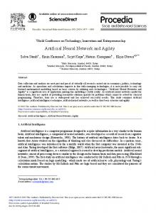
FIG. 1. Alignment of the deduced amino acid sequences of Xenopus laevis metalloprotease– disintegrins with the most highly related mammalian protein sequences. xMDC9 is aligned with human and mouse MDC9 in A, xMDC11a and xMDC11b are aligned with human MDC/ADAM11 in B, and xMDC13 is aligned with ADAM13 in C. Residues present in at least two of the aligned sequences are boxed. Predicted signal sequence cleavage sites (von Heijne, 1986) are marked with an arrowhead. Predicted domain boundaries are marked by a vertical bar with an arrowhead, and the metalloprotease catalytic site consensus sequence HEXXH is surrounded by a hatched box in A and C. The putative integrin binding sequence which is found in lieu of the RGD sequence in snake venom disintegrins is underlined (Wolfsberg and White, 1996). Examples of cytoplasmic proline-rich regions which may function as SH3 ligand domains (Alexandropoulos et al., 1995) are boxed, although it should be noted that other potential signaling motifs and sequences predicting cytoplasmic protein–protein interactions can be found. The cDNA and protein sequences have been deposited with GenBank under the following Accession Ns.: xMDC9, AF032382; xMDC11a, AF032384; xMDC11b, AF032383; xMDC13, AF032385.
Copyright © 1998 by Academic Press. All rights of reproduction in any form reserved.
512
Cai et al.
FIGURE 1
Metalloprotease–Disintegrins in Early Xenopus Development
FIG. 1—Continued Copyright © 1998 by Academic Press. All rights of reproduction in any form reserved.
513
514
Cai et al.
FIG. 1—Continued
515
Metalloprotease–Disintegrins in Early Xenopus Development
TABLE 1 Predicted Features of xMDC9, xMDC11a, xMDC11b, and xMDC13
HEXXH *
Signal Prosequence domain
Metalloprotease domain
Disintegrin domain
Cysteine-rich region
EGF repeat
Amino acid residues
Predicted MW
Metalloprotease catalytic site
Predicted integrin binding sequence
Potential SH-3 binding sequences
xMDC9
873
95.255
+ (HELGH)
ANEC
3
xMDC11a xMDC11b xMDC13
n/a 938 910
n/a 104.135 99.084
µ (QTLGQ) µ (QSLAH) + (HEIGH)
LNEC VNDC AGSC
0 1 4
Xenopus laevis MDCs
TM
Cytoplasmic domain
% Sequence similarity to: hMDC9, 64.1%; mMDC9, 59.6% hMDC11, 75% hMDC11, 51.3% ADAM 13, 90%
Note. The domain organization of a typical metalloprotease– disintegrin is shown above. A catalytic site consensus sequence (HEXXH) is present in xMDC9 and xMDC13, but not in xMDC11a and xMDC11b. The position of the predicted integrin binding sequence in the disintegrin domain is marked by an asterisk. The cytoplasmic domains of xMDC9, xMDC11b, and xMDC13 contain proline-rich sequences that may function as potential SH-3 ligand domains.
approximately 3 kb, which is not detectable in muscle and liver. As a control for equal loading, both Northern blots were probed with a fibronectin probe. It should be noted that expression of the fibronectin gene cannot be detected in A6 kidney epithelial cells, but since xmdc9 and xmdc13 are expressed in these cells, these two probes served to verify the presence of intact mRNA in the A6 cell sample.
In Situ mRNA Hybridization with xmdc9, xmdc11a, xmdc11b, and xmdc13 Probes An important goal of this study was to elucidate the expression pattern of xmdc9, xmdc11a, xmdc11b, and xmdc13 mRNA in early X. laevis embryos. To this end, the localization of these transcripts was examined by wholemount in situ mRNA hybridization in gastrula, neurula and tailbud stage embryos. At the gastrula stage (stage 13), xmdc9 mRNA is localized uniformly in the whole embryo (Fig. 3C). In later stages, xmdc9 transcripts continue to be widely expressed, although increased levels of expression are visible in the somites, notochord, and the head region of the tailbud stage embryos (Figs. 3D–3F). In contrast, xmdc11a expression could not be detected in embryos until neurulation (stage 18/20). At this stage (Fig. 3I) staining is present in a symmetrical pattern on either side of the neural tube. In the anterior part of the embryo, the xmdc11a mRNA is present in three groups of cells (Fig. 3G). This localization is similar to the expression of xap2 (Schuh et al., 1993), xslug (Mancilla and Mayor, 1996), and adam13 (Alfandari et al., 1997), all of which are expressed in the cranial neural crest. In the trunk, xmdc11a differs from
xslug and adam13 in that it extends into more dorsal parts of the embryo. On a section of the trunk of an early tailbud stage embryo (Fig. 3K, stage 22) the staining appears localized to a subset of neuronal cells within the neural tube. This mRNA staining pattern is very similar, but slightly more dorsal compared to that described for the a6 integrin (Lallier et al., 1996). During later development of the tailbud, expression of the xmdc11a mRNA is found in the spaces surrounding the brain and optic vesicle as well as in the mandibular, hyoid and branchial arches (Fig. 3L). These areas are known to be colonized by neural crest cells. xmdc11a mRNA is low or absent in the optic vesicle, as well as between the epidermis (Figs. 3H, 3J, and 3L). At this later stage, xmdc11a expression in the trunk is still restricted to two rows of cells in the dorsal part of the neural tube. The appearance of the strong neural staining pattern at stage 30 correlates with the presence of the xmdc11a band on stage-specific Northern blots (Fig. 2A). Expression of the closely related xmdc11b could not be detected by in situ hybridization or by Northern blot analysis in any of the embryonic stages examined here (data not shown). In contrast to adam13, which is expressed in somitic mesoderm and neural crest cells (Alfandari et al., 1997), expression of the highly related xmdc13 was relatively weak and ubiquitous (data not shown). Several control experiments were undertaken to confirm the specificity of the xmdc9, xmdc11a, and xmdc13 antisense probes. These included sense transcripts as negative control probes and adam13 antisense transcripts as a positive control probe. Staining with the control sense transcript of xmdc9 is shown in Figs. 3A and 3B.
Copyright © 1998 by Academic Press. All rights of reproduction in any form reserved.
516
Cai et al.
FIG. 2. Northern blot analysis of xMDC9, 11a, 11b, and 13 expression during X. laevis development and in adult tissues and A6 cells. (A) A Northern blot containing RNA extracted from 10 embryos at different stages of development (from the unfertilized egg through the swimming tadpole stage), was probed with [32P]dCTP-labeled xmdc9, xmdc11a, and xmdc13 cDNA, as indicated. (B) Northern blots containing 15 mg of total RNA per lane from adult X. laevis testis (lane 1), heart (lane 2), muscle (lane 3), liver (liver 4), and A6 cells (lane 5) were probed sequentially with [32P]dCTP-labeled cDNA fragments of xmdc9, xmdc11a, xmdc11b, and xmdc13, and with X. laevis fibronectin cDNA as control.
Analysis of xMDC9 Protein Expression and Processing Polyclonal antibodies were raised against GST–fusion proteins with the cytoplasmic tail of xMDC9, xMDC11a, xMDC11b to analyze the expression and processing of these MDC proteins in X. laevis tissues and in A6 cells. Preliminary screening for reactivity of the different antisera with the corresponding protein by Western blot analysis was performed using Con A-purified glycoproteins from adult X. laevis heart. Although all antisera reacted well with the
fusion proteins that had been used as antigens, the only MDC protein that could reliably be detected on a tissue Western blot was xMDC9. The failure to detect xMDC11a and 11b may be due to relatively low expression of these proteins and/or an insufficient titer of the antiserum. To further characterize the xMDC9 antiserum, three different antibody samples were generated (see Materials and Methods for details). Protein A-purified IgG were first depleted of antibodies reacting with GST alone, and are referred to as xMDC9 IgG. After depletion of GST-
Copyright © 1998 by Academic Press. All rights of reproduction in any form reserved.
517
Metalloprotease–Disintegrins in Early Xenopus Development
reactive antibodies, the protein A-purified IgG was further depleted of all antibodies reacting with GST– xMDC9 – cytotail fusion protein, and this sample is referred to as control IgG. Finally, antibodies binding to the GST–xMDC9 – cytotail were eluted with 0.1 M glycine, pH 3.0, and these antibodies are referred to as affinity-purified xMDC9 IgG. Blots of Con A-purified glycoproteins from a X. laevis heart lysate were probed with xMDC9 IgG (Fig. 4, lanes 1 and 2), with affinitypurified xMDC9 IgG (lanes 3 and 4), with control IgG (lanes 5 and 6), or with the secondary antibody alone (lane 7). Both the xMDC9 IgG and affinity-purified xMDC9 IgG, but not the control IgG or the secondary antibody alone, recognized a band of 75 kDa under nonreducing conditions, and a band of 95 kDa in samples separated under reducing conditions. A significant increase in the apparent MW after reduction is consistent with the relatively high cysteine content of the extracellular sequence of xMDC9, and is a general feature of metalloprotease– disintegrin proteins. To further confirm the specificity of the xMDC9 antibodies, Western blots of nonreduced extracts of COS-7 cells transfected with xMDC9, or with the expression vector alone, were probed with either anti-xMDC9 IgG or control IgG. In the extract of xMDC9-transfected COS-7 cells, the anti-xMDC9 IgG recognized a 75-kDa band (Fig. 4B, lane 1), which was not present in the vector transfected cells (lane 2), and was also not recognized by the control IgG, (Fig. 4B, lane 3). In Western blots of different X. laevis tissues and A6 cells, the xMDC9 protein was visible as a 75-kDa protein under nonreducing condition (Fig. 4C). The expression level correlated well with the mRNA expression level seen in the Northern blot analysis, with the highest expression in testis, heart, and A6 cells, and relatively low expression in muscle and liver. Immunoblot analysis of various embryonic stages under nonreducing conditions showed that the xMDC9 protein is expressed as a 75-kDa glycoprotein at all stages examined including unfertilized eggs (Fig. 4E), in agreement with the temporal expression pattern of xMDC9 RNA. To determine whether xMDC9 is present on the surface of A6 cells, these cells were cell-surface biotinylated with a non-membrane-permeable biotinylation reagent, and xMDC9 was subsequently immunoprecipitated using xMDC9 IgG. Figure 4D (lane 1) shows that the biotinylated material which can be immunoprecipitated with the xMDC9 IgG consists mainly of a 95-kDa and a weaker 65-kDa band under reducing conditions, and of a 75- and a 48-kDa band under nonreducing conditions. These bands appear to be specific for xMDC9 since they are not immunoprecipitated by the control antibodies. This result indicates that xMDC9 is present on the cell surface of A6 cells, and is consistent with the observation that mouse MDC9 can be cell-surface biotinylated in NIH 3T3 fibroblasts (Weskamp et al., 1996).
DISCUSSION The main goal of this study was to gain a better understanding of the potential functions of four metalloprotease– disintegrins (xmdc9, xmdc11a, xmdc11b, and xmdc13) through an analysis of their expression patterns in developing and adult X. laevis. Our results indicate that expression of xmdc11a is restricted to neural crest derivatives such as cranial and truncal neural crest cells during early X. laevis development, suggesting a role for xmdc11a in the migration or differentiation of cells or tissues derived from the neural crest. xmdc9 is expressed at all stages of development and in all adult tissues analyzed. These results indicate that xmdc9 may have a more general function that is utilized by most or all cells. xmdc11b expression was not observed in developing embryos and was relatively weak in the adult tissues examined, whereas xmdc13 expression was low yet ubiquitous in early embryos and thus differed from the expression pattern of the highly related adam13 (Alfandari et al., 1997) in somitic mesoderm and in cranial neural crest cells. The first step of this study was to attempt to isolate and sequence full-length cDNAs for four metalloprotease– disintegrin PCR sequence tags which had previously been identified in X. laevis testis (Shilling et al., 1997). The protein sequences deduced from the longer cDNA sequences presented here essentially confirm the initial comparison between the PCR sequence tags and other known metalloprotease– disintegrins (Shilling et al., 1997). The present and previous analyses both suggest that two of the cDNA fragments (xMDC9, 11a) have a known putative mammalian orthologue, that a third one is closely related to the human MDC11 protein (xMDC11b), and that one is highly related to X. laevis ADAM13 (Alfandari et al., 1997). Following is a discussion of the noteworthy features and expression pattern of each of these four metalloproteasedisintegrins.
xmdc9 Like human and mouse MDC9, the putative X. laevis xMDC9 contains a metalloprotease domain with a catalytic site consensus sequence HEXXH, and cytoplasmic signaling motifs, including proline-rich putative SH3–ligand domains. In the case of mouse MDC9, these proline-rich sequences have been shown to bind the SH3 domain of src in a blot overlay assay, suggesting that in principle these domains may be able to bind to SH3–ligand domains (Weskamp et al., 1996). The strong conservation of these proline-rich sequences between mouse, human, and X. laevis MDC9 relative to less conserved adjacent cytoplasmic sequences lends further support to the idea that the proline-rich sequences might be important for the function of MDC9. Northern blot analysis revealed expression of xmdc9 at all stages examined, including the unfertilized egg. In situ hybridization further established that xmdc9 mRNA is
518
Cai et al.
519
Metalloprotease–Disintegrins in Early Xenopus Development
uniformly expressed in the whole embryo at the gastrula and neurula stages. In later tailbud stage embryos, xmdc9 mRNA is detectable throughout the embryo, although an elevated level of expression is seen in the somite, head, and notochord region. The pattern of xmdc9 expression in X. laevis embryos is similar to that reported for the metalloprotease– disintegrin kuz, a maternal gene which is first expressed widely in gastrula embryos, whereas at later stages an increased level of expression is detected in neural tissues (Pan and Rubin, 1997). It should be noted that although kuz expression is not restricted to developing neural tissues, it nevertheless has a specific role in neurogenesis. The expression pattern of xmdc9 in all stages of development, including the unfertilized egg, suggests that xMDC9 plays a general role that is utitilized or required by all cells and tissues, although such a general expression does not rule out specific roles in different tissues or developmental stages. Since mouse MDC9 has been shown to have catalytic activity (Roghani et al., manuscript submitted), it is possible that xMDC9 may have a role in protein ectodomain processing, in cell– cell interactions, or both (Blobel, 1997; Weskamp et al., 1996). Antibodies raised against xMDC9 were used to extend the analysis of xMDC9 expression to the protein level and essentially confirmed the results of the Northern blot analysis. Expression of xMDC9 in the unfertilized egg suggests a maternal mRNA contribution, which must be replenished by endogenous expression at later stages of development. In a previous study, a peptide corresponding to the predicted integrin binding sequence of xMDC9, which differs from the sequence of mouse and human MDC9, has been shown to block X. laevis fertilization (Shilling et al., 1997). Since xMDC9 is apparently present on both sperm and egg, this finding raises that possibility that a metalloprotease– disintegrin on the egg might also be involved in binding an integrin-type receptor on the sperm,
in addition to the predicted role of sperm metalloprotease– disintegrins in binding integrins on the egg. In Northern blots, different xmdc9 mRNA transcripts are found in somitic tissues and in testis, indicating that xmdc9 mRNA might be alternatively spliced or have divergent 39 untranslated regions. Yet in all tissues and developmental stages examined here, there was no evidence for different polypeptide species. The observed MW of xMDC9 is similar to that of mouse and human MDC9, and suggests that the predominant form of xMDC9 contains a membrane anchored metalloprotease and disintegrin domain, but lacks a prodomain (Weskamp et al., 1996). In the case of mouse MDC9, furin or a related pro-protein convertase appears to be responsible for removal of the pro-domain in the transGolgi network (Roghani et al., manuscript submitted).
xmdc11a and xmdc11b The high degree of sequence identity between xMDC11a and human MDC11 (Emi et al., 1993; Katagiri et al., 1995), including short and nearly identical cytoplasmic sequences, suggests that these two proteins are orthologues. xMDC11b is clearly more related to xMDC11a and human MDC11 than to other presently known MDC proteins, but has a longer cytoplasmic domain with at least one potential SH3 ligand domain. While the xMDC11a cDNA clone isolated in this study does not contain a full-length open reading frame (it lacks the signal sequence, pro-domain, and parts of the metalloprotease domain), the deduced xMDC11b sequence appears to be full-length because a hydrophobic signal sequence and a pro-domain are present in the open reading frame. The alignment shown in Fig. 1B further suggests that the full-length human MDC/ADAM11 sequence has not yet been determined, since the predicted initial methionine in the reported human protein sequence (Emi et al., 1993; Katagiri et al., 1995) is not followed by a
FIG. 3. Whole mount in situ mRNA hybridization of embryos at different developmental stages with xmdc9 and xmdc11a cRNA, and hybridization of a tailbud stage embryo section with xmdc11a cRNA. Albino X. laevis embryos at various developmental stages were hybridized with an xmdc9 sense probe (A, B), an xmdc9 antisense probe (C–F), or an xmdc11a antisense probe (G–L). All embryos are oriented with their anterior toward the left of the figure. A shows a lateral view of a late neurula embryo (stage 20), and B shows a tailbud embryo (stage 30). Both embryos were hybridized with the sense xmdc9 control probe. C shows that xmdc9 expression can be detected in the entire gastrula stage embryo. In a neurula embryo (E, stage 20) xmdc9 mRNA is present along the entire anteroposterior axis with a more pronounced staining on the dorsal side. At stage 25 (D), an essentially similar localization is observed in the head, the dorsal trunk structures and the tail bud. In addition, a more pronounced staining of the somites is visible. In a dorsal view of a tailbud embryo (F, stage 25), expression of xmdc9 appears strongest in dorsal structures. G and I show a lateral and dorsal view, respectively, of a late neurula embryo (stage 20) hybridized with an xmdc11a antisense probe. The most anterior signal is divided into three populations of cells (arrows) extending from the neural tube. In the trunk, a thin line of cells on either side of the neural tube express xmdc11a (black arrowheads). K shows a transverse section through the trunk of a tailbud embryo (stage 22). Two rows of xmdc11a-expressing cells (small black arrowhead) extend along the sides of the neural tube (nt). The somites (s), gut (g), and notochord structures (nc) are also indicated. In a lateral view of a tailbud stage embryo (H, stage 25), xmdc11a staining is present in four groups of cells (arrows) within the head and branchial arches. J demonstrates that expression in the same anterior structures as in H can be seen in a dorsal view of a tailbud embryo. No xmdc11a expression is detected in the optic vesicle (J, red arrowhead). In the trunk, the staining clearly appears along two stripes on either side of the midline. L depicts a magnification of the anterior region of a tailbud stage embryo. xmdc11a mRNA is localized to the four segments derived from the cranial neural crest. From posterior to anterior, these structures correspond to the posterior branchial crest, the anterior branchial crest, the hyoid crest, and the mandibular crest. The optic vesicle is marked by a red arrowhead, and the brain is indicated (b).
Copyright © 1998 by Academic Press. All rights of reproduction in any form reserved.
520
Cai et al.
hydrophobic signal sequence, and since parts of the prodomain are missing compared to xMDC11b (see Fig. 1B), or other known MDC proteins (data not shown). By Northern blot analysis, xmdc11a expression could first be detected in stage 18 embryos. It is also present in adult testis, heart, and muscle, but not in liver and A6 cells. In contrast, xmdc11b expression was not observed during early development, in A6 cells or adult liver, and was highest in adult testis, followed by heart and muscle. By in situ hybridization, xmdc11a RNA is first detected during neurulation in cells derived from the neuroectoderm. In the head of the tailbud stage embryo, xmdc11a is only detected in the cranial neural crest. The cranial neural crest structures are thought to emerge from the anterior neural tube and migrate ventrally to fill spaces between the neural tube and laterally under the epidermis (Bronner-Fraser, 1993, 1994; Chang and Hemmati-Brivanlou, 1998; Krotoski et al., 1988; Mayor et al., 1995; Sadaghiani and Thiebaud, 1987). These cells give rise to facial structures including cartilage, facial muscles, the sclera of the eye, and the sensory ganglia. Interestingly, the cranial neural crest staining pattern of xmdc11a resembles that of adam13 (Alfandari et al., 1997). This raises the possibility that the two metalloprotease– disintegrins xMDC11a and ADAM13 may interact, as has been described for the a and b subunit of the heterodimeric sperm protein fertilin (Blobel et al., 1990, 1992). In the trunk, transversal sections show that xmdc11a is expressed in the dorsal half of the neural tube on either side of the midline. While we are not aware of a detailed description of the cell types in this region in X. laevis, it seems unlikely that these cells represent neural crest cell precursors because at this stage the truncal neural crest cells have already emerged and begun migrating ventrally. Since xMDC11a does not have a catalytic site in its metalloprotease domain, any role in the neural crest should be quite different from that of catalytically active proteins such as KUZ. xMDC11a may therefore function in cell– cell interactions, perhaps as an integrin ligand, or it could engage other proteins or receptors on the cell surface or
extracellular matrix. It is thus conceivable that xMDC11a has a role in certain aspects of neural crest cell migration, which is thought to require dynamic changes in cell– cell and cell–matrix interactions (Bronner-Fraser, 1993, 1994; Erickson and Perris, 1993; Mayor et al., 1995; MonierGavelle and Duband, 1997).
xmdc13 The fourth cDNA presented here is highly related to X. laevis ADAM13. Because X. laevis is tetraploid, it is not unusual to isolate highly related, but distinct cDNA clones from X. laevis cDNA libraries (DeSimone and Hynes, 1988). While there is a high degree of sequence conservation between ADAM13 and xMDC13, the respective mRNAs have different sizes on a Northern blot, and there are clear differences between the expression of these two genes in embryos and adult tissues. Both observations suggest that adam13 and xmdc13 are not pseudoalleles (DeSimone and Hynes, 1988). During early development, adam13 expression begins at the midblastula transition and is localized to the cranial neural crest, while xmdc13 expression first appears at stage 13, and is weak but ubiquitous in embryos. Furthermore, xmdc13 is expressed in adult testis, whereas adam13 is not. One interpretation of these observations is that xMDC13 and ADAM13 may have distinct functions despite their strong sequence similarity. While ADAM13 most likely is important in early development (Alfandari et al., 1997), peptides corresponding to the predicted integrinbinding site of xMDC13 block X. laevis fertilization in a concentration-dependent manner, suggesting a potential role of xMDC13 in fertilization (Shilling et al., 1997). As described previously (Alfandari et al., 1997), ADAM 13 and xMDC13 belong to a subfamily of metalloprotease– disintegrins that also includes Meltrin a (Yagami-Hiromasa et al., 1995) and Meltrin b (Inoue et al., 1998), but do not appear to be orthologues of either of these two mammalian proteins. In summary, the identification of cDNA clones for
FIG. 4. Western blot analysis of xMDC9 in different tissues, and at different developmental stages. A and B show the characterization of the xMDC9 cytotail antibodies. All lanes in A contain glycoproteins isolated from adult X. laevis heart as described under Materials and Methods, which were treated with or without DTT prior to electrophoresis as indicated. Lanes 1 and 2 were probed with xMDC9 – cyto IgG, lanes 3 and 4 with affinity-purified xMDC9 – cyto IgG, lanes 5 and 6 with control IgG depleted of xMDC9 reactive antibodies, and lane 7 with the secondary antibody alone. The generation of the antibody samples used here is described in more detail under Materials and Methods. In B, a Western blot of glycoproteins from an extract of COS-7 cells expressing xMDC9 (lanes 1 and 3) or of COS-7 cells transfected with the pcDNA3 vector alone (lanes 2 and 4) was probed with xMDC9 – cyto IgG (lanes 1 and 2) or control IgG (lanes 2 and 4). In C, glycoproteins purified from extracts of Xenopus testis (lane 1), heart (lane 2), muscle (lane 3), and liver (lane 4) and from extracts of A6 cells (lane 5) were probed with xMDC9 – cytotail IgG (top) or control IgG (bottom). The 75-kDa band appars to be the only band that is specifically recognized by the xMDC9 –IgG, but not by the control IgG. In D, extracts of cell-surface-biotinylated A6 cells were immunoprecipitated with xMDC9 – cyto IgG (lanes 1 and 2), or with control IgG (lanes 3 and 4), and treated with 10 mM DTT (lanes 1 and 3) or left nonreduced (lanes 2 and 4) prior to SDS–PAGE and then transferred to nitrocellulose. The immunoprecipitated biotinylated material was detected with horseradish peroxidase-coupled streptavidin (see Materials and Methods). E shows a Western blot of glycoprotein extracts from eggs and from embryos at different stages of development. Only the 75-kDa protein was specifically recognized by the xMDC9 –IgG (see arrow), but not by the control antibody (not shown).
Copyright © 1998 by Academic Press. All rights of reproduction in any form reserved.
Metalloprotease–Disintegrins in Early Xenopus Development
521
522
Cai et al.
metalloprotease– disintegrins in X. laevis presented here has unveiled apparent orthologues to mammalian members of this protein family, and has provided new insights into the spatial and temporal expression of these genes in X. laevis. The expression of xmdc11a is highly localized to developing neural tissues, suggesting a role for this gene in the migration or differentiation of tissues derived from the neural crest. The expression of xmdc9 in developing X. laevis is consistent with a general role which may be required in all cells and tissues, but may nevertheless manifest itself differently in diverse tissues. Since X. laevis can be used as a system to evaluate protein function in early development, this study represents an important first step toward analyzing the role of xMDC9, xMDC11a, and xMDC13 in vertebrate development.
ACKNOWLEDGMENTS We thank Ms. Selina Noramly for isolating the xMDC11a and b clones from a cDNA library, H. Cousin and C. Montmory for their excellent assistance in sectioning embryos, Drs. F. Fagotto, L. Jaffe, B. Gumbiner, D. DeSimone, J. L Duband, T. Wolfsberg, L. Howard, L. Lum and J. Schlo¨ndorff for helpful discussions and advice, and Dr. W.-D. Schleuning for his continued interest and encouragement. This work was supported by NIH Grant R55GM51988 to C.P.B., and the Cancer Center Support Grant NCI-P30-CA-08748.
REFERENCES Alexandropoulos, K., Cheng, G., and Baltimore, D. (1995). Prolinerich sequences that bind to Src homology 3 domains with individual specificities. Proc. Natl. Acad. Sci. USA 92, 3110 – 3114. Alfandari, D., Whittaker, C. A., DeSimone, D. W., and Darribere, T. (1995). Integrin alpha v subunit is expressed on mesodermal cell surfaces during amphibian gastrulation. Dev Biol 170, 249 – 61. Alfandari, D., Wolfsberg, T. G., White, J. M., and DeSimone, D. W. (1997). ADAM13: A novel ADAM expressed in somitic mesoderm and neural crest cells during Xenopus laevis development. Dev. Biol. 182, 314 –330. Almeida, E. A. C., Huovila, A.-P. J., Sutherland, A. E., Stephens, L. E., Calarco, P. G., Shaw, L. M., Mercurio, A. M., Sonnenberg, A., Primakoff, P., Myles, D. G., and White, J. M. (1995). Mouse egg integrin a6b1 functions as a sperm receptor. Cell 81, 1095– 1104. Black, R., Rauch, C. T., Kozlosky, C. J., Peschon, J. J., Slack, J. L., Wolfson, M. F., Castner, B. J., Stocking, K. L., Reddy, P., Srinivasan, S., Nelson, N., Boiani, N., Schooley, K. A., Gerhart, M., Davis, R., Fitzner, J. N., Johnson, R. S., Paxton, R. J., March, C. J., and Cerretti, D. P. (1997). A metalloprotease disintegrin that releases tumour-necrosis factor-a from cells. Nature 385, 729 – 733. Blobel, C. P. (1997). Metalloprotease-disintegrins: Links to cell adhesion and cleavage of TNFa and Notch. Cell 90, 589 –592. Blobel, C. P., Myles, D. G., Primakoff, P., and White, J. W. (1990). Proteolytic processing of a protein involved in sperm-egg fusion correlates with acquisition of fertilization competence. J. Cell Biol. 111, 69 –78.
Blobel, C. P., Wolfsberg, T. G., Turck, C. W., Myles, D. G., Primakoff, P., and White, J. M. (1992). A potential fusion peptide and an integrin ligand domain in a protein active in sperm– egg fusion. Nature 356, 248 –252. Bronner-Fraser, M. (1993). Mechanisms of neural crest cell migration. Bioessays 15, 221–30. Bronner-Fraser, M. (1994). Neural crest cell formation and migration in the developing embryo. FASEB J. 8, 699 –706. Brower, D. L., Brabant, M. C., and Bunch, T. A. (1995). Role of the PS integrins in Drosophila development. Immunol. Cell Biol. 73, 558 –564. Chang, C., and Hemmati-Brivanlou, A. (1998). Neural crest induction by Xwnt7B in Xenopus. Dev Biol 194, 129 –34. Chomczynski, P., and Sacchi, N. (1987). Single-step method of RNA isolation by acid guanidium thiocyanate–phenol– chloroform extraction. Anal. Biochem. 162, 156 –159. DeSimone, D. W., and Hynes, R. O. (1988). Xenopus laevis integrins. Structural conservation and evolutionary divergence of integrin beta subunits. J. Biol. Chem. 263, 5333– 40. DeSimone, D. W., Norton, P. A., and Hynes, R. O. (1992). Identification and characterization of alternatively spliced fibronectin mRNAs expressed in early Xenopus embryos. Dev Biol 149, 357– 69. Emi, M., Katagiri, T., Harada, Y., Saito, H., Inazawa, J., Ito, I., Kasumi, F., and Nakamura, Y. (1993). A novel metalloprotease/ disintegrin-like gene at 17q21.3 is somatically rearranged in two primary breast cancers. Nature genetics 5, 151–157. Erickson, C. A., and Perris, R. (1993). The role of cell– cell and cell–matrix interactions in the morphogenesis of the neural crest. Dev Biol 159, 60 –74. Evans, J. P., Kopf, G. S., and Schultz, R. M. (1997a). Characterization of the binding of recombinant mouse sperm fertilin beta subunit to mouse eggs: Evidence for adhesive activity via an egg b1 integrin-mediated interaction. Dev. Biol. 187, 79 –93. Evans, J. P., Schultz, R. M., and Kopf, G. S. (1995). Mouse sperm-egg plasma membrane interactions: Analysis of roles of egg integrins and the mouse homologue of PH-30 (fertilin) b. J. Cell Sci. 108, 3267–3278. Evans, J. P., Schultz, R. M., and Kopf, G. S. (1997b). Characterization of the binding of recombinant mouse sperm fertilin alpha subunit to mouse eggs: evidence for function as a cell adhesion molecule in sperm– egg binding. Dev. Biol. 187, 94 –106. Fambrough, D., Pan, D., Rubin, G. M., and Goodman, C. S. (1996). The cell surface metalloprotease/disintegrin kuzbanian is required for axonal extension in Drosophila. Proc. Natl. Acad. Sci. USA 93, 13233–13238. Fa¨ssler, R., Georges-Labouesse, E., and Hirsch, E. (1996). Genetic analysis of integrin function in mice. Curr. Opin. Cell Biol. 8, 641– 646. Finelli, A. L., Bossie, C. A., Xie, T., and Padgett, R. W. (1994). Mutational analysis of the Drosophila tolloid gene, a human BMP-1 homolog. Development 120, 861– 870. Gumbiner, B. M. (1996). Cell adhesion: The molecular basis of tissue architecture and morphogenesis. Cell 84, 345–357. Harland, R. M. (1991). In situ hybridization: An improved wholemount method for Xenopus embryos. Methods Cell Biol. 36, 685–95. Harlow, E., and Lane, D. (1988). “Antibodies: A Laboratory Manual.” Cold Spring Harbor Laboratories, Cold Spring Harbor, NY.
Copyright © 1998 by Academic Press. All rights of reproduction in any form reserved.
523
Metalloprotease–Disintegrins in Early Xenopus Development
Harris, H. A., Murrills, R. J., and Komm, B. S. (1997). Expression of meltrin-alpha mRNA is not restricted to fusagenic cells. J. Cell Biochem. 67, 136 – 42. Heinlein, U. A. O., Wallat, S., Senftleben, A., and Lemaire, L. (1994). Male germ cell-expressed mouse gene TAZ83 encodes a putative, cysteine rich transmembrane protein (cyritestin) sharing homologies with snake venom toxins and sperm egg fusion proteins. Dev. Growth Differ. 36, 49 –58. Hooper, N. M., Karran, E. H., and Turner, A. J. (1997). Membrane protein secretases. Biochem. J. 321, 265–279. Howard, L., and Glynn, P. (1995). Membrane-associated metalloproteinase recognized by characterisitic cleavage of myelin basic protein: assay and isolation. In “Proteolytic Enzymes: Aspartic and Metalloproteases” (A. J. Barrett, Ed.), Vol. 248, pp. 388 –395. Academic Press, San Diego, CA. Howard, L., Lu, X., Mitchell, S., Griffiths, S., and Glynn, P. (1996). Molecular cloning of MADM: A catalytically active disintegrinmetalloprotease expressed in various cell types. Biochem. J. 317, 45–50. Huovila, A. P. J., Almeida, E. A., and White, J. M. (1996). ADAMs and cell fusion. Curr. Opin. Cell Biol. 8, 692–9. Hynes, R. O. (1996). Targeted mutations in cell adhesion genes: What have we learned from them? Dev. Biol. 180, 402– 412. Inoue, D., Reid, M., Lum, L., Kra¨tzschmar, J., Weskamp, G., Myung, Y. M., Baron, R., and Blobel, C. P. (1998). Cloning and initial characterization of mouse meltrin beta and analysis of the expression of four metalloprotease– disintegrins in bone cells. J. Biol. Chem. 273, 4180 – 4187. Katagiri, T., Harada, Y., Emi, M., and Nakamura, Y. (1995). Human metalloprotease/disintegrin-like (MDC) gene: Exon–intron organization and alternative splicing. Cytogenet. Cell Genet. 68, 39 – 44. Krotoski, D. M., Fraser, S. E., and Bronner-Fraser, M. (1988). Mapping of neural crest pathways in Xenopus laevis using interand intraspecific cell markers. Dev Biol 127, 119 –32. Lallier, T. E., Whittaker, C. A., and DeSimone, D. W. (1996). Integrin a6 expression is required for early nervous system development in Xenopus laevis. Development 122, 2539 –2554. Linder, B., and Heinlein, U. A. (1997). Decreased in vitro fertilization efficiencies in the presence of specific cyritestin peptides. Dev. Growth Differ. 39, 243–7. Lunn, C. A., Fan, X., Dalie, B., Miller, K., Zavodny, P. J., Narula, S. K., and Lundell, D. (1997). Purification of ADAM10 from bovine spleen as TNFalpha convertase. FEBS Lett. 400, 333–335. Mancilla, A., and Mayor, R. (1996). Neural crest formation in Xenopus laevis: Mechanisms of Xslug induction. Dev Biol 177, 580 –9. Marques, G., Musacchio, M., Shimell, M. J., WunnenbergStapleton, K., Cho, K. W., and O’Connor, M. B. (1997). Production of a DPP activity gradient in the early Drosophila embryo through the opposing actions of the SOG and TLD proteins. Cell 91, 417–26. Mayor, R., Morgan, R., and Sargent, M. G. (1995). Induction of the prospective neural crest of Xenopus. Development 121, 767–77. Monier-Gavelle, F., and Duband, J. L. (1997). Cross talk between adhesion molecules: control of N-cadherin activity by intracellular signals elicited by beta1 and beta3 integrins in migrating neural crest cells. J. Cell Biol. 137, 1663– 81. Moss, M. L., Jin, S.-L. C., Milla, M. E., Burkhart, W., Cartner, H. L., Chen, W.-J., Clay, W. C., Didsbury, J. R., Hassler, D., Hoffman, C. R., Kost, T. A., Lambert, M. H., Lessnitzer, M. A., McCauley, P., McGeehan, G., Mitchell, J., Moyer, M., Pahel, G., Rocque, W.,
Overton, L. K., Schoenen, F., Seaton, T., Su, J.-L., Warner, J., Willard, D., and Becherer, J. D. (1997). Cloning of a disintegrin metalloproteinase that processes precursor tumour-necrosis factor-a. Nature 385, 733–736. Myles, D. G., Kimmel, L. H., Blobel, C. P., White, J. M., and Primakoff, P. (1994). Identification of a binding site in the disintegrin domain of fertilin required for sperm– egg fusion. Proc. Natl. Acad. Sci. USA 91, 4195– 4198. Pan, D., and Rubin, J. (1997). KUZBANIAN controls proteolytic processing of NOTCH and mediates lateral inhibition during Drosophila and vertebrate neurogenesis. Cell 90, 271–280. Piccolo, S., Agius, E., Lu, B., Goodman, S., Dale, L., and De Robertis, E. M. (1997). Cleavage of Chordin by Xolloid metalloprotease suggests a role for proteolytic processing in the regulation of Spemann organizer activity. Cell 91, 407–16. Podbilewicz, B. (1996). ADM-1, a protein with metalloproteaseand disintegrin-like domains, is expressed in syncitial organs, sperm and sheath cells of sensory organs in Caenorhabditis elegans. Mol. Biol. Cell 7, 1877–1893. Primakoff, P., Hyatt, H., and Tredick-Kline, J. (1987). Identification and purification of a sperm surface protein with a potential role in sperm– egg membrane fusion. J. Cell Biol. 104, 141–149. Ramos, J. W., and DeSimone, D. W. (1996). Xenopus embryonic cell adhesion to fibronectin: position-specific activation of RGD/ synergy site-dependent migratory behavior at gastrulation. J Cell Biol 134, 227– 40. Ramos, J. W., Whittaker, C. A., and DeSimone, D. W. (1996). Integrin-dependent adhesive activity is spatially controlled by inductive signals at gastrulation. Development 122, 2873– 83. Roodman, G. D. (1996). Advances in bone biology: the osteoclast. Endocrin. Rev. 17, 308 –332. Rooke, J., Pan, D., Xu, T., and Rubin, G. M. (1996). KUZ, a conserved metalloprotease– disintegrin protein with two roles in Drosophila neurogenesis. Science 273, 1227–1230. Rosendahl, M. S., Ko, S. C., Long, D. L., Brewer, M. T., Rosenzweig, B., Hedl, E., Anderson, L., Pyle, S. M., Moreland, J., Meyers, M. A., Kohno, T., Lyons, D., and Lichenstein, H. S. (1997). Identification and characterization of a pro-tumor necrosis factor- alpha-processing enzyme from the ADAM family of zinc metalloproteases. J. Biol. Chem. 272, 24588 –93. Sadaghiani, B., and Thiebaud, C. H. (1987). Neural crest development in the Xenopus laevis embryo, studied by interspecific transplantation and scanning electron microscopy. Dev. Biol. 124, 91–110. Schuh, T. J., Hall, B. L., Kraft, J. C., Privalsky, M. L., and Kimelman, D. (1993). v-erbA and citral reduce the teratogenic effects of all-trans retinoic acid and retinol, respectively, in Xenopus embryogenesis. Development 119, 785–98. Shilling, F. M., Kra¨tzschmar, J., Cai, H., Weskamp, G., Gayko, U., Leibow, J., Myles, D. G., Nuccitelli, R., and Blobel, C. P. (1997). Identification of metalloprotease/disintegrins in Xenopus laevis testis with a potential role in fertilization. Dev. Biol. 186, 155–164. Shimell, M. J., Ferguson, E. L., Childs, S. R., and O’Connor, M. B. (1991). The Drosophila dorsal–ventral patterning gene tolloid is related to human bone morphogenic protein 1. Cell 67, 469 – 481. Sotillos, S., Roch, F., and Campuzano, S. (1997). The metalloprotease-disintegrin Kuzbanian participates in Notch activation during growth and patterning of Drosophila imaginal discs. Development 124, 4769 – 4779. von Heijne, G. (1986). A new method for predicting signal sequence cleavage sites. Nucleic Acids Res. 14, 4683– 4690. Wen, C., Metzstein, M. M., and Greenwald, I. (1997). SUP-17, a Caenorhabditis elegans ADAM protein related to Drosophila
Copyright © 1998 by Academic Press. All rights of reproduction in any form reserved.
524
Cai et al.
KUZBANIAN, and its role in LIN-12/NOTCH signaling. Development 124, 4759 – 4767. Weskamp, G., Kra¨tzschmar, J. R., Reid, M., and Blobel, C. P. (1996). MDC9, a widely expressed cellular disintegrin containing cytoplasmic SH3 ligand domains. J. Cell Biol. 132, 717–726. Wolfsberg, T. G., Bazan, J. F., Blobel, C. P., Myles, D. G., Primakoff, P., and White, J. M. (1993). The precursor region of a protein active in sperm– egg fusion contains a metalloprotease and a disintegrin domain: Structural, functional and evolutionary implications. Proc. Natl. Acad. Sci. USA 90, 10783–10787. Wolfsberg, T. G., and White, J. M. (1996). ADAMs in fertilization and development. Dev. Biol. 180, 389 – 401.
Yagami-Hiromasa, T., Sato, T., Kurisaki, T., Kamijo, K., Nabeshima, Y., and Fujisawa-Sehara, A. (1995). A metalloprotease– disintegrin participating in myoblast fusion. Nature 377, 652– 656. Yuan, R., Primakoff, P., and Myles, D. G. (1997). A role for the disintegrin domain of cyritestin, a sperm surface protein belonging to the ADAM family, in mouse sperm– egg plasma membrane adhesion and fusion. J. Cell Biol. 137, 105–112. Received for publication May 7, 1998 Revised July 13, 1998 Accepted July 14, 1998
Copyright © 1998 by Academic Press. All rights of reproduction in any form reserved.