frame into the CNN. Mapping cortical activations with natural movie stimuli. Each segment of the training movie was pre-
Neural Encoding and Decoding with Deep Learning for Dynamic Natural Vision Haiguang Wen
2,3 †
, Junxing Shi
2,3 †
How does the brain represent visual information from the outside world? Here, we approach this question with a deep convolutional neural network that mimics neuronal circuitry and coding, and learns to solve computer vision tasks. Using this network as a computational model of the visual cortex, we develop novel encoding and decoding models to describe the bi-directional relationships between visual input and cortical activity measured with functional magnetic resonance imaging. Testing these models with imaging data from humans watching natural movies, we show that the encoding model can predict cortical responses and retrieve visual representations at individual brain locations, and that the decoding model can decipher the measured cortical activity to reconstruct the visual and semantic experiences. Both the encoding and decoding models utilize cortical representations of hierarchical, invariant, and nonlinear visual features. Being self-contained, efficient, and generalizable, these models constitute a computational workbench for high-throughput investigation of all stages of visual processing. We also anticipate that the general strategy for neural encoding and decoding via deep-learning models will be applicable to other sensory or cognitive experiences, e.g. speech, imagery, memories and dreams. Neural encoding | brain decoding | deep learning | natural vision
Significance Statement: this study brings major advances in encoding and decoding cortical activity that supports human natural vision. For encoding, we demonstrate the unique promise of using deep learning to model and visualize the functional representations at the level of single cortical locations along the entire visual pathway, and to create a computational workbench for high-throughput vision research. For decoding, we present a stand-alone, efficient, reliable, and generalizable strategy to decode cortical fMRI activity to directly reconstruct the visual and semantic experiences during natural vision. These unique capabilities highlight a promising emerging direction of using the artificial brain to understand the biological brain.
Introduction For centuries philosophers and scientists have tried to speculate, observe, understand, and eventually decode †
2,3
2,3
, Yizhen Zhang , Kun-Han Lu , Zhongming Liu
*1,2,3
the workings of the brain that enables the human natural vision. The central questions are how the brain represents visual information from the outside world, and whether one may decode brain activity to reconstruct what a person is seeing. These questions, generally known as neural encoding and decoding, have been mostly approached with overly simplified strategies that use artificial patterns or static pictures as visual stimuli. However, it remains largely unknown how dynamic and realistic visual experiences are represented along the entire visual pathway. What is needed is an alternative strategy that embraces the complexity of vision to fully uncover and decode the visual representations of distributed cortical activity. Despite its diversity and complexity, the visual world is composed of a finite number of hierarchical and invariant visual features (Zeiler and Fergus, 2014; LeCun et al., 2015; Russ and Leopold, 2015), including those in the low-level visual space (e.g. orientation and color), the middle levels (e.g. shape and texture), and the highlevel semantic space (e.g. face and house). In the brain, these features emerge from cascaded stages of visual processing (Felleman and Van Essen, 1991; Dicarlo et al., 2012; Russakovsky et al., 2015) via complex neural circuits. To decipher natural vision, a more effective strategy is to identify and decode neural representations of visual features at all levels, rather than attempting to relate brain activity to infinite pixel combinations. In this regard, we model neural representations using visual features extracted by brain-inspired deep learning (LeCun et al., 2015): a class of deep artificial neural networks trained to emulate or surpass human performance in computer-vision tasks (Russakovsky et al., 2015). Recent studies also show that such network models are well aligned to and predictive of cascaded cortical processes underlying visual perception (Khaligh-Razavi and Kriegeskorte, 2014; Yamins et al., 2014; Güçlü and van Gervan, 2015a,b; Cichy et al., 2016). In addition, the model is fully observable and computable both forward and backward (Zeiler and Fergus, 2014), such that the extracted features can be transformed, either top-down or bottom-up, to visualize their internal representations, to reconstruct the visual input, as well as to deduce its semantic categorization.
Haiguang Wen and Junxing Shi contributed equally to the work. *Correspondence:
[email protected] 2 3 Weldon School of Biomedical Engineering, School of Electrical and Computer Engineering, Purdue Institute for Integrative Neuroscience, Purdue University, West Lafayette, Indiana, 47906, USA. 1
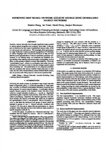
Figure. 1. Neural encoding and decoding through a deep learning model. When a person is seeing a film (a), information is processed through a cascade of cortical areas (b), generating fMRI activity patterns (c). A deep convolutional neural network is used here to model cortical visual processing (d). This model transforms every movie frame into multiple layers of features, st th ranging from orientations and colors in the visual space (the 1 layer) to object categories in the semantic space (the 8 layer). For encoding, this network serves to model the nonlinear relationship between the movie stimuli and the response at each cortical st th location. For decoding, cortical responses are combined across locations to estimate the feature outputs from the 1 and 8 layer. The former is deconvolved to reconstruct every movie frame, while the latter outputs the semantic descriptions.
Here, we explore this new strategy for encoding and decoding natural vision. We acquired functional magnetic resonance imaging (fMRI) data from three healthy subjects watching natural movies with their eyes fixated at the screen center. The stimuli included two different sets of video clips: one for training the encoding and decoding models, and the other for testing them. Our goals were 1) to develop the encoding model to predict fMRI responses and retrieve visual representations at individual cortical locations, and 2) to develop the decoding model to reconstruct the visual and semantic experiences based on fMRI activity. Towards these goals, we used a deep convolutional neural network (CNN) (Krizhevsky et al., 2012) as a fully accessible model of the human visual cortex. For encoding, the CNN-extracted visual features were projected onto cortical activity; for decoding, cortical activity was converted to image and semantic feature representations. Fig. 1 illustrates our encoding and decoding strategy.
Materials and Methods Experiments. Three healthy volunteers (female, age: 22-25) watched natural color video clips (20.3o×20.3o) with a central fixation cross (0.8o×0.8o). All subjects
were healthy volunteers with normal vision. Informed written consent was obtained from every subject according to the research protocol approved by the Institutional Review Board at Purdue University. In total 276 video clips were included in a 2.4-hour training movie, randomly split into 18 8-min segments; 38 different video clips were included in an 8-min testing movie. Each subject watched the training movie twice and the testing movie ten times with their eyes fixated to a central cross. Experiments were done during several days in ~2 weeks; each day included multiple sessions; in each session, an 8-min movie segment was presented as the visual stimulation. The order of the movie segments was random and counter-balanced across subjects. Visual stimuli were delivered through a goggle system (NordicNeuroLab NNL Visual System). The display resolution was 800×600. The stimulus presentation was controlled by using Psychophysics Toolbox 3 (http://psychtoolbox.org). The movies were displayed on a black background and scaled to 600×600 pixel arrays.
Data Acquisition and Preprocessing. T1 and T2weighted MRI and fMRI data were acquired in a 3 tesla MRI system (Signa HDx, General Electric, Milwaukee) with a 16-channel receive-only phase-array surface coil (NOVA Medical, Wilmington). The fMRI data were acquired at 3.5-mm isotropic spatial resolution and 2-s temporal resolution by using a single-shot, gradientrecalled echo-planar imaging sequence (38 interleaved axial slices with 3.5 mm thickness and 3.5×3.5 mm2 inplane resolution, TR/TE=2000/35ms, flip angle=78°, field of view=22×22 cm2). All fMRI images were coregistered, preprocessed, and transformed onto the cortical surfaces by using the processing pipeline developed for the Human Connectome Project, as described in (Glasser et al., 2013). When training and testing the encoding and decoding models (as described later), the cortical fMRI signals were averaged over multiple repetitions of the same movie segment: two repetitions for the training movie and 10 repetitions for the testing movie. Convolutional Neural Network (CNN). We used a deep CNN (also known as the AlexNet) as a model of the visual cortex to extract hierarchical visual features from the movie stimuli. This model has been pretrained to achieve the best-performing object recognition in Large Scale Visual Recognition Challenge 2012 (Krizhevsky et al., 2012). This CNN includes eight layers of artificial neurons stacked into a hierarchical architecture: the first five are convolutional layers, and the last three layers are fully connected for object classification. In each convolutional layer, artificial neurons encode a number of features, each of which represents a kernel convolved over its inputs. Layer 1 through 5 consists of 96, 256, 384, 384, and 256 kernels, respectively. Each convolutional layer is composed of some or all of the following four stages: linear filtering, nonlinear transformation, max-pooling, and divisive normalization. For classification, layer 6 and 7 are fully connected networks with a rectified linear threshold; layer 8 uses a softmax function to output a vector of probabilities by which the input image belongs to individual categories. The numbers of artificial neurons in layer 6 to 8 are 4096, 4096, and 15. Unlike the original model, we reduced the number of neurons in the output layer from 1000 to 15. The modified categories were indoor, outdoor, people, face, bird, insect, water animal, land animal, flower, fruit, natural scene, car, airplane, ship, exercise. We retrained the last classification layer to have achieved a top-1 test error rate of 14.8%. The retraining images were a subset of ImageNet with ~22,500 training images and 3,000 testing images within the 15 categories. The parameters were optimized using gradient descent with weight decay to minimize the multinomial classification error.
Passing a natural image into the CNN yields an activation value from each artificial neuron; the artificial neurons with a same kernel collectively output a feature map. In this study, we extracted the time-varying feature maps by passing all movie segments frame by frame into the CNN. Mapping cortical activations with natural movie stimuli. Each segment of the training movie was presented twice to each subject. To find all cortical locations activated by such natural stimuli, for each voxel we computed the cross correlation between the fMRI voxel time series when each subject watched the same movie segment for the first vs. second time. The correlation coefficient was converted to a z score by using the Fisher z-transformation. The voxel-wise z scores were averaged across all 18 segments and tested for significance by using one-sample t-test (p