Neuropsychologia 45 (2007) 32–41
Neural systems for recognition of familiar faces M. Ida Gobbini a,b,∗ , James V. Haxby a a
Department of Psychology, Princeton University, Princeton, NJ 08544, United States b Department of Psychology, Medical School, University of Bologna, Italy Available online 22 June 2006
Abstract Immediate access to information about people that we encounter is an essential requirement for effective social interactions. In this manuscript we briefly review our work and work of others on familiar face recognition and propose a modified version of our model of neural systems for face perception with a special emphasis on processes associated with recognition of familiar faces. We argue that visual appearance is only one component of successful recognition of familiar individuals. Other fundamental aspects include the retrieval of “person knowledge” – the representation of the personal traits, intentions, and outlook of someone we know – and the emotional response we experience when seeing a familiar individual. Specifically, we hypothesize that the “theory of mind” areas, that have been implicated in social and cognitive functions other than face perception, play an essential role in the spontaneous activation of person knowledge associated with the recognition of familiar individuals. The amygdala and the insula, structures that are involved in the representation of emotion, also are part of the distributed network of areas that are modulated by familiarity, reflecting the role of emotion in face recognition. © 2006 Elsevier Ltd. All rights reserved.
1. Introduction The recognition of familiar individuals is critical for appropriate social interactions. The capacity to readily access information about a person we encounter determines how we should interact with that particular individual. It is a common experience that our behavior changes rapidly based on whether we are interacting with a friend, a son or daughter, or the boss, and that this “changing of gears” is totally automatic. In the present manuscript, we propose a model for the distributed neural systems that participate in the recognition of familiar faces, highlighting that this spatially distributed process involves not only visual areas but also areas that primarily have cognitive and social functions other than visual perception. We hypothesize that visual familiarity is only a partial aspect of how we recognize familiar individuals and that person knowledge and emotional responses are also essential requirements for the successful identification of someone we know. We will focus our attention on different aspects of person knowledge that are activated during face recognition. “Person knowledge” refers to a broad class of information about an individual that encompasses subjective characteristics, such as personal traits, intentions, attitudes, and transient mental states, and ∗
Corresponding author. Tel.: +1 609 258 1168; fax: +1 609 258 1113. E-mail address:
[email protected] (M.I. Gobbini).
0028-3932/$ – see front matter © 2006 Elsevier Ltd. All rights reserved. doi:10.1016/j.neuropsychologia.2006.04.015
objective information, such as biographical facts and episodic memories. In particular, we will highlight the role of a set of areas that have been associated with the performance of tasks involving “theory of mind”. Theory of mind refers to the capacity to represent the mental state of others and to interpret and predict someone else’s behavior based on that representation (Gallagher & Frith, 2003; Leslie, 1994). The anterior paracingulate cortex, the posterior superior temporal sulcus (pSTS)/temporoparietal junction (TPJ), and the precuneus have been associated with “theory of mind” tasks (Frith & Frith, 1999). We are proposing a new function of this set of areas: the spontaneous retrieval of some aspects of person knowledge in the act of face recognition. Finally, we propose that the emotional response to a familiar individual plays a key role in person recognition. The modulation of activity in the amygdala and in the insula based on familiarity supports the hypothesis that these structures play a fundamental role during social interactions and familiarity recognition. Numerous neuroimaging experiments on familiar face recognition exist in the literature, but the findings in the face-responsive regions of the ventral extrastriate cortex have been inconsistent (Dubois et al., 1999; Gobbini, Leibenluft, Santiago, & Haxby, 2004; Gorno-Tempini et al., 1998; Henson, Shallice, & Dolan, 2000; Leibenluft, Gobbini, Harrison, & Haxby, 2004; Leveroni et al., 2000; Nakamura et al., 2000; Rossion, Schiltz, Robaye, Pirenne, & Crommelinck, 2001; Rotshtein, Henson, Treves, Driver, & Dolan, 2005). In some
M.I. Gobbini, J.V. Haxby / Neuropsychologia 45 (2007) 32–41
cases, familiar faces evoked a stronger response (Henson et al., 2000; Leveroni et al., 2000; Rotshtein et al., 2005), in other cases a weaker response (Dubois et al., 1999; Rossion et al., 2001), and in others no modulation at all (Gorno-Tempini et al., 1998; Shah et al., 2001). The inconsistency of these findings could be due to the different types of familiar faces that were used in these experiments. In some experiments, the effect of simple, experimentally learned visual familiarity has been investigated (Dubois et al., 1999; Leveroni et al., 2000; Rossion et al., 2001), in others the familiarity associated with individuals known through the media (Gorno-Tempini et al., 1998; Henson et al., 2000; Leveroni et al., 2000; Sergent, Ohta, & MacDonald, 1992) and in other cases the familiarity associated with personal acquaintances (Bartels & Zeki, 2000; Gobbini et al., 2004; Leibenluft et al., 2004; Nakamura et al., 2000). In contrast to the inconsistent effect of familiarity on responses in the extrastriate visual areas, anterior temporal regions consistently show stronger responses to a variety of familiar stimuli, including faces (Gorno-Tempini et al., 1998; Leveroni et al., 2000; Nakamura et al., 2000; Rotshtein et al., 2005; Sergent et al., 1992), names (Gorno-Tempini et al., 1998; Grabowski et al., 2001) and landscapes (Nakamura et al., 2000), suggesting a possible role for these anatomical structures in the retrieval of biographical or autobiographical information. Medial temporal regions also respond more strongly to familiar, as compared to unfamiliar, faces and names, probably reflecting the general role of these regions in long-term memory retrieval (Douville et al., 2005; Haist, Bowden Gore, & Mao, 2001; Leveroni et al., 2000). We argue that visual familiarity and biographical information are only two aspects of the recognition of familiar individuals. We propose that the emotional response that we experience when we meet someone we know and the spontaneous retrieval of information about that person’s personality, outlook, and intentions are also integral components of the representation of a familiar individual that play key roles in recognition. Our hypotheses are based largely on the results of two fMRI experiments that we have conducted to investigate the effect of the type of familiarity that accrues naturally with years of social interactions and long exposure. In the present manuscript, we briefly describe the findings of these two fMRI experiments and the relevant related literature; then we describe the areas we identified and amplify our hypothesis about their role in the representation of familiar individuals. Finally, we propose a modified and updated version of our model for the human face perception neural system (Haxby, Hoffman, & Gobbini, 2000) with a specific focus on the components that play a role in familiar face recognition.
33
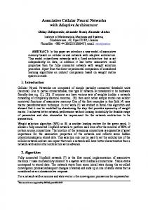
and to faces of strangers (see Gobbini et al., 2004 for details on the experimental design). In the second fMRI experiment, we studied mothers looking at pictures of their own child, at pictures of familiar unrelated children, and at pictures of unfamiliar children (see Leibenluft et al., 2004 for details on the experimental design). The task used by our participants during the fMRI sessions was a “one back repetition detection task” based on the identity of different pictures of the same individual. The purpose of this task was to maintain attention to the stimuli without explicitly requiring the retrieval of semantic information about the faces being viewed. The results of these experiments revealed that activity in the extrastriate visual cortex demonstrated a complex modulation by the type of familiarity. The faces of strangers evoked a stronger response in face-responsive regions of the fusiform gyrus than did famous familiar faces, but the most familiar faces (the faces of family members and friends) evoked a stronger response than did the famous familiar faces. The responses evoked by faces of personally familiar individuals and strangers in the fusiform gyrus did not differ. Similarly, a stronger response in fusiform cortex was observed in mothers viewing faces of unfamiliar children as compared to viewing faces of familiar unrelated children, but the face of one’s own child evoked a stronger response than the faces of familiar unrelated children. Thus, the strength of familiarity did not have a simple monotonic effect on neural activity in this region. By contrast, we observed a more direct relationship between the strength of familiarity and the neural response in the anterior paracingulate cortex, the pSTS/TPJ, and the precuneus (Fig. 1), with stronger responses for more familiar faces. Finally, the different types of familiar faces modulated neural activity in areas associated with emotional response, such as the amygdala and the insula. Surprisingly, even if the more familiar faces are associated with more emotion, the amygdala showed a weaker response to the faces of family members and friends as compared to the famous familiar faces, and the famous familiar faces evoked a weaker response as compared to the faces of strangers. In the experiment with the mothers, the familiar unrelated children evoked a weaker response in the amygdala as compared to the faces of unfamiliar children, but the face of one’s own child evoked a stronger response. Moreover, looking at the face of one’s own child also evoked a stronger response in the insula. Thus, in general the effect of familiarity is a decrease of neural activity in the amygdala with the notable exception of mothers looking at their own child. Stronger responses in the amygdala and insula may reflect the special intensity and protectiveness of maternal attitudes toward one’s own children. 3. Retrieval of person knowledge
2. fMRI studies of recognition of personally familiar and famous faces To investigate the roles of social attachment and emotion in the representation of familiar individuals, we designed two fMRI experiments employing different types of familiar faces. In one fMRI experiment we compared the response to personally familiar faces (faces of friends and family), to famous familiar faces,
A key component of the neural representation of a familiar individual concerns information about that person, such as personal traits, intentions, attitudes, transient mental states, biographical information and episodic memory. We use the term “person knowledge” to refer to this broad class of information. Our hypothesis is that, the “theory of mind” areas encode aspects related to personal traits, intentions and transient mental
34
M.I. Gobbini, J.V. Haxby / Neuropsychologia 45 (2007) 32–41
Fig. 1. Example of activation of the anterior paracingulate cortex, posterior superior temporal sulcus (“theory of mind” areas) and precuneus for the contrast “personally familiar faces” vs. “famous familiar faces (A) and for the contrast “one’s own child” vs. “familiar unrelated children” (B). The more familiar faces (faces of family members and friends and faces of familiar unrelated children and the face of one’s own child) evoked a stronger response in these areas suggesting their role in retrieval of person knowledge.
states (Allison, Puce, & McCarthy, 2000; Mitchell, Heatherton, & Macrae, 2002), while the precuneus and the anterior temporal areas are involved in retrieval of episodic memory (Burgess, Maguire, Spiers, & O’Keefe, 2001; Fletcher et al., 1995) and biographical information (Damasio, Grabowski, Tranel, Hichwa, & Damasio, 1996; Ellis, Young, & Critchley, 1989; GornoTempini et al., 1998; Grabowski et al., 2001; Leveroni et al., 2000; Nakamura et al., 2000; Rotshtein et al., 2005; Sergent et al., 1992). 3.1. Personal traits and intentions: role of the “theory of mind” areas in person knowledge We found that personally familiar faces evoked a stronger response in areas associated with the representation of the mental states of others (Gobbini et al., 2004; Leibenluft et al., 2004). The anterior paracingulate cortex and the posterior superior temporal sulcus are consistently activated by tasks involving theory of mind (Castelli, Happe, Frith, & Frith, 2000; Frith & Frith, 1999; Gallagher et al., 2000, 2003; Saxe & Kanwisher, 2003). Moreover, tasks that require monitoring one’s own mental state and self-reflection (Gusnard & Raichle, 2001; Kelley et al., 2002) also evoke a stronger hemodynamic response in these areas. Activity in these areas seems to be independent of the modality of input. For example, movements of geometrical figures that evoke the attribution of specific mental states in the classic Heider and Simmel animations elicit activity in these areas (Castelli et al., 2000). “Theory of mind” areas also are activated during competitive games, when the
participants believe they are playing against a human partner rather than against a computer (Gallagher, Jack, Roepstorff, & Frith, 2002; McCabe, Houser, Ryan, Smith, & Trouard, 2001; Rilling, Sanfey, Aronson, Nystrom, & Cohen, 2004), and when making moral decisions that involve awareness of the direct consequences of these decisions on a victim who is clearly represented as an individual (Greene, Sommerville, Nystrom, Darley, & Cohen, 2001). In two other studies of strong personal attachment, namely romantic and maternal love, however, weaker activity in theory of mind areas was observed (Bartels & Zeki, 2000, 2004). The inconsistency between this finding in mothers viewing their children and our results (Leibenluft et al., 2004) may be due to their use of infant pictures, whereas our stimuli were pictures of children age 6–12 years. The anterior paracingulate cortex and the pSTS/TPJ seem to play different roles in interpreting the mental states of others. The anterior paracingulate may be involved more in the representation of the personal traits (Mitchell et al., 2002) and mental states of others (Calder et al., 2002; Frith & Frith, 1999), whereas the pSTS/TPJ may play a more general role in social cognition, especially the evaluation of the intentions of others (Allison et al., 2000; Hoffman & Haxby, 2000; Puce & Perrett, 2003; Winston, Strange, O’Doherty, & Dolan, 2002). Gaze perception can also be an indicator of the mental state and of the intentions of someone else, and gaze perception evokes activity in both the pSTS (Hoffman & Haxby, 2000; Puce, Allison, Bentin, Gore, & McCarthy, 1998) and in the anterior paracingulate cortex (Calder et al., 2002).
M.I. Gobbini, J.V. Haxby / Neuropsychologia 45 (2007) 32–41
Functional imaging research has highlighted the engagement of the anterior paracingulate cortex in tasks that involve self-referential processing (Fossati et al., 2003; Kelley et al., 2002), during monitoring of the current affective state (Gusnard, Raichle & Raichle, 2001) or during the retrieval of autobiographical memories (Maguire, 2001). A recent study has addressed the role of the anterior paracingulate cortex during self reflection and while inferring the mental state of others (Mitchell, Banaji, & Macrae, 2005). In this study a dorsal region of the anterior paracingulate cortex was associated with “mentalizing” about others (in a location similar to the areas that we identified in our fMRI experiments on recognition of familiar faces) whereas a ventral portion of the anterior paracingulate cortex was associated with self reflection. The task that we used in our experiments was an implicit task that did not explicitly require the retrieval of semantic information about the faces being viewed. Nevertheless, the type of familiarity modulated the neural response to faces in the theory of mind areas. Behavioral studies of social cognition show evidence for the spontaneous activation of traits and attitudes associated with perceived individuals (Andersen, Reznik, & Manzella, 1996; Bargh, Chen, & Burrows, 1996; Greenwald & Banaji, 1995; Todorov & Uleman, 2002). Viewing someone familiar, therefore, is associated with the spontaneous retrieval of an elaborate representation of personal knowledge about that individual. The spontaneous retrieval of information about the personal traits, intentions, mental state and attitudes of someone and about that person’s relationships to oneself and others prepares one to interact appropriately and effectively with that person. 3.2. Episodic memory and biographical knowledge: role of precuneus and anterior temporal areas in person knowledge In both of our fMRI experiments (Gobbini et al., 2004; Leibenluft et al., 2004), the more familiar faces evoked a stronger response in the precuneus. Others also have reported a response to familiar faces in the anterior temporal cortex (Gorno-Tempini et al., 1998; Leveroni et al., 2000; Nakamura et al., 2000; Rotshtein et al., 2005; Sergent et al., 1992) or impaired face recognition or naming of familiar faces after lesions in these areas (Damasio, Tranel, Grabowski, Adolphs, & Damasio, 2004; Tranel, Damasio, & Damasio, 1997). The precuneus is activated in tasks requiring long-term memory retrieval (Burgess et al., 2001) and tasks requiring imagery (Fletcher et al., 1995; Ishai, Ungerleider, & Haxby, 2000). The posterior cingulate and the precuneus were also activated in several studies comparing stimuli that are familiar to novel ones (Gorno-Tempini et al., 1998; Nakamura et al., 2001; Shah et al., 2001). Activity in the posterior cingulate is evoked by perception of emotionally salient stimuli (Maddock, 1999) and by self-generated emotions (Damasio et al., 2000), suggesting that the stronger response to familiar stimuli in this region might be related to their higher emotional content. Several imaging studies have also demonstrated an increased hemodynamic response in the anterior temporal poles and the anterior middle temporal gyrus during the exposure to familiar
35
stimuli such as familiar faces (Gorno-Tempini et al., 1998; Leveroni et al., 2000; Nakamura et al., 2000; Rotshtein et al., 2005; Sergent et al., 1992; Sugiura et al., 2001), familiar names (Gorno-Tempini et al., 1998; Grabowski et al., 2001), and familiar landscapes (Nakamura et al., 2000), suggesting that these areas may be involved in the representation of biographical or autobiographical information. Rotshtein et al. (2005) reported strong correlation between the degree of recall for famous familiar faces and the magnitude of the response in these areas. Lesion studies of the anterior temporal areas have demonstrated an impairment of the access to semantic information about people (Damasio et al., 1996; Ellis et al., 1989). We did not observe modulation of activity in these anterior temporal regions in our experiments, but this negative finding might be due to the high number of repetitions of stimuli, which may have induced adaptation of the hemodynamic response in these regions (Sugiura et al., 2001). 4. Emotional response Another key component of the neural representation of familiar individuals that we are emphasizing in our model concerns the emotional response that we experience when we meet someone we know. Simple visual familiarity with faces that are learned in an experimental setting is sufficient to induce a weaker response in the amygdala as compared to faces that are completely new (Dubois et al., 1999; Schwartz et al., 2003). In our fMRI experiments, we detected a weaker response in the amygdala for personally familiar faces compared to famous faces and to the faces of strangers (Gobbini et al., 2004; Leibenluft et al., 2004) (Fig. 2). There are several lines of evidence from imaging studies demonstrating that the amygdala plays a role in the evaluation of emotionally relevant stimuli with either a positive or a negative valence (Breiter et al., 1996; Canli, Sivers, Whitfield, Gotlib, & Gabrieli, 2002; Morris et al., 1996; Zalla et al., 2000). Data from patients also highlight the role of the amygdala in social judgment. Patients with bilateral amygdala lesions rate faces as trustworthy that normal subjects rate as unapproachable and untrustworthy (Adolphs, Tranel, & Damasio, 1998). Similarly, mature macaque monkeys with bilateral amygdala lesions exhibit “socially uninhibited” behavior, suggesting that the amygdala functions as a “social brake” that plays a role in the evaluation of sources of potential threat when approaching a new environment (Amaral, 2002; Kl¨uver & Bucy, 1938). In normal volunteers, perception of untrustworthy faces elicits activity in the amygdala (Winston et al., 2002). The amygdala is central for inducing a state of alertness that plays a central role in the appraisal of new individuals (Davis & Whalen, 2001). The reduced activity in the amygdala while viewing personally familiar faces might reflect a lower level of vigilance when encountering someone we know. Therefore, the increased activity in the amygdala in response to faces of strangers could reflect the role of this anatomical structure in mediating a cautious or wary attitude when encountering unfamiliar people. Although weaker activity in the amydgala was found in the response to the most familiar faces (relatives and friends) as com-
36
M.I. Gobbini, J.V. Haxby / Neuropsychologia 45 (2007) 32–41
Fig. 2. Example of activation in the amygdala for the contrast “personally familiar faces” vs. “famous familiar faces” (A), and for the contrast “one’s own child” vs. “familiar unrelated children” (B). While faces of family members and friends evoke a weaker response in the amygdala as compared to the famous familiar faces, the face of one’s own child evoke a stronger response as compared to the face of a familiar unrelated child. The amygdala might mediate the feeling of vigilance when encountering someone new and might mediate the protective vigilant attitude that characterizes maternal attachment.
pared to famous familiar faces or faces of strangers (Gobbini et al., 2004) and in the response to the face of a lover (Bartels & Zeki, 2000), our study of maternal attachment revealed a different modulation of the hemodynamic response in this structure (Leibenluft et al., 2004). Although viewing familiar unrelated children evoked a weaker amygdala response than viewing unfamiliar children (similar to what we found in the first fMRI experiment in the responses to personally familiar faces as compared to the faces of strangers), the amygdala responded more strongly when viewing the face of one’s own child as compared to viewing familiar but unrelated children. Moreover, perceiving the face of one’s own child also evoked a stronger hemodynamic response in the insula. The insula responds strongly to negatively valenced stimuli such as expressions of disgust (Calder, Lawrence, & Young, 2001; Phillips, Drevets, Rauch, & Lane, 2003), during autonomic arousal (Critchley, Melmed, Featherstone, Mathias, & Dolan, 2002), and during negotiation games when one feels treated unfairly (Sanfey, Rilling, Aronson, Nystrom, & Cohen, 2003). But the insula also responds more strongly during imitation of facial expressions (Carr, Iacoboni, Dubeau, Mazziotta, & Lenzi, 2003), when viewing the face of one’s beloved (Bartels & Zeki, 2000) or when the loved one is experiencing pain (Singer et al., 2004) suggesting that this structure might be part of the neural substrate for empathic experiences. Increased activity in the amygdala and insula when mothers view pictures of their children, stands in contrast to results obtained in other neuroimaging experiments on familiarity when the participants viewed other individuals with whom they have a close personal relationship (Gobbini et al., 2004) suggesting that the stronger response in the amygdala and the insula in response to one’s own child may reflect the mixture of intense attachment and vigilant protectiveness that characterizes the maternal relationship but not other close personal relationships.
4.1. Dissociation of emotional response and visual recognition The emotional response to a familiar face can be dissociated from recognition of the familiar visual appearance. In a study of amnesic patients with Korsakoff’s syndrome (Johnson, Kim, & Risse, 1985), patients rated faces that were previously associated with positive characteristics as more pleasant than faces associated with negative characteristics, despite the fact that these patients could not recollect having seen these faces before or any associated biographical information. In a case study by Tranel & Damasio (1993), a patient with severe anterograde amnesia due to herpes encephalitis, which damaged both medial and lateral temporal structures, could not learn the identity of new faces but, nevertheless, was capable of discriminating newly met people according to the positive or negative interactions he had had with them. These results suggest that amnesic patients who cannot retrieve any specific information about faces and do not explicitly recall having seen those faces, nevertheless, maintain an implicit form of recognition that allowed them to rate as more pleasant those that were associated with positive traits. Similarly, in an imaging experiment conducted by Todorov, Gobbini, Evans, & Haxby (2007) with healthy participants, response to faces could be modulated based on the type of traits associated with those faces independently from explicit memory for those individuals. Other neurological disorders also demonstrate the dissociation of the emotional response to faces from explicit recognition. Tranel, Damasio, & Damasio (1995) reported the case of four patients with ventromedial frontal lesions who could recognize the identity of familiar faces but failed to generate skin conductance responses to those same familiar faces (Tranel et al., 1995). Two other neurological conditions, prosopagnosia and Capgras’ syndrome also demontrate this dissociation. Prosopagnosia is
M.I. Gobbini, J.V. Haxby / Neuropsychologia 45 (2007) 32–41
the inability to recognize familiar faces. Acquired prosopagnosia is usually the consequence of a bilateral lesion in the occipitotemporal junction (Damasio, Damasio, & Van Hoesen, 1982). These patients are unable to recognize familiar faces when asked to name those faces, but several lines of evidence suggest these patients are able to recognize the identity of a face implicitly. When prosopagnosics see familiar faces that they do not recognize explicitly, skin conductance reactions show an increased response relative to the response when seeing unfamiliar faces (Bauer, 1984, 1986; De Haan, Bauer, & Greve, 1992; Tranel & Damasio, 1985). Implicit recognition of faces also has been demonstrated in prosopagnosic patients with behavioral measures. For example, these patients perform better in priming experiments when the task involves familiar faces as compared to faces of strangers (De Haan, Young, & Newcombe, 1987; Ellis, Young, & Flude, 1990; Young, Hellawell, & De Haan, 1988). Capgras’ syndrome (Capgras & Reboul-Lachaux, 1923; Ellis & Young, 1990; Hirstein & Ramachandran, 1997) is characterized by the certainty that familiar people, most frequently, relatives or friends, have been replaced by a duplicate, a robot, an alien or an impostor. The peculiar aspect of this syndrome is that patients with Capgras’ syndrome are able to recognize the identity of a familiar face but deny the “authenticity” of such a face. Capgras’ syndrome is usually seen in patients affected by other psychiatric (e.g. schizophrenia) or neurological disorders (e.g. Alzheimer disease). While in prosopagnosic patients the skin conductance reaction shows an increased autonomic response to familiar faces, patients with Capgras’ syndrome do not show any difference in their autonomic response to familiar faces as compared to the faces of strangers (Ellis, Young, Quayle, & De Pauw, 1997). These findings suggest that when recognition of the visual appearance is accompanied by an altered emotional response, it causes an impairment of the ability to identify a face as familiar. 5. Visual familiarity As mentioned above, the effect of familiarity on the neural response to faces in the ventral occipital temporal cortex has not been consistent across different experiments reported in the literature. We found that activation in face-responsive regions of the extrastriate cortex did not show a simple monotonic modulation of the response based on the type of familiarity. Comparing personally familiar faces to famous familiar faces showed a stronger response to the more familiar faces (the ones of relatives and friends). Similarly, the face of one’s own child, as compared to the face of a friend of one’s own child has evoked a stronger response in these areas. By contrast, the faces of strangers evoked a stronger hemodynamic response than the famous faces and the faces of familiar children in these areas. This complex modulation of the response to faces based on the type of familiarity suggests that the effect of familiarity in these areas may be mediated by feedback from other areas (Gobbini et al., 2004; Vuilleumier, Armony, Driver, & Dolan, 2001; Wojciulik, Kanwisher, & Driver, 1998). ERP studies in
37
humans provide some support for this interpretation. ERP studies in humans have shown that the earliest face-specific evoked response (the N170) is not modulated by familiarity or stimulus repetition, but later responses (between 200 and 500 ms), recorded in parietal and frontal locations, are sensitive to familiarity (Bentin & Deouell, 2000; Eimer, 2000; Henson et al., 2003; Paller, Hutson, Miller, & Boehm, 2003; Schweinberger, Pickering, Burton, & Kaufmann, 2002). Therefore, the early response may reflect a rapid feed-forward process that does not carry information about familiarity, and later responses are modulated by the interactions of multiple face-responsive areas. In particular, we propose that the recruitment of neural structures associated with the spontaneous activation of person knowledge and with emotional responses play a major role in modulating responses in ventral extrastriate face-responsive cortex. 6. New model for face recognition Recognizing a familiar individual entails recognition of his or her visual appearance, the spontaneous activation of person knowledge and an appropriate emotional response. These components are all integral to successful recognition of a familiar individual. Neuropsychological studies demonstrate that these different components are dissociable and that the failure to access one of these components can lead to impaired recognition. Bruce and Young’s cognitive model on face perception and recognition (1986) proposed that visual recognition (mediated by face recognition units, or FRU) necessarily precedes the access to person knowledge (mediated by personal identity nodes, or PIN) (Fig. 3). Neuropsychological studies have suggested that the emotional response to a familiar face is independent from visual recognition. Therefore, alternatives to Bruce and Young’s model have been proposed. These models posit an independent pathway for the emotional response that is parallel to the pathway to access person identity (Bauer, 1984; Ellis & Lewis, 2001). We proposed a general model of the distributed neural systems for face perception that included a core system for the analysis of the visual appearance of faces and an extended system involved with extracting further information that a face can convey (Haxby et al., 2000). Here, we present a modified version of that model that emphasizes the components that are integral for the representation of a familiar individual, namely systems that participate in visual analysis, the representation of person knowledge and the emotional response (Fig. 4). The inferior occipital gyrus, the lateral fusiform gyrus and the pSTS are part of the core system. The core system encodes the visual appearance of a face. The face-responsive region in the fusiform gyrus (the fusiform face area or FFA, Kanwisher, McDermott, & Chun, 1997) is involved more in the representation of invariant features of faces and, therefore, presumably plays a central role in the recognition of familiar identities. The face-responsive region in the STS is involved more in the representation of dynamic features of faces (but see also Calder & Young, 2005). Its role in the recognition of the familiar visual appearance of a face may be related to the perception of dynamic features that are characteristic of an
38
M.I. Gobbini, J.V. Haxby / Neuropsychologia 45 (2007) 32–41
Fig. 3. The cognitive model proposed by Bruce and Young (1986) highlights the sequential order of face processing operations for recognition and retrieval of person knowledge. Data from patients with prosopagnosia and Capgras’ syndrome suggest that activation of emotional responses may be mediated by a separate pathway that is independent of explicit recognition of visual appearance and retrieval of person knowledge.
individual, such as a facial expression (O’Toole, Roark, & Abdi, 2002). As part of the extended system, we have listed a new set of areas that we propose participate in encoding person knowledge. We grouped these areas in separate subcategories that define different aspects of person knowledge. The anterior paracingulate and the pSTS/TPJ participate in the retrieval of personal traits, intentions, attitudes, and mental states of familiar individuals.
The anterior temporal areas participate in the representation of semantic and biographical information. The precuneus is involved with the retrieval of episodic memories (Fig. 4). The relationship between the STS region that is involved in the representation of dynamic visual features of faces and the region in the pSTS/TPJ that is involved in the representation of mental states is uncertain. This list of areas that are involved in the representation of person knowledge is not meant to be exhaustive,
Fig. 4. A model of the distributed set of areas that mediate familiar face recognition. The core system deals with the encoding of the visual appearance of a familiar face while the extended system extracts further information from a face (Haxby et al., 2000). In this version of our functional model for face perception, the structures that participate in the retrieval of different aspects of person knowledge (such as biographical information and personality traits) and in the emotional response are highlighted.
M.I. Gobbini, J.V. Haxby / Neuropsychologia 45 (2007) 32–41
nor is the list of the different kinds of information that are part of person knowledge. Similarly, our model lists three neural structures that have been associated with the representation of different emotions and with responses that are modulated by the familiarity of faces. The amygdala shows a complex modulation based on the type of familiarity that may be explained by its role in maintaining a vigilant or wary attitude towards strangers but not personal acquaintances, but increased vigilance for one’s own children. The insula shows an increased response for certain faces with whom one has a particularly intense emotional relationship. Again, this list of neural structures that are associated with appropriate emotional responses to familiar faces is not meant to be complete. 7. Conclusions Perception of a familiar individual modulates neural activity in a distributed network of areas that extends beyond the visual extrastriate cortex. Spontaneous retrieval of semantic information and information about the personality, attitude and outlook of a familiar individual is tightly linked to the recognition of the visual appearance of his or her face. The appropriate emotional response to a familiar face is also an essential component of successful face recognition. The immediate availability of this social, personal, and emotional information is essential for appropriate social interactions. References Adolphs, R., Tranel, D., & Damasio, A. R. (1998). The human amygdala in social judgment. Nature, 393, 470–474. Allison, T., Puce, A., & McCarthy, G. (2000). Social perception from visual cues: Role of the STS region. Trends in Cognitive Science, 4, 267–278. Amaral, D. G. (2002). The primate amygdala and the neurobiology of social behavior: Implications for understanding social anxiety. Biological Psychiatry, 51, 11–17. Andersen, S. M., Reznik, I., & Manzella, L. M. (1996). Eliciting facial affect, motivation, and expectancies in transference: Significant-other representations in social relations. Journal of Personality and Social Psychology, 71, 1108–1129. Bargh, J. A., Chen, M., & Burrows, L. (1996). Automaticity of social behavior: Direct effects of trait construct and stereotype-activation on action. Journal of Personality and Social Psychology, 71, 230–244. Bartels, A., & Zeki, S. (2000). The neural basis of romantic love. Neuroreport, 11, 3829–3834. Bartels, A., & Zeki, S. (2004). The neural correlates of maternal and romantic love. Neuroimage, 21, 1155–1166. Bauer, R. M. (1984). Autonomic recognition of names and faces in prosopagnosia: A neuropsychological application of the guilty knowledge test. Neuropsychologia, 22, 457–469. Bauer, R. M. (1986). The cognitive psychophysiology of prosopagnosia. In H. Ellis, M. Jeeves, F. Newcombe, & A. Young (Eds.), Aspects of face processing (pp. 253–267). Dordrecht, The Netherlands: Martinus Nijhoff. Bentin, S., & Deouell, L. Y. (2000). Structural encoding and identification in face processing: ERP evidence for separate mechanisms. Cognitive Neuropsychology, 17, 35–54. Breiter, H. C., Etcoff, N. L., Whalen, P. J., Kennedy, W. A., Rauch, S. L., Buckner, R. L., et al. (1996). Response and habituation of the human amygdala during visual processing of facial expression. Neuron, 17, 875–887. Bruce, V., & Young, A. (1986). Understanding face recognition. British Journal of Psychology, 77, 305–327.
39
Burgess, N., Maguire, E. A., Spiers, H. J., & O’Keefe, J. (2001). A temporoparietal and prefrontal network for retrieving the spatial context of lifelike events. Neuroimage, 14, 439–453. Calder, A. J., Lawrence, A. D., Keane, J., Scott, S. K., Owen, A. M., Christoffels, I., et al. (2002). Reading the mind from eye gaze. Neuropsychologia, 40, 1129–1138. Calder, A. J., Lawrence, A. D., & Young, A. W. (2001). Neuropsychology of fear and loathing. Nature Reviews. Neuroscience, 2, 352–363. Calder, A. J., & Young, A. W. (2005). Understanding the recognition of facial identity and facial expression. Nature Reviews. Neuroscience, 6, 641–651. Canli, T., Sivers, H., Whitfield, S. L., Gotlib, I. H., & Gabrieli, J. D. (2002). Amygdala response to happy faces as a function of extraversion. Science, 296, 2191. Capgras, J. M. J., & Reboul-Lachaux, J. (1923). L’illusion des “sosies” dans un d´elire syst´ematis´e chronique. Bulletin in Society and Clinical Medicine Mental, 11, 6–16. Carr, L., Iacoboni, M., Dubeau, M. C., Mazziotta, J. C., & Lenzi, G. L. (2003). Neural mechanisms of empathy in humans: A relay from neural systems for imitation to limbic areas. Proceedings of the National Academy of Sciences of the United States of America, 100, 5497–5502. Castelli, F., Happe, F., Frith, U., & Frith, C. (2000). Movement and mind: A functional imaging study of perception and interpretation of complex intentional movement patterns. Neuroimage, 12, 314–325. Critchley, H. D., Melmed, R. N., Featherstone, E., Mathias, C. J., & Dolan, R. J. (2002). Volitional control of autonomic arousal: A functional magnetic resonance study. Neuroimage, 16, 909–919. Damasio, A. R., Damasio, H., & Van Hoesen, G. W. (1982). Prosopagnosia: Anatomic basis and behavioral mechanisms. Neurology, 32, 331–341. Damasio, A. R., Grabowski, T. J., Bechara, A., Damasio, H., Ponto, L. L., Parvizi, J., et al. (2000). Subcortical and cortical brain activity during the feeling of self-generated emotions. Nature Neuroscience, 3, 1049–1056. Damasio, H., Grabowski, T. J., Tranel, D., Hichwa, R. D., & Damasio, A. R. (1996). A neural basis for lexical retrieval. Nature, 380, 499–505. Damasio, H., Tranel, D., Grabowski, T., Adolphs, R., & Damasio, A. (2004). Neural systems behind word and concept retrieval. Cognition, 92, 179–229. Davis, M., & Whalen, P. J. (2001). The amygdala: Vigilance and emotion. Molecular Psychiatry, 1, 13–34. De Haan, E. H., Bauer, R. M., & Greve, K. W. (1992). Behavioural and physiological evidence for covert face recognition in a prosopagnosic patient. Cortex, 28(1), 77–95. De Haan, E. H., Young, A., & Newcombe, F. (1987). Faces interfere with name classification in a prosopagnosic patient. Cortex, 23, 309–316. Douville, K., Woodard, J. L., Seidenberg, M., Miller, S. K., Leveroni, C. L., Nielson, K. A., et al. (2005). Medial temporal lobe activity for recognition of recent and remote famous names: An event-related fMRI study. Neuropsychologia, 43, 693–703. Dubois, S., Rossion, B., Schiltz, C., Bodart, J. M., Michel, C., Bruyer, R., et al. (1999). Effect of familiarity on the processing of human faces. Neuroimage, 9, 278–289. Eimer, M. (2000). The face-specific N170 component reflects late stages in the structural encoding of faces. Neuroreport, 11, 2319–2324. Ellis, A., Young, A., & Flude, B. (1990). Repetition priming and face processing: Priming occurs within the system that responds to the identity of a face. Quarterly Journal of Experimental Psychology, A, Human Experimental Psychology, 42, 495–512. Ellis, A. W., Young, A. W., & Critchley, E. M. (1989). Loss of memory for people following temporal lobe damage. Brain, 112, 1469–1483. Ellis, H. D., & Lewis, M. B. (2001). Capgras delusion: A window on face recognition. Trends in Cognitive Science, 5, 149–156. Ellis, H. D., & Young, A. W. (1990). Accounting for delusional misidentifications. British Journal of Psychiatry, 157, 239–248. Ellis, H. D., Young, A. W., Quayle, A. H., & De Pauw, K. W. (1997). Reduced autonomic responses to faces in Capgras delusion. Proceedings of the Royal Society of London. Series B. Biological Sciences, 264, 1085– 1092. Fletcher, P. C., Frith, C. D., Baker, S. C., Shallice, T., Frackowiak, R. S., & Dolan, R. J. (1995). The mind’s eye: Precuneus activation in memory-related imagery. Neuroimage, 2, 195–200.
40
M.I. Gobbini, J.V. Haxby / Neuropsychologia 45 (2007) 32–41
Fossati, P., Hevenor, S. J., Graham, S. J., Grady, C., Keightley, M. L., Craik, F., et al. (2003). In search of the emotional self: An FMRI study using positive and negative emotional words. American Journal of Psychiatry, 160, 1938–1945. Frith, C. D., & Frith, U. (1999). Interacting minds: A biological basis. Science, 286, 1692–1695. Gallagher, H. L., & Frith, C. D. (2003). Functional imaging of ‘theory of mind’. Trends in Cognitive Science, 7, 77–83. Gallagher, H. L., Happe, F., Brunswick, N., Fletcher, P. C., Frith, U., & Frith, C. D. (2000). Reading the mind in cartoons and stories: An fMRI study of ‘theory of mind’ in verbal and nonverbal tasks. Neuropsychologia, 38, 11–21. Gallagher, H. L., Jack, A. I., Roepstorff, A., & Frith, C. D. (2002). Imaging the intentional stance in a competitive game. Neuroimage, 16, 814–821. Gobbini, M. I., Leibenluft, E., Santiago, N., & Haxby, J. V. (2004). Social and emotional attachment in the neural representation of faces. Neuroimage, 22, 1628–1635. Gorno-Tempini, M. L., Price, C. J., Josephs, O., Vandenberghe, R., Cappa, S. F., Kapur, N., et al. (1998). The neural systems sustaining face and proper-name processing. Brain, 121, 2103–2118. Grabowski, T. J., Damasio, H., Tranel, D., Ponto, L. L. B., Hichwa, R. D., & Damasio, A. R. (2001). A role for left temporal pole in the retrieval of words for unique entities. Human Brain Mapping, 13, 199–212. Greene, J. D., Sommerville, R. B., Nystrom, L. E., Darley, J. M., & Cohen, J. D. (2001). An fMRI investigation of emotional engagement in moral judgment. Science, 293, 2105–2108. Greenwald, A. G., & Banaji, M. R. (1995). Implicit social cognition: Attitudes, self-esteem, and stereotypes. Psychological Review, 102, 4–27. Gusnard, D. A., & Raichle, M. E. (2001). Searching for a baseline: Functional imaging and the resting human brain. Nature Reviews. Neuroscience, 2, 685–694. Haist, F., Bowden Gore, J., & Mao, H. (2001). Consolidation of human memory over decades revealed by functional magnetic resonance imaging. Nature Neuroscience, 4, 1139–1145. Haxby, J. V., Hoffman, E. A., & Gobbini, M. I. (2000). The distributed human neural system for face perception. Trends in Cognitive Science, 46, 223–233. Henson, R., Shallice, T., & Dolan, R. (2000). Neuroimaging evidence for dissociable forms of repetition priming. Science, 287, 1269–1272. Henson, R. N., Goshen-Gottstein, Y., Ganel, T., Otten, L. J., Quayle, A., & Rugg, M. D. (2003). Electrophysiological and haemodynamic correlates of face perception, recognition and priming. Cerebral Cortex, 13, 793–805. Hirstein, W., & Ramachandran, V. S. (1997). Capgras’ syndrome: A novel probe for understanding the neural representation of the identity and familiarity of persons. Proceedings of the Royal Society of London. Series B. Biological Sciences, 264, 437–444. Hoffman, E. A., & Haxby, J. V. (2000). Distinct representations of eye gaze and identity in the distributed human neural system for face perception. Nature Neuroscience, 3, 80–84. Ishai, A., Ungerleider, L. G., & Haxby, J. V. (2000). Distributed neural systems for the generation of visual images. Neuron, 28, 979–990. Johnson, M. K., Kim, J. K., & Risse, G. (1985). Do alcoholic Korsakoff’s syndrome patients acquire affective reactions? Journal of Experimental Psychology. Learning, Memory, and Cognition, 11, 22–36. Kanwisher, N., McDermott, J., & Chun, M. M. (1997). The fusiform face area: A module in human extrastriate cortex specialized for face perception. Journal of Neuroscience, 17, 4302–4311. Kelley, W. M., Macrae, C. N., Wyland, C. L., Caglar, S., Inati, S., & Heatherton, T. F. (2002). Finding the self? An event-related fMRI study. Journal of Cognitive Neuroscience, 14, 785–794. Kl¨uver, H., & Bucy, P. C. (1938). An analysis of certain effects of bilateral temporal lobectomy in the rhesus monkey with special reference to ‘psychic blindness’. Journal of Psychology, 5, 33–54. Leibenluft, E., Gobbini, M. I., Harrison, T., & Haxby, J. V. (2004). Mothers’ neural activation in response to pictures of their children and other children. Biological Psychiatry, 56, 225–232. Leslie, A. M. (1994). Pretending and believing: Issues in the theory of ToMM. Cognition, 50, 211–238. Leveroni, C. L., Seidenberg, M., Mayer, A. R., Mead, L. A., Binder, J. R., & Rao, S. M. (2000). Neural systems underlying the recognition
of familiar and newly learned faces. Journal of Neuroscience, 20, 878– 886. Maddock, R. J. (1999). The retrosplenial cortex and emotion: New insights from functional neuroimaging of the human brain. Trends in Neuroscience, 22, 310–316. Maguire, E. A. (2001). Neuroimaging studies of autobiographical event memory. Philosophical Transactions of the Royal Society of London. Series B: Biological Sciences, 356, 1441–1451. McCabe, K., Houser, D., Ryan, L., Smith, V., & Trouard, T. (2001). A functional imaging study of cooperation in two-person reciprocal exchange. Proceedings of the National Academy of Sciences of the United States of America, 98, 11832–11835. Mitchell, J. P., Banaji, M. R., & Macrae, C. N. (2005). The link between social cognition and self-referential thought in the medial prefrontal cortex. Journal of Cognitive Neuroscience, 17, 1306–1315. Mitchell, J. P., Heatherton, T. F., & Macrae, C. N. (2002). Distinct neural systems subserve person and object knowledge. Proceedings of the National Academy of Sciences of the United States of America, 99, 15238–15243. Morris, J. S., Frith, C. D., Perrett, D. I., Rowland, D., Young, A. W., Calder, A. J., et al. (1996). A differential neural response in the human amygdala to fearful and happy facial expressions. Nature, 383, 812–815. Nakamura, K., Kawashima, R., Sato, N., Nakamura, A., Sugiura, M., Kato, T., et al. (2000). Functional delineation of the human occipito-temporal areas related to face and scene processing. A PET study. Brain, 123, 1903–1912. Nakamura, K., Kawashima, R., Sugiura, M., Kato, T., Nakamura, A., Hatano, K., et al. (2001). Neural substrates for recognition of familiar voices: A PET study. Neuropsychologia, 39, 1047–1054. O’Toole, A. J., Roark, D. A., & Abdi, H. (2002). Recognizing moving faces: A psychological and neural synthesis. Trends in Cognitive Science, 6, 261–266. Paller, K. A., Hutson, C. A., Miller, B. B., & Boehm, S. G. (2003). Neural manifestations of memory with and without awareness. Neuron, 38, 507–516. Phillips, M. L., Drevets, W. C., Rauch, S. L., & Lane, R. (2003). Neurobiology of emotion perception. I: The neural basis of normal emotion perception. Biological Psychiatry, 54, 504–514. Puce, A., Allison, T., Bentin, S., Gore, J. C., & McCarthy, G. (1998). Temporal cortex activation in humans viewing eye and mouth movements. Journal of Neuroscience, 18, 2188–2199. Puce, A., & Perrett, D. (2003). Electrophysiology and brain imaging of biological motion. Philosophical Transactions of the Royal Society of London. Series B: Biological Sciences, 358, 435–445. Rilling, J. K., Sanfey, A. G., Aronson, J. A., Nystrom, L. E., & Cohen, J. D. (2004). The neural correlates of theory of mind within interpersonal interactions. Neuroimage, 22, 1694–1703. Rossion, B., Schiltz, C., Robaye, L., Pirenne, D., & Crommelinck, M. (2001). How does the brain discriminate familiar and unfamiliar faces? A PET study of face categorical perception. Journal of Cognitive Neuroscience, 13, 1019–1034. Rotshtein, P., Henson, R. N., Treves, A., Driver, J., & Dolan, R. J. (2005). Morphing Marilyn into Maggie dissociates physical and identity face representations in the brain. Nature Neuroscience, 8, 107–113. Sanfey, A. G., Rilling, J. K., Aronson, J. A., Nystrom, L. E., & Cohen, J. D. (2003). The neural basis of economic decision-making in the Ultimatum Game. Science, 300, 1755–1758. Saxe, R., & Kanwisher, N. (2003). People thinking about thinking people. The role of the temporo-parietal junction in “theory of mind”. Neuroimage, 19, 1835–1842. Schwartz, C. E., Wright, C. I., Shin, L. M., Kagan, J., Whalen, P. J., McMullin, K. G., et al. (2003). Differential amygdalar response to novel versus newly familiar neutral faces: A functional MRI probe developed for studying inhibited temperament. Biological Psychiatry, 53, 854– 862. Schweinberger, S. R., Pickering, E. C., Burton, A. M., & Kaufmann, J. M. (2002). Human brain potential correlates of repetition priming in face and name recognition. Neuropsychologia, 40, 2057– 2073. Sergent, J., Ohta, S., & MacDonald, B. (1992). Functional neuroanatomy of face and object processing. A positron emission tomography study. Brain, 115, 15–36.
M.I. Gobbini, J.V. Haxby / Neuropsychologia 45 (2007) 32–41 Shah, N. J., Marshall, J. C., Zafiris, O., Schwab, A., Zilles, K., Markowitsch, H. J., et al. (2001). The neural correlates of person familiarity. A functional magnetic resonance imaging study with clinical implications. Brain, 124, 804–815. Singer, T., Seymour, B., O’Doherty, J., Kaube, H., Dolan, R. J., & Frith, C. D. (2004). Empathy for pain involves the affective but not sensory components of pain. Science, 303, 1157–1162. Sugiura, M., Kawashima, R., Nakamura, K., Sato, N., Nakamura, A., Kato, T., et al. (2001). Activation reduction in anterior temporal cortices during repeated recognition of faces of personal acquaintances. Neuroimage, 13, 877–890. Todorov, A., & Uleman, J. S. (2002). Spontaneous trait inferences are bound to actors’ faces: Evidence from a false recognition paradigm. Journal of Personality and Social Psychology, 83, 1051–1065. Todorov, A., Gobbini, M. I., Evans, K. K., & Haxby, J. V. (2007). Spontaneous retrieval of affective person knowledge in face perception. Neuropsychologia, 45, 163–173. Tranel, D., & Damasio, A. R. (1985). Knowledge without awareness: An autonomic index of facial recognition by prosopagnosics. Science, 228, 1453–1454. Tranel, D., & Damasio, A. R. (1993). The covert learning of affective valence does not require structures in hippocampal system or amygdala. Journal of Cognitive Neuroscience, 5, 79–88.
41
Tranel, D., Damasio, H., & Damasio, A. R. (1995). Double dissociation between overt and covert face recognition. Journal of Cognitive Neuroscience, 7, 425–432. Tranel, D., Damasio, H., & Damasio, A. R. (1997). A neural basis for the retrieval of conceptual knowledge. Neuropsychologia, 35, 1319–1327. Vuilleumier, P., Armony, J. L., Driver, J., & Dolan, R. J. (2001). Effects of attention and emotion on face processing in the human brain: An eventrelated fMRI study. Neuron, 30, 829–841. Winston, J. S., Strange, B. A., O’Doherty, J., & Dolan, R. J. (2002). Automatic and intentional brain responses during evaluation of trustworthiness of faces. Nature Neuroscience, 5, 277–283. Wojciulik, E., Kanwisher, N., & Driver, J. (1998). Covert visual attention modulates face-specific activity in the human fusiform gyrus: fMRI study. Journal of Neurophysiology, 79, 1574–1578. Young, A. W., Hellawell, D., & De Haan, E. H. (1988). Cross-domain semantic priming in normal subjects and a prosopagnosic patient. The Quarterly Journal of Experimental Psychology. A, 40, 561–580. Zalla, T., Koechlin, E., Pietrini, P., Basso, G., Aquino, P., Sirigu, A., et al. (2000). Differential amygdala responses to winning and losing: A functional magnetic resonance imaging study in humans. European Journal of Neuroscience, 12, 1764–1770.