DEVELOPMENTAL DYNAMICS 238:2103–2114, 2009
SPECIAL ISSUE REVIEWS–A PEER REVIEWED FORUM
New Meaning in the Message: Noncoding RNAs and Their Role in Retinal Development Nicole A. Rapicavoli and Seth Blackshaw*
Recent studies have indicated that non-protein-coding RNAs (ncRNAs) may play prominent and diverse roles in the development of the nervous system. These ncRNAs are now known to perform a broad range of cellular functions, and in particular appear to be prominent players in the regulation of transcription and translation. In this review, we discuss recent advances in our understanding of the role of ncRNAs in vertebrate retinal development. Noncoding RNAs that are known or suspected to play a functional role in the specification and maturation of retinal cell subtypes include miRNAs, long noncoding opposite-strand transcripts (OSTs), and other long ncRNAs such as Tug1 and RNCR2. Though the mechanism of action of most of these ncRNAs is still largely unclear, it is likely that these molecules represent a major, and thus far largely unappreciated, component of the molecular machinery involved in retinal cell fate specification. Developmental Dynamics 238:2103–2114, 2009. © 2009 Wiley-Liss, Inc. Key words: noncoding RNA; microRNA; retina; cell fate; homeodomain; opposite strand transcript Accepted 15 November 2008
INTRODUCTION Large-scale DNA sequencing projects are beginning to drastically alter our concept of the gene. Since vertebrate model organisms show a substantially greater diversity of cell types than do invertebrates, it comes as no surprise to learn that the complexity of the mammalian transcriptional landscape is immense, and substantially more diverse that of invertebrates. Over 120,000 distinct human transcriptional units are listed in the latest version of the Unigene database, for instance, while the corresponding numbers for Drosophila and the roundworm Caenorhabditis elegans are each less than 22,000. The sequencing in the last decade of over
fifty animal genomes, however, has unexpectedly revealed that the number of protein-coding sequences in vertebrate and invertebrates is nearly identical. C. elegans is thought to have roughly 19,000 protein-coding genes, while the most recent estimate places the number of human protein– coding genes at roughly 20,500, despite humans having a genome that is over thirty-fold larger (Clamp et al., 2007). The notion that a worm with fewer than 1,000 cells would employ a scarcely larger number of genes than an organism with over 1015 cells, hundreds of distinct cell types, and a selfaware brain would have seemed laughable only a decade ago. What explains the so-called gene
number paradox posed by these findings? Mechanisms ranging from an increase in the complexity of regulation of mRNA transcription to a greater capacity for alternative mRNA splicing are undoubtedly partially involved in mediating the increased transcriptional complexity seen in vertebrates. However, several lines of evidence have accumulated in the last five years suggest that a diverse population of non-protein-coding RNAs (ncRNAs) may be a key player as well. Projects aimed at generating a full catalogue of human and mouse cDNAs have identified at least several thousand polyadenylated transcripts that contain no predicted open reading frames (ORFs) and that are conserved
Additional Supporting Information may be found in the online version of this article. Department of Neuroscience, Neurology and Ophthalmology, Center for High-Throughput Biology and Institute for Cell Engineering, Johns Hopkins University School of Medicine, Baltimore, Maryland Grant sponsor: Whitehall Foundation *Correspondence to: Seth Blackshaw, Department of Neuroscience, Johns Hopkins University School of Medicine, BRB 329, 733 N. Broadway Avenue, Baltimore, MD 21287. E-mail:
[email protected] DOI 10.1002/dvdy.21844 Published online 2 February 2009 in Wiley InterScience (www.interscience.wiley.com).
© 2009 Wiley-Liss, Inc.
2104 RAPICAVOLI AND BLACKSHAW
even in very closely related species (Carninci et al., 2005; Frith et al., 2006; Furuno et al., 2006; Carninci and Hayashizaki, 2007). Many of these ncRNAs are spliced, 5⬘capped and transcribed by RNA polymerase II, thus resembling mRNAs in every way except for the fact that they do not appear to code for proteins. Furthermore, screens using tiling arrays that cover entire mammalian chromosomes have found that a large fraction of all intergenic regions are transcribed (Kapranov et al., 2002). Indeed, by all measures and in all tested organisms from C. elegans to humans, the majority of the genomes is transcribed (Kapranov et al., 2007). Though little is known about what, if any, role is played by the great majority of these transcripts, recent years have seen a steady increase in the number of ncRNAs for which a specific cellular function has been identified (Cao et al., 2006; Mehler and Mattick, 2006, 2007; Pang et al., 2007; Pheasant and Mattick, 2007; Prasanth and Spector, 2007; Taft et al., 2007). Unsurprisingly, the categories of ncRNAs with demonstrated function are highly diverse. Classes include RNAs that play a structural and/or catalytic function, often as part of a ribonucleoprotein complex, a category that includes rRNAs, tRNAs, snoRNAs, and transcripts such as BC1 that may regulate protein subcelluar targeting. Nuclear localized RNAs such as Xist and Air are targeted to specific chromosomal domains, and through largely unknown mechanisms, trigger changes in chromatin structure associated with dosage compensation and imprinting. MicroRNAs, the ncRNA subtype to attract the most attention recently, typically regulate translation or mRNA stability by base-pairing with target sequences in the 3⬘UTRs of target genes and recruiting effector proteins to the dsRNA complex. Other ncRNAs, despite showing all the structural features of an mRNA, show effects on target cells that are more typical of regulatory proteins. These include transcription coregulators such as SRA and Evf-2 and regulators of development and differentiation such as TUG1. Still other ncRNAs are short (⬍200 bp) and unpolyadenylated. Tiling microarray analysis has
indicated that the majority of intergenic transcripts, which represent by far the most diverse category of ncRNAs, fall into this category, though their function remains unclear. Numerous ncRNAs have been implicated in the regulation of retinal development. This review will focus on two main classes—miRNAs and long, mRNA-like ncRNAs of unknown mechanism of action—and discuss what is known about the mechanism of action and cellular targets of these molecules. Several recent reviews have covered related topics, and it is recommended that the reader refer to these for additional perspective on the topic of retinal ncRNAs (Amaral and Mattick, 2008; Huang et al., 2008).
MIRNAS Retinal Cell–Specific miRNAs Several recent studies have characterized miRNAs expressed in the mammalian retina using microarray-based expression profiling and/or in situ hybridization (Deo et al., 2006; Ryan et al., 2006; Arora et al., 2007; Karali et al., 2007; Loscher et al., 2007; Xu et al., 2007). Studies in zebrafish have also identified several miRNAs with cellspecific expression in retina (Kapsimali et al., 2007). Hundreds of different miRNAs are expressed in the retina, with several dozen showing substantially enriched expression in retina relative to the brain or peripheral tissues (Table 1). The majority of these retina-enriched miRNAs show substantially stronger expression postnatally, with peak expression occurring after the end of neurogenesis. The most retinally-enriched transcripts are miR-96, miR-182, and miR183, which are transcribed as a single precursor pri-RNA (Xu et al., 2007). In the mouse retina, these mature miRNAs are strongly expressed in photoreceptors, bipolar cells, and amacrine cells of the inner nuclear layer (INL) (Xu et al., 2007), while in zebrafish expression is more strongly photoreceptor-specific (Kapsimali et al., 2007). Expression of these miRNAs is reduced several-fold in rd1 mice where rod photoreceptors have fully degenerated, confirming that they are photoreceptor-enriched, though not entirely photoreceptor-specific. MiR-96, miR-182, and miR-183 are strongly ex-
pressed in other primary sensory receptor cells, including inner hair cells, olfactory epithelia, and dorsal root ganglia. Expression is barely detectable in brain, however. This expression pattern is quite remarkable given the immense diversity in morphology, signal transduction pathways, and gene expression profiles of these primary receptor neurons, and raises the question of what functional feature the targets of this miRNA cluster might share. Some insight into these targets comes from the finding that translation of microphthalmia-associated transcription factor (MITF), a transcription factor necessary for development and function of the retinal pigment epithelium (RPE), is directly inhibited by miR-96 and miR-182 (Xu et al., 2007). Since MITF is initially expressed in the optic primordium and later downregulated in the neuroretina following the onset of neurogenesis, it has been suggested that miRNAs in this cluster may directly repress genes that are expressed in cell types that are lineal precursors of sensory receptors during development (Xu et al., 2007). Since MITF expression is undetectable in the neuroretina after the onset of neurogenesis (Nguyen and Arnheiter, 2000), and expression of the miR-96/ 182/183 cluster is very low in embryonic retina, this implies that other mRNAs are likely to be more relevant developmental targets. Interestingly, transcription of the miR-96/182/183 cluster is under light control in the adult retina (Xu et al., 2007). Other miRNAs are less clearly retinal-enriched than the miR-96/182/183 cluster but still show strong, cell-specific expression within the retina. MiR-204 and miR-181c are both selectively expressed in amacrine cells of the adult mouse retina, and miR-181b is expressed in a similar pattern in zebrafish. MiR-124a, one of the most highly expressed of all retinal miRNAs, is expressed in all neuronal subtypes of the adult retina, though it is found at higher levels in photoreceptor cells (Deo et al., 2006). Translation of Rdh10, a retinol dehydrogenase that is selectively expressed in Muller glia and RPE, was found to be directly repressed by miR-124a in rats (Arora et al., 2007). This finding fits well with the previously demonstrated role of
NONCODING RNAS IN RETINAL DEVELOPMENT 2105
TABLE 1. Selected miRNAs Expressed in Vertebrate Retinaa
Tissue expression
Developmental expression (mouse)
Adult expression (mouse)
Adult expression (zebrafish)
Retinal targets
Overlap with target mRNA
Expression in rd1 retina
miRNA
Species
mmu-1
Retina⫽brain
ND
ND
ND
ND
NA
Up
mmu-124a
Mm, Mm, Rn, Hs, Dr
Retina⫽brain
ONBL⬎INBL
None
NC
Mm Mm, Dr
ND ND
NA NA
Up NC
mmu-182
Mm, Dr
Retina⫽brain ND Retina⬎brain High postnatal Retina⫹sensory organs⬎brain High postnatal
ONL⫹INL ⬎GCL ND AC
Rdh10
mmu-133 mmu-181a/b/c
ONL⫹INL ⬎GCL ND AC ONL⫹INL
ONL
Adcy 6 /MITF
Limited
Down
mmu-183 mmu-185 mmu-194 mmu-204 mmu-219 mmu-29
Mm, Dr Mm Mm Mm Mm Mm, Rn, Hs
High postnatal Embryonic Embryonic ND Embryonic None detected
ONL⫹INL ND ND AC ND ONL ⫹ BC
ONL ND ND ND ND ND
ND ND ND ND ND ND
NA NA NA NA NA NA
Down NC NC NC NC NC
mmu-9
Mm, Dr
ND
CMZ
ND
NA
NC
mmu-92
Mm, Dr
Early postnatal Embryonic ⫹postnatal
ND
CMZ
ND
NA
NC
mmu-96
Mm, Dr
ONL⫹INL
ONL
Limited
NC
mmu-98/let7d mmu-335
Mm, Rn, Hs Mm
AC⫹GC
CMZ
Adcy 6 /MITF ND
NA
NC
ND
ND
ND
NA
NC
Retina⫹sensory organs⬎brain Retina⬎brain Retina⬎brain Retina⬎brain Retina⬎brain Retina⬎brain Retina⫽brain
Retina⬎brain
Retina⫹sensory organs⬎brain High in P8 ONL Widespread ND Retina⬎brain
Embryonic
a
Selected retinal-expressed miRNAs for which cellular expression or functional data are available. References used are as follows: tissue specificity (Loscher et al., 2007; Xu et al., 2007), species in which retinal expression is observed (Arora et al., 2007; Kapsimali et al., 2007; Karali et al., 2007; Loscher et al., 2007; Xu et al., 2007), developmental expression pattern (Xu et al., 2007), mouse cellular expression pattern (Karali et al., 2007; Loscher et al., 2007; Xu et al., 2007), zebrafish cellular expression pattern (Kapsimali et al., 2007), cellular targets (Arora et al., 2007; Xu et al., 2007), expression change in rd animals (Loscher et al., 2007). AC, amacrine cells; CMZ, ciliary marginal zone; GC, ganglion cells; GCL, ganglion cell layer; HC, horizontal cells; HD, homeodomain; INL, inner nuclear layer; MG, Muller glia; NA, not applicable; ND, not determined; NHR, nuclear hormone receptor; ONL, outer nuclear layer; Prog, retinal progenitors; s(prefix) ⫽ subset (cell type).
this miRNA in repressing non-neuronal transcripts (Conaco et al., 2006).
Retinal miRNAs in Cell Fate Determination Retinal progenitors move through successive stages of developmental competence during the course of neurogenesis, a largely cell-autonomous process in which the ability to give rise to specific cell types is restricted with time (Cepko et al., 1996). While large-scale efforts at gene expression profiling in retinal progenitors (Blackshaw et al., 2004; Livesey et al., 2004) have identified a considerable diversity of gene expression patterns among progenitors, very few
genes show coordinated, temporally restricted expression across large subsets of progenitors, as would be predicted from the competence model. This suggests that changes in mRNA levels of effector genes may not be sufficient to drive changes in developmental competence. MicroRNAs have emerged as an attractive candidate for driving changes in developmental competence, since they usually exert their effects by translational repression rather than degradation of mRNA, and thus would account for a failure to see coordinated stage-specific changes in mRNA across the majority of progenitors. Moreover, they have long been known to regulate
developmental timing and cell lineage specification in C. elegans and other model organisms. Several miRNAs, including miR-185, miR-194, miR-219, and miR-335 (Xu et al., 2007), show strong expression in the embryonic mouse retina, while miR-9 and miR-92 are strongly expressed in neonatal retina. In zebrafish, miR-9 and miR-92 are highly expressed in the mitotically active ciliary marginal zone (CMZ), and are likely to be progenitor-enriched in mouse (Kapsimali et al., 2007). The targets of these embryonic and early postnatally-enriched mouse miRNAs have yet to be investigated, and no key developmental regulatory genes have yet
2106 RAPICAVOLI AND BLACKSHAW
been shown to be targets of miRNAdependent regulation, although this is certain to change in the near future. A particularly striking example of possible miRNA regulation of developmentally important proteins was recently highlighted in Xenopus by Cremesi and colleagues (Decembrini et al., 2006). In this study, it was found that although mRNA for the homeodomain transcription factors xOtx2, xOtx5b, and xVsx1 were expressed in both mitotic retinal progenitors and postmitotic precursors of photoreceptors and bipolar cells, the proteins encoded by these mRNAs were only found in postmitotic cells. The 3⬘UTR sequences of these transcripts were both necessary and sufficient to mediate translation in precursor but not in progenitor cells, a feature that is typical of miRNA-regulated transcripts. Translation of these full-length mRNAs requires cell-cycle progression, while cellcycle blockade results in arrest of photoreceptor and bipolar cell differentiation. However, this developmental arrest can be rescued by transfection of coding sequences for either xOtx5b or xVsx1, demonstrating the importance of this posttranscriptional regulation in retinal cell fate specification. Two questions are posed by these findings. First, what is the function of this posttranscriptional regulation? Second, what miRNAs might mediate this process, and are they under direct cell cycle control, as is suggested by these results? With respect to the first question, no discordance in retinal protein and mRNA localization has been reported for Otx2, Vsx1, or Crx (the mammalian Otx5b orthologue) in the mouse (Baas et al., 2000; Hayashi et al., 2005; Rath et al., 2007), suggesting that these effects may be directly related to aspects of retinal development not shared between frogs and mammals. One potentially relevant feature is the far shorter duration of retinal neurogenesis in Xenopus than in mouse, roughly 24 hr as opposed to 22 days. Retinal progenitors must move through multiple competence states and finally exit the cell cycle during the course of neurogenesis. Transcription-mediated changes in mRNA level, however, can require many hours to fully take effect. As a result, it seems plausible to suppose that miRNA-based mechanisms may
play their greatest role in progenitorspecific gene expression in the species that take the shortest time to complete retinal neurogenesis. Xenopus and possibly zebrafish orthologues of developmentally important transcription factors would then be targeted by progenitor-expressed miRNAs that do not target the mammalian orthologues of these genes. Support for this hypothesis, and insight into the identity of the miRNAs that might mediate this process, comes from predicted miRNA target sites in the 3⬘UTRs of xOtx2, xVsx1, or xOtx5b by Cremisi and co-workers. They reported that numerous miRNAs (although none of the known or putative mammalian retinal progenitor-enriched miRNAs listed in Table 1) were predicted to target these transcripts. Four of these miRNAs, including miR17-5p, miR-34, miR-138, and miR-432, were predicted to target all three transcripts. None of these target sites, however, were conserved in the mammalian homologues of these genes. Ample precedence, moreover, exists for cell cycle– dependent action of miRNAs, with several mammalian miRNAs reported to be under the direct transcriptional control of p53 and c-myc (Mendell, 2005; He et al., 2007). Attractive though this model is, it is still not known whether any of the putative regulatory miRNAs are expressed in Xenopus retinal progenitors, let alone whether they are involved in translational control of retinal homeodomain factors.
Regulated Processing of Retinal pri-miRNAs? Unexpectedly, certain retinally-expressed miRNAs are found at abundant levels as unprocessed precursors. MicroRNAs are initially transcribed as primiRNA by RNA polymerase II, and spliced and polyadenylated much like protein coding mRNAs. These native primiRNAs, however, are usually rapidly cleaved to 60-bp hairpin pre-miRNAs by the nuclear Drosha complex. With the exception of a limited number of intron-encoded miRNAs (Ruby et al., 2007), most pri-miRNAs are only readily detectable in cells that are deficient in Drosha (Mendell, 2005). Finally, pre-miRNAs are cleaved in the cytoplasm by Dicer. In the relatively few cases where primiRNAs are easily detected, such as
BIC/miR-155 in immune cells, the primiRNA is far less abundant than the corresponding mature miRNA (Eis et al., 2005; Kluiver et al., 2005). By contrast, pri-miR-124a-1 (previously reported as RNCR3) represents over 0.2% of total polyadenylated transcripts in the postnatal retina, making it more abundant than beta-actin mRNA (Blackshaw et al., 2004). Examination of public SAGE data indicates that this pri-miRNA is equally abundant in the human retina. Analysis of SAGE data also indicates that the pri forms of miR-124a-2 and miR-9 are abundant in both human and mouse retinas, though less so than pri-miR124a-1 (Sharon et al., 2002; Blackshaw et al., 2004). By contrast, the pri-form of the highly retinal-enriched miR-96/182/ 183 cluster is barely detectable even by RT-PCR, so abundance of some primiRNAs is unlikely to reflect general deficiencies in miRNA processing in retina (Xu et al., 2007). It is not yet clear what role, if any, these abundant primiRNAs may be playing in the retina, although high levels of other unprocessed pri-miRNAs have also recently been observed in embryonic tissue and cancer cells (Thomson et al., 2006), and this has led to the proposal that primiRNA processing by Drosha may be regulated in certain circumstances (Wulczyn et al., 2007). The recent discovery that activated Smads can associate with the Drosha complex and regulate its activity suggests a possible mechanism by which this could occur. Given the known role of BMP/TGFbeta signaling in regulating various aspects of retinal development (Davis et al., 2000; Sakuta et al., 2001; Kim et al., 2005), this may be a possible mechanism of action for these factors that bears investigating.
MRNA-LIKE NCRNAS Opposite Strand Transcripts (OSTs) Homeodomain-associated opposite strand transcripts (HOSTs) It is common for protein-coding genes to be transcribed in head-to-head orientation, where the transcriptional start sites of the genes in question are separated by less than a few thousand base pairs and transcription occurs in oppo-
NONCODING RNAS IN RETINAL DEVELOPMENT 2107
site directions. Large-scale microarray analysis in both yeast and humans indicates that such genes are often coregulated, as might be expected given that they share common 5⬘ cis-regulatory sequences (Cohen et al., 2000; Trinklein et al., 2004). A surprising finding to emerge from large-scale cDNA sequencing efforts is that noncoding, mRNA-like transcripts are also often transcribed in a head-to-head, or discordant, orientation with protein-coding genes (Carninci et al., 2005; Katayama et al., 2005). Though in some such cases the unspliced and the processed mature mRNAs from these pairs overlap, there is usually no obvious mechanism for homologous base-pairing between the two transcripts. It is thus more appropriate to refer to these mRNA-like ncRNAs more broadly as opposite-strand transcripts (OST) rather than antisense transcripts, as they have been sometimes termed. The first OST identified that corresponded to a protein-coding gene that plays an important role in retinal development was the Six3-associated OST, initially termed RNCR1 (Blackshaw et al., 2004). Analysis of SAGE tags for Six3 and RNCR1 revealed that these two transcripts showed an almost identical temporal expression pattern, with expression of both RNAs high in retinal progenitors and decreasing dramatically with the end of retinal neurogenesis. A systematic investigation of OSTs associated with retinal homeodomain factors confirmed the prominent expression of the Six3OS transcript and identified seven other homeodomainassociated opposite-strand transcripts (or HOSTs) partners (Alfano et al., 2005). These HOSTs are named Pax6OS, Six3OS, Six6OS, Vax2OS, CrxOS, Otx2OS, Pax2OS, and RaxOS for their partnered transcription factors Pax6, Six3, Six6, Vax2, Crx, Otx2, Pax2, and Rax, respectively. Retinal expression of each HOST was confirmed by reverse transcriptase (RT)-PCR analysis. Overlap in exonic or intronic sequence is sometimes seen between the 5⬘ end of the HOSTs and some isoforms of the associated coding mRNA, although this is clearly not the case for all HOSTs or homeodomain transcripts. For instance, though one alternative 5⬘ isoform of Six3 is found in an intronic sequence of SixOS, it is clear from fulllength EST and CAGE tag analysis
(Geng et al., 2007; Rapicavoli and Blackshaw, unpublished data) that this is a relatively uncommon isoform of Six3 and that the most commonly used transcriptional start site for Six3 lies several kilobases downstream of the common start site for Six3OS. Examination of EST data implied that the HOSTs are usually spliced as well as polyadenylated, and elaborate alternative splicing is sometimes seen; this is particularly pronounced for Six3OS, which shows at least 10 different splice forms (Alfano et al., 2005; Geng et al., 2007). The majority of these HOSTs lack any clear protein coding potential, and have relatively little primary sequence homology among mammalian species, despite often being found in syntenic positions. Many, such as Six3OS, undergo such extensive alternative splicing that little common sequence is found among the various isoforms. It is thus highly plausible that they represent genuine ncRNAs. The longest transcript of Six6OS, however, does appear to encode an evolutionarily conserved protein of unknown function, although several full-length alternative isoforms of this OST appear to be noncoding (Alfano et al., 2005). One other prominent exception is CrxOS, which encodes a highly divergent homeodomain protein, though one predicted to have a reduced likelihood of protein coding potential relative to experimentally verified ORFs (Alfano et al., 2005). In humans, the atypical homeodomain Tprx1 is found as an OST for Crx (Booth and Holland, 2007). These two putative protein coding genes have no primary homology to each other aside from belonging to the homeodomain superfamily. It is thus possible that some HOSTs might also contain ORFs, but that these ORFs are under a high degree of divergent selection and not detectable via the phylogenetic analysis typically used to identify protein coding genes. It is also possible that some OSTs may encode both biologically active ncRNAs and proteins, as is the case for the RNAbased steroid receptor coactivator Sra-1 (Chooniedass-Kothari et al., 2004) Functional studies designed to differentiate these two possibilities will be needed to directly address this possibility.
Both the cellular expression pattern and the RNA expression level of a given HOST RNA and its partnered homeodomain transcription factor can be either concordant or discordant in the retina. RNA expression levels of both the HOST and its associated coding sequence can be practically identical, as is demonstrated for Six3 and Six3OS (Fig. 1). RNA levels of the HOST transcript can also be far less abundant than the coding transcript, as is seen for Six6 and Six6OS. Cellular expression patterns of HOSTs and coding sequences can likewise converge or diverge. Six3OS is essentially coexpressed with Six3 in retinal and diencephalic progenitor cells, while several other regions of the developing brain that express Six3 do not express Six3OS (Geng et al., 2007). In the mature retina, different Six3OS splice forms show differing expression, with some isoforms coexpressed with Six3 in retinal ganglion cells while others are restricted to Muller glia, which do not express Six3 (Blackshaw et al., 2004; Geng et al., 2007). Other HOSTs are expressed in very different cellular patterns from their partnered homeodomain transcription factors. Both CrxOS and Otx2OS are expressed primarily in amacrine and ganglion cells in the adult retina, while Crx and Otx2 are expressed in photoreceptor and bipolar cells, and thus show essentially complementary patterns of expression (Alfano et al., 2005). The only other HOST whose cellular expression pattern has been examined in retina is Vax2OS, which was independently identified as a transcriptional target of both Crx and Nrl in a recent microarray-based screen (Corbo et al., 2007; Hsiau et al., 2007). Vax2OS is selectively expressed in rod photoreceptors and, uniquely for rod-enriched transcripts, at higher levels in ventral than in dorsal retina. This is reminiscent of the embryonic expression pattern of Vax2, which is confined to ventral retinal progenitors (Barbieri et al., 1999; Ohsaki et al., 1999). Vax2 is weakly expressed in the outer nuclear layer of the adult retina, with RNA accumulating in the outer plexiform layer, and thus is also likely to overlap with Vax2OS in adult retina. In some cases, there appears to be either a mutually reinforcing or a re-
2108 RAPICAVOLI AND BLACKSHAW
ciprocal relationship between expression levels of the HOSTs and their partnered homeodomain transcription factor. Mutually reinforcing relationships are seen for Vax2 and Vax2OS, as mice bearing a targeted deletion of Vax2 also showed a decrease in Vax2OS RNA (Alfano et al., 2005). This same study, however, reported that reciprocal relationships are seen for Crx and CrxOS, as overexpression of CrxOS by adenoviral transduction in postnatal retina leads to a decrease in Crx mRNA levels. A caveat applies here though, because the isoform of CrxOS selected contains an open reading frame and may be translated into protein, as discussed above, and these experiments did not directly determine whether these effects are mediated by CrxOS-encoded protein or by the CrxOS RNA itself. In the case of Six3 and Six3OS, neither relationship was observed, as mice mutant for Six3 showed no change in Six3OS expression (Geng et al., 2007).
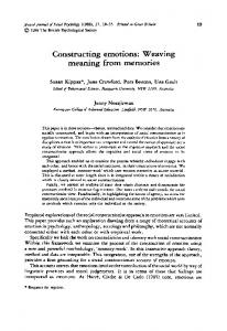
Novel OSTs associated with retinal transcription factors We further investigated the extent to which OSTs are found in association with transcription factors that are prominently expressed in the developing mouse retina. We compiled a list of 100 transcription factors that have been either been previously shown to regulate retinal cell fate specification or to be expressed in specific retinal cell types during development (Blackshaw et al., 2004; Gray et al., 2004). We found that 35 of these transcription factors had ESTs in Genbank corresponding to a putative OST, including 18 out of a total 34 homeodomaincontaining transcription factors (see Supp. Table ST1, which is available online). This implies that the phenomenon of OSTs is relatively widespread for developmentally important transcription factors, but not exclusively for homeodomain factors. Though some of these novel OSTs have been previously reported in other studies, their retinal expression has not been examined (Engstrom et al., 2006). To identify OSTs expressed at readily detectable levels in the retina, we investigated whether SAGE tags corresponding to any of these OSTs were expressed and found that 10 of
Fig. 2. Novel noncoding OSTs expressed in retina and associated with retinally-expressed transcription factors. See Supp. Tables ST1 and ST2 for more details on the OSTs.
the 34 OSTs identified were detected, all but one of which were spliced. Five of these OSTs had been previously reported in the retina (Six3OS, Six6OS, CrxOS, Otx2OS, and Vax2OS) (Alfano et al., 2005), while five others are partnered with mRNAs for Lhx1, Prox1, Hmx1, Zfhx4, and Hes5 (Fig. 2). Several of these, including Lhx1OS, Prox1OS, and Zfhx4OS are present at substantially higher levels than the previously identified HOSTs, with the exception of the abundant Six3OS transcript. RaxOS and Pax6OS are not detected at all in the SAGE data set, while OSTs such as CrxOS and Otx2OS present only once each in a pool of over 500,000 total retinal SAGE tags (see Supp. Table ST2 for a list of all SAGE tag counts for the retinally expressed OSTs and associated transcription factors). The cellular expression patterns of these newly identified OSTs remains to be investigated. The extent to which OSTs are found in association with retinal transcrip-
tion factors in species other than mice is not clear. Previous studies reported that OSTs are associated with retinalexpressed transcription factors in humans as well as mice (Alfano et al., 2005). These human transcripts often share little primary sequence homology with their murine counterparts, with the position of exon-intron boundaries varying substantially. More surprisingly, the genomic sequence transcribed by the human OSTs often only partially overlaps the equivalent mouse OST, or in some cases does not overlap at all (Alfano et al., 2005; Babak et al., 2005). Clearly, the evolutionary constraints on OSTs are far more relaxed than for their associated coding transcripts, a fact that makes identifying true homologues challenging, particularly in nonmammalian vertebrates. Nonetheless, identifying OSTs associated with retinal transcription factors is quite straightforward if one simply asks whether OSTs of any sort
NONCODING RNAS IN RETINAL DEVELOPMENT 2109
in nonmammalian vertebrates (see Supp. Table ST1). In nearly half of the nonmammalian OSTs, the associated OST was spliced, implying that these transcripts represent bona fide mRNAs. These data imply that OSTs are found throughout the vertebrate lineage, though the question of whether there is a difference in the number or complexity of OSTs among different vertebrates awaits further clarification.
How might OSTs work?
Fig. 1. SAGE tag level for Six3, Six6, Vax2, Prox1, and their associated noncoding OSTs in developing mouse retina. Data obtained from Blackshaw et al. (2004).
A. Transcription Dependent Mechanism POL II II POL Associated Protein Coding Gene
OST gene
A
OR
B
POL II Enhancer
Facilitation HOST ncRNA transcript
Or Interference Degradation
POL II
A
B
POL II
B. Transcript Dependent Mechanism Factor X
POL II
Factor X
POL II
Fig. 3. Potential transcript and transcription-dependent mechanism of action of ncOSTs. A: The act of transcription through the upstream cis-regulatory sequences of the protein coding gene associated with the ncOST acts to promote or prevent recruitment of transcription factors to regulatory elements in the transcribed region. The ncRNA itself is irrelevant to the regulation of the associated gene, and is degraded after transcription. B: The ncRNA itself interacts with protein factors directly to modulate expression and/or activity of the associated protein-coding gene.
are present within 5 kb of the transcriptional start site of the relevant coding transcript. Using these criteria to search the genomes of chick, frog, and zebrafish, we determined that OSTs are found associated with 22 out
of 37 of the transcription factors that had associated OSTs in mouse in at least one of these three species (Supp. Table ST1). Nine out of the 10 retinally expressed mouse OSTs listed in Supp. Table ST2 had counterparts
What might the function and mechanism of action of these OSTs be? Given their close proximity to developmentally important protein coding genes, the most obvious hypothesis would be that they act in cis to regulate expression of their associated protein coding gene via transcriptional facilitation or interference (Fig. 3A). Numerous examples of naturally occurring transcriptional interference or transcriptional gene silencing are known, where the act of transcription through an enhancer or promoter region of a nearby gene reduces or eliminates expression of that gene. Some cases, such as the SRG1 transcript in yeast that blocks activation of the SER3 gene, involve transcriptional regulation of a protein coding gene by an ncRNA (Martens et al., 2004, 2005). The converse, transcriptional facilitation, where the act of transcription of an ncRNA through the regulatory region of a nearby gene enables access of transcriptional activators to enhancer elements in the transcribed region, can also occur (Ho et al., 2006). At least one retinal noncoding OST may act in trans to regulate retinal cell fate. We have recently observed that ectopic overexpression and knockdown of Six3OS/RNCR1 in neonatal retina result in changes in cell fate. Interestingly, these changes partially phenocopy the changes seen with Six3 overexpression and dominate negative expression, indicating that Six3 and Six3OS/RNCR1 may also interact in vivo to regulate cell fate (Rapicavoli and Blackshaw, unpublished data). Other studies performed outside the retina suggest that OSTs may at least in part function in trans to regulate transcription of nearby genes (Fig. 3B). In this case, the ncRNA itself may have functions that are distinct
2110 RAPICAVOLI AND BLACKSHAW
from the simple act of transcription through the genomic locus covered by the transcript (Fig. 2). This has been perhaps most clearly demonstrated by work on Evf-2, a ncRNA associated with the Dlx5/6 locus (Feng et al., 2006), which is itself strongly expressed in retinal progenitors (Rapicavoli and Blackshaw, unpublished data). Evf-2 can act in trans to activate transcription at the Dlx5/6 locus by interacting with Dlx2, another Dlx family member. Dlx5 and Dlx6 are members of a homeodomain protein family that are related to the Drosophila Distalless genes. The Dlx genes play important roles in the neuronal differentiation and craniofacial and limb patterning in development. The Dlx 5/6 genes are transcribed in convergent orientation, and important intergenic enhancer elements have been identified for the Dlx5/6 loci (Zerucha et al., 2000). Evf-2 is transcribed from ei, the ultraconserved region of the Dlx5/6 locus, which is located between Dlx5 and Dlx6. Evf-2 interacts directly with Dlx-2 to increase the activity of the Dlx5/6 enhancer by binding to the enhancer element, ei. Evf-2 thus appears to be acting at least partially as a RNA-based transcriptional coactivator. This potential function is not unprecedented, as the ncRNA SRA has been known for some time to act as a transcriptional coactivator by directly interacting with steroid receptors (Lanz et al., 1999), while the ncRNAs NRSE and HSR have been shown to regulate the activity of REST and HSF-1, respectively (Kuwabara et al., 2004; Shamovsky et al., 2006). However, the story is likely more complex for Evf-2. The region of Evf-2 that is essential for transcriptional activation by ei by Dlx2 corresponds precisely to the ei sequence itself. It thus seems plausible that the Evf-2 RNA may directly base pair with the ei DNA sequence, thereby altering chromatin structure or facilitating recruitment of transcription factors and leading to activation of transcription in conjunction with Dlx2. Other work from both Drosophila and mammals has linked ncRNAs transcribed from the Hox gene cluster to changes in chromatin structure that in turn lead to altered regulation of Hox gene transcription. One such
Drosophila ncRNA is bxd, which contains enhancer elements for the nearby Ubx gene. Though there is agreement that transcription of bxd does indeed regulate chromatin conformation in these enhancer elements, which in turn regulate expression of Ubx, there is disagreement as to the effect of bxd and its mechanism of action. It has been suggested that bxd acts in trans to recruit histone methyltransferases to Ubx enhancer elements and thus promotes stable activation of transcription (SanchezElsner et al., 2006). Other groups, however, have claimed that bxd has no effect in trans, but instead acts in cis via transcriptional interference to prevent recruitment of trithorax proteins to the enhancer elements, and thus represses expression of Ubx (Petruk et al., 2006). Human HOX complex-associated OSTs have also been shown to regulate chromatin structure. HOTAIR is an OST located at the boundary of two chromatin domains at the HOXC locus (Rinn et al., 2007). Depletion of HOTAIR had no effect on transcription of genes in the HOXC cluster, but did lead to transcriptional activation at the HOXD cluster, which lies on a separate chromosome. HOTAIR was found to be directly associated with the Polycomb Repressive Complex 2 (PCR2), which mediates transcriptional silencing. HOTAIR depletion, moreover, resulted in loss of both Suz12 (a component of PRC2) and H3K27me3 at the HOXD locus, implying HOTAIR ncRNA acts in trans to recruit PRC2 to the HOXD locus to promote transcriptional silencing. Finally, John Mattick’s group has recently demonstrated that two novel OSTs, Hoxb5/6as and Evx1as, can associate with transcriptionally active chromatin in mouse embryonic stem cells. However, the genomic regions with which these OSTs associate remains to be determined (Dinger et al., 2008). Finally, five of the ncOSTs listed in Supp. Table ST1 overlap the 5⬘ sequences of their associated protein coding transcript, potentially allowing the possibility of homologous base pairing and double-stranded RNA formation between the two transcripts. This is shown for Prox1 and Hes5 in Figure 2. While it is unclear whether
such duplexes actually form in vivo, naturally occurring sense-antisense pairs of this sort can inhibit translation (Werner and Berdal, 2005), and can also serve as substrates for the generation of endogenous siRNAs directed against the coding transcripts (Tam et al., 2008; Watanabe et al., 2008). ncRNAs expressed from homeodomain transcription factor loci may thus act to regulate transcription of either a partnered opposite strand transcription factor or, alternatively, a family member located at a separate locus. ncRNAs may also act in cis to block transcription by transcriptional interference or perhaps activate transcription through transcriptional facilitation. They may also act in trans to repress transcription by binding to polycomb proteins or activate transcription by binding to another homeodomain transcription factor or trithorax family members. Cis and transacting mechanisms of action for OSTs, moreover, need not be mutually exclusive. Interestingly, a number of chromatin-modifying proteins, including both components of the PRC2 DNA methyltransferase complexes, directly bind to RNA (Zhang et al., 2004; Bernstein et al., 2006; Jeffery and Nakielny, 2004). Perhaps ncRNAs direct histone modifications in chromatin to influence expression of transcription factors. ncRNAs may thus act as critical regulators of transcription factor expression in higher organisms, at least in part by facilitating chromatin remodeling.
Other Retinal mRNA-Like ncRNAs Sox2OT In a study of a child with bilateral anophthalmia, it was discovered that the Sox2 gene lies with the intron of an ncRNA, which was named Sox2 overlapping transcript (Sox2OT) (Fantes et al., 2003) just as the Dlx6 gene lies within an intron of the ncRNA Evf-2 (Feng et al., 2006). In contrast to OSTs, Sox2OT is transcribed from the same strand as Sox2 and splices around its partnered protein coding gene. The functional relationship between Sox2 and Sox2OT is unclear. However, Sox2 is expressed throughout the developing nervous system embryonically and
NONCODING RNAS IN RETINAL DEVELOPMENT 2111
postnatally, and Sox2OT is found abundantly in EST and SAGE libraries from developing retina and brain, implying that Sox2 and Sox2OT may be coexpressed. Further investigation is necessary to determine if this is indeed the case.
TUG1 Taurine upregulated gene (TUG1) was discovered and characterized as a novel retinal ncRNA that appears to act through a mechanism distinct from both miRNA and OSTs (Young et al., 2005). TUG1 was found in a screen to identify genes that are upregulated in response to taurine, which induces rod photoreceptor production (Altshuler et al., 1993; Young and Cepko, 2004). When TUG1 was knocked down, developing rod photoreceptors showed a defect in migration into the outer nuclear layer, ectopic expression of cone-specific markers, and increased apoptosis in transfected cells. The mechanism of action of TUG1 remains unclear.
Xist and Tsix The twin nuclear-localized ncRNAs Xist and Tsix have been studied for a number of years now as critical regulators of X chromosome inactivation (Plath et al., 2002; Wutz and Gribnau, 2007). Xist and Tsix, both of which are mRNA-like and tens of kilobases long, are transcribed from the Xic (X-inactivation center) in antisense orientation and act antagonistically to specify which X chromosome undergoes inactivation. Xist is then stably expressed from the inactive X-chromosome and silences the inactive X-chromosome in cis by binding all along the chromosome and then recruiting, via mechanisms that are still uncharacterized, polycomb family proteins that lead to heterochromatinization of the inactive X chromosome. This process of random X inactivation occurs prior to implantation, and the pattern of X inactivation is inherited stably in daughter cells. Unexpectedly, both Xist and Tsix are expressed dynamically during mouse retinal development in subsets of cells in both the outer and inner neuroblastic layers embryonically. Postnatal expression is restricted to the inner nuclear layer around the end of the first postnatal week, with minimal expression in photoreceptors and ganglion cells (Blackshaw et al., 2004). This might imply
that those cells negative for Xist and Tsix might escape X-inactivation, although genetic evidence suggests that this is not the case (Reese et al., 1999). An alternate cell-specific pathway of Xinactivation may exist, or dramatic cellspecific variations in Xist levels may be required to mediate X-inactivation. Interestingly, a number of exceptions to the canonical mechanism of action of Xist detailed above have been recently documented. In certain circumstances, an inactive X-chromosome can be maintained without modifications and Xist can be selectively associated with the X-chromosome without it becoming inactivated or modified (Wutz and Gribnau, 2007). Whether this is the mechanism of action for retinal Xist awaits further investigation.
RNCR2/MIAT/Gomafu Finally, another long, nuclear mRNAlike noncoding RNA has recently been shown to be prominently expressed in the developing retina. Variously termed RNCR2 (Blackshaw et al., 2004), MIAT (Ishii et al., 2006), and Gomafu (Sone et al., 2007), this RNA that is evolutionarily conserved from amphibians to mammals (Rapicavoli and Blackshaw, unpublished data) is 9 kb long, spliced, and like Xist is a nuclear-retained transcript. It is not, however, associated with chromatin but instead binds to specific, though uncharacterized, subdomains of the nuclear matrix (Sone et al., 2007). It is expressed selectively in the developing central and peripheral nervous system (Ishii et al., 2006; Sone et al., 2007), and is also expressed in focal regions of the adult brain (Mercer et al., 2008). The transcript is highly abundant in the developing retina, comprising a maximum of 0.2% of polyadenylated RNA at embryonic day 18 in mouse (Blackshaw et al., 2004). It is strongly expressed in a subset of both progenitor and postmitotic cells, with pronounced and consistent expression in a subset of immature amacrine cells, yet is virtually undetectable in other amacrine cells of the same developmental stage (Blackshaw et al., 2004). Recent data from our lab have indicated that, while overexpression produces no obvious retinal phenotype, knockdown of RNCR2 in developing retina promotes development of both amacrine cells and Muller glia (Rapicavoli and Blackshaw, unpublished data), suggest-
ing a role for this ncRNA in selectively inhibiting differentiation of specific retinal cell lineages. How RNCR2 performs this function at a molecular level is still unclear, however.
CONCLUSIONS AND FUTURE PROSPECTS The field of retinal ncRNAs is certain to expand dramatically in the years ahead. The use of high-throughput sequencing in combination with microarray-based approaches should lead to the identification of a comprehensive catalog of both miRNAs and other ncRNAs that are dynamically expressed in developing retina. Electroporation and retroviral infection make it quite straightforward to overexpress and knockdown ncRNA expression in vivo, and we should soon obtain proof that ncRNAs play a role in retinal cell fate specification. Likewise, reporter assays allow ready identification of miRNA target sequences, and within a few years we should begin to get the outlines of the miRNA regulatory network in retinal progenitors. This will determine whether these RNAs actually play a major role in regulating progenitor competence. The bigger challenge ahead lies in identifying the biochemical targets and mechanisms of action of biologically active mRNA-like ncRNAs. Of specific interest is unraveling what is likely to be the complex interplay of both trans-acting RNAmediated effects and cis-acting transcription-dependent effects of these ncRNAs in modulating the activity and expression of developmentally important protein coding genes. Noncoding RNAs are likely to be an extremely functionally heterogeneous class of biomolecules, and it is likely to be a long time before we know how far and wide this unexplored country stretches.
ACKNOWLEDGMENTS We thank Akishi Onishi and Jimmy de Melo for comments on the manuscript.
REFERENCES Alfano G, Vitiello C, Caccioppoli C, Caramico T, Carola A, Szego MJ, McInnes RR, Auricchio A, Banfi S. 2005. Natural antisense transcripts associated
2112 RAPICAVOLI AND BLACKSHAW
with genes involved in eye development. Hum Mol Genet 14:913–923. Altshuler D, Lo Turco JJ, Rush J, Cepko C. 1993. Taurine promotes the differentiation of a vertebrate retinal cell type in vitro. Development 119:1317–1328. Amaral PP, Mattick JS. 2008. Noncoding RNA in development. Mamm Genome. Arora A, McKay GJ, Simpson DA. 2007. Prediction and verification of miRNA expression in human and rat retinas. Invest Ophthalmol Vis Sci 48:3962–3967. Baas D, Bumsted KM, Martinez JA, Vaccarino FM, Wikler KC, Barnstable CJ. 2000. The subcellular localization of Otx2 is cell-type specific and developmentally regulated in the mouse retina. Brain Res Mol Brain Res 78:26 –37. Babak T, Blencowe BJ, Hughes TR. 2005. A systematic search for new mammalian noncoding RNAs indicates little conserved intergenic transcription. BMC Genomics 6:104. Barbieri AM, Lupo G, Bulfone A, Andreazzoli M, Mariani M, Fougerousse F, Consalez GG, Borsani G, Beckmann JS, Barsacchi G, Ballabio A, Banfi S. 1999. A homeobox gene, vax2, controls the patterning of the eye dorsoventral axis. Proc Natl Acad Sci USA 96:10729 –10734. Bernstein E, Duncan EM, Masui O, Gil J, Heard E, Allis CD. 2006. Mouse polycomb proteins bind differentially to methylated histone H3 and RNA and are enriched in facultative heterochromatin. Mol Cell Biol 26:2560 –2569. Blackshaw S, Harpavat S, Trimarchi J, Cai L, Huang H, Kuo WP, Weber G, Lee K, Fraioli RE, Cho SH, Yung R, Asch E, Ohno-Machado L, Wong WH, Cepko CL. 2004. Genomic analysis of mouse retinal development. PLoS Biol 2:E247. Booth HA, Holland PW. 2007. Annotation, nomenclature and evolution of four novel homeobox genes expressed in the human germ line. Gene 387:7–14. Cao X, Yeo G, Muotri AR, Kuwabara T, Gage FH. 2006. Noncoding RNAs in the mammalian central nervous system. Annu Rev Neurosci 29:77–103. Carninci P, Hayashizaki Y. 2007. Noncoding RNA transcription beyond annotated genes. Curr Opin Genet Dev 17:139 –144. Carninci P, Kasukawa T, Katayama S, Gough J, Frith MC, Maeda N, Oyama R, Ravasi T, Lenhard B, Wells C, Kodzius R, Shimokawa K, Bajic VB, Brenner SE, Batalov S, Forrest AR, Zavolan M, Davis MJ, Wilming LG, Aidinis V, Allen JE, Ambesi-Impiombato A, Apweiler R, Aturaliya RN, Bailey TL, Bansal M, Baxter L, Beisel KW, Bersano T, Bono H, Chalk AM, Chiu KP, Choudhary V, Christoffels A, Clutterbuck DR, Crowe ML, Dalla E, Dalrymple BP, de Bono B, Della Gatta G, di Bernardo D, Down T, Engstrom P, Fagiolini M, Faulkner G, Fletcher CF, Fukushima T, Furuno M, Futaki S, Gariboldi M, Georgii-Hemming P, Gingeras TR, Gojobori T, Green RE, Gustincich S, Harbers M, Hayashi Y, Hensch TK, Hirokawa N, Hill D, Huminiecki L, Iacono M, Ikeo K, Iwama A, Ishikawa T, Jakt M, Kanapin A, Katoh M, Kawasawa Y,
Kelso J, Kitamura H, Kitano H, Kollias G, Krishnan SP, Kruger A, Kummerfeld SK, Kurochkin IV, Lareau LF, Lazarevic D, Lipovich L, Liu J, Liuni S, McWilliam S, Madan Babu M, Madera M, Marchionni L, Matsuda H, Matsuzawa S, Miki H, Mignone F, Miyake S, Morris K, Mottagui-Tabar S, Mulder N, Nakano N, Nakauchi H, Ng P, Nilsson R, Nishiguchi S, Nishikawa S, Nori F, Ohara O, Okazaki Y, Orlando V, Pang KC, Pavan WJ, Pavesi G, Pesole G, Petrovsky N, Piazza S, Reed J, Reid JF, Ring BZ, Ringwald M, Rost B, Ruan Y, Salzberg SL, Sandelin A, Schneider C, Schonbach C, Sekiguchi K, Semple CA, Seno S, Sessa L, Sheng Y, Shibata Y, Shimada H, Shimada K, Silva D, Sinclair B, Sperling S, Stupka E, Sugiura K, Sultana R, Takenaka Y, Taki K, Tammoja K, Tan SL, Tang S, Taylor MS, Tegner J, Teichmann SA, Ueda HR, van Nimwegen E, Verardo R, Wei CL, Yagi K, Yamanishi H, Zabarovsky E, Zhu S, Zimmer A, Hide W, Bult C, Grimmond SM, Teasdale RD, Liu ET, Brusic V, Quackenbush J, Wahlestedt C, Mattick JS, Hume DA, Kai C, Sasaki D, Tomaru Y, Fukuda S, Kanamori-Katayama M, Suzuki M, Aoki J, Arakawa T, Iida J, Imamura K, Itoh M, Kato T, Kawaji H, Kawagashira N, Kawashima T, Kojima M, Kondo S, Konno H, Nakano K, Ninomiya N, Nishio T, Okada M, Plessy C, Shibata K, Shiraki T, Suzuki S, Tagami M, Waki K, Watahiki A, Okamura-Oho Y, Suzuki H, Kawai J, Hayashizaki Y. 2005. The transcriptional landscape of the mammalian genome. Science 309:1559 –1563. Cepko CL, Austin CP, Yang X, Alexiades M, Ezzeddine D. 1996. Cell fate determination in the vertebrate retina. Proc Natl Acad Sci USA 93:589 –595. Chooniedass-Kothari S, Emberley E, Hamedani MK, Troup S, Wang X, Czosnek A, Hube F, Mutawe M, Watson PH, Leygue E. 2004. The steroid receptor RNA activator is the first functional RNA encoding a protein. FEBS Lett 566: 43– 47. Clamp M, Fry B, Kamal M, Xie X, Cuff J, Lin MF, Kellis M, Lindblad-Toh K, Lander ES. 2007. Distinguishing protein-coding and noncoding genes in the human genome. Proc Natl Acad Sci USA 104:19428 –19433. Cohen BA, Mitra RD, Hughes JD, Church GM. 2000. A computational analysis of whole-genome expression data reveals chromosomal domains of gene expression. Nat Genet 26:183–186. Conaco C, Otto S, Han JJ, Mandel G. 2006. Reciprocal actions of REST and a microRNA promote neuronal identity. Proc Natl Acad Sci USA 103:2422–2427. Corbo JC, Myers CA, Lawrence KA, Jadhav AP, Cepko CL. 2007. A typology of photoreceptor gene expression patterns in the mouse. Proc Natl Acad Sci USA 104:12069 –12074. Davis AA, Matzuk MM, Reh TA. 2000. Activin A promotes progenitor differentiation into photoreceptors in rodent retina. Mol Cell Neurosci 15:11–21.
Decembrini S, Andreazzoli M, Vignali R, Barsacchi G, Cremisi F. 2006. Timing the generation of distinct retinal cells by homeobox proteins. PLoS Biol 4:e272. Deo M, Yu JY, Chung KH, Tippens M, Turner DL. 2006. Detection of mammalian microRNA expression by in situ hybridization with RNA oligonucleotides. Dev Dyn 235:2538 –2548. Dinger ME, Amaral PP, Mercer TR, Pang KC, Bruce SJ, Gardiner BB, AskarianAmiri ME, Ru K, Solda G, Simons C, Sunkin SM, Crowe ML, Grimmond SM, Perkins AC, Mattick JS. 2008. Long noncoding RNAs in mouse embryonic stem cell pluripotency and differentiation. Genome Res 18:1433–1445. Eis PS, Tam W, Sun L, Chadburn A, Li Z, Gomez MF, Lund E, Dahlberg JE. 2005. Accumulation of miR-155 and BIC RNA in human B cell lymphomas. Proc Natl Acad Sci USA 102:3627–3632. Engstrom PG, Suzuki H, Ninomiya N, Akalin A, Sessa L, Lavorgna G, Brozzi A, Luzi L, Tan SL, Yang L, Kunarso G, Ng EL, Batalov S, Wahlestedt C, Kai C, Kawai J, Carninci P, Hayashizaki Y, Wells C, Bajic VB, Orlando V, Reid JF, Lenhard B, Lipovich L. 2006. Complex Loci in human and mouse genomes. PLoS Genet 2:e47. Fantes J, Ragge NK, Lynch SA, McGill NI, Collin JR, Howard-Peebles PN, Hayward C, Vivian AJ, Williamson K, van Heyningen V, FitzPatrick DR. 2003. Mutations in SOX2 cause anophthalmia. Nat Genet 33:461– 463. Feng J, Bi C, Clark BS, Mady R, Shah P, Kohtz JD. 2006. The Evf-2 noncoding RNA is transcribed from the Dlx-5/6 ultraconserved region and functions as a Dlx-2 transcriptional coactivator. Genes Dev 20:1470 –1484. Frith MC, Bailey TL, Kasukawa T, Mignone F, Kummerfeld SK, Madera M, Sunkara S, Furuno M, Bult CJ, Quackenbush J, Kai C, Kawai J, Carninci P, Hayashizaki Y, Pesole G, Mattick JS. 2006. Discrimination of non-protein-coding transcripts from protein-coding mRNA. RNA Biol 3:40 – 48. Furuno M, Pang KC, Ninomiya N, Fukuda S, Frith MC, Bult C, Kai C, Kawai J, Carninci P, Hayashizaki Y, Mattick JS, Suzuki H. 2006. Clusters of internally primed transcripts reveal novel long noncoding RNAs. PLoS Genet 2:e37. Geng X, Lavado A, Lagutin OV, Liu W, Oliver G. 2007. Expression of Six3 Opposite Strand (Six3OS) during mouse embryonic development. Gene Expr Patterns 7:252–257. Gray PA, Fu H, Luo P, Zhao Q, Yu J, Ferrari A, Tenzen T, Yuk DI, Tsung EF, Cai Z, Alberta JA, Cheng LP, Liu Y, Stenman JM, Valerius MT, Billings N, Kim HA, Greenberg ME, McMahon AP, Rowitch DH, Stiles CD, Ma Q. 2004. Mouse brain organization revealed through direct genome-scale TF expression analysis. Science 306:2255–2257. Hayashi T, Huang J, Deeb SS. 2005. Expression of rinx/vsx1 during postnatal eye development in cone-bipolar, differ-
NONCODING RNAS IN RETINAL DEVELOPMENT 2113
entiating ganglion, and lens fiber cells. Jpn J Ophthalmol 49:93–105. He L, He X, Lowe SW, Hannon GJ. 2007. microRNAs join the p53 network—another piece in the tumour-suppression puzzle. Nat Rev Cancer 7:819 – 822. Ho Y, Elefant F, Liebhaber SA, Cooke NE. 2006. Locus control region transcription plays an active role in long-range gene activation. Mol Cell 23:365–375. Hsiau TH, Diaconu C, Myers CA, Lee J, Cepko CL, Corbo JC. 2007. The cis-regulatory logic of the mammalian photoreceptor transcriptional network. PLoS ONE 2:e643. Huang KM, Dentchev T, Stambolian D. 2008. MiRNA expression in the eye. Mamm Genome 19:510 –516. Ishii N, Ozaki K, Sato H, Mizuno H, Saito S, Takahashi A, Miyamoto Y, Ikegawa S, Kamatani N, Hori M, Nakamura Y, Tanaka T. 2006. Identification of a novel non-coding RNA, MIAT, that confers risk of myocardial infarction. J Hum Genet 51:1087–1099. Jeffery L, Nakielny S. 2004. Components of the DNA methylation system of chromatin control are RNA-binding proteins. J Biol Chem 279:49479 – 49487. Katayama S, Tomaru Y, Kasukawa T, Waki K, Nakanishi M, Nakamura M, Nishida H, Yap CC, Suzuki M, Kawai J, Suzuki H, Carninci P, Hayashizaki Y, Wells C, Frith M, Ravasi T, Pang KC, Hallinan J, Mattick J, Hume DA, Lipovich L, Batalov S, Engstro¨m PG, Mizuno Y, Faghihi MA, Sandelin A, Chalk AM, Mottagui-Tabar S, Liang Z, Lenhard B, Wahlestedt C; RIKEN Genome Exploration Research Group; Genome Science Group (Genome Network Project Core Group); FANTOM Consortium. 2005. Antisense transcription in the mammalian transcriptome. Science 309:1564 –1566 Kapranov P, Cawley SE, Drenkow J, Bekiranov S, Strausberg RL, Fodor SP, Gingeras TR. 2002. Large-scale transcriptional activity in chromosomes 21 and 22. Science 296:916 –919. Kapranov P, Cheng J, Dike S, Nix DA, Duttagupta R, Willingham AT, Stadler PF, Hertel J, Hackermuller J, Hofacker IL, Bell I, Cheung E, Drenkow J, Dumais E, Patel S, Helt G, Ganesh M, Ghosh S, Piccolboni A, Sementchenko V, Tammana H, Gingeras TR. 2007. RNA maps reveal new RNA classes and a possible function for pervasive transcription. Science 316:1484 –1488. Kapsimali M, Kloosterman WP, de Bruijn E, Rosa F, Plasterk RH, Wilson SW. 2007. MicroRNAs show a wide diversity of expression profiles in the developing and mature central nervous system. Genome Biol 8:R173. Karali M, Peluso I, Marigo V, Banfi S. 2007. Identification and characterization of microRNAs expressed in the mouse eye. Invest Ophthalmol Vis Sci 48:509 – 515. Kim J, Wu HH, Lander AD, Lyons KM, Matzuk MM, Calof AL. 2005. GDF11 controls the timing of progenitor cell
competence in developing retina. Science 308:1927–1930. Kluiver J, Poppema S, de Jong D, Blokzijl T, Harms G, Jacobs S, Kroesen BJ, van den Berg A. 2005. BIC and miR-155 are highly expressed in Hodgkin, primary mediastinal and diffuse large B cell lymphomas. J Pathol 207:243–249. Kuwabara T, Hsieh J, Nakashima K, Taira K, Gage FH. 2004. A small modulatory dsRNA specifies the fate of adult neural stem cells. Cell 116:779 –793. Lanz RB, McKenna NJ, Onate SA, Albrecht U, Wong J, Tsai SY, Tsai MJ, O’Malley BW. 1999. A steroid receptor coactivator, SRA, functions as an RNA and is present in an SRC-1 complex. Cell 97:17–27. Livesey FJ, Young TL, Cepko CL. 2004. An analysis of the gene expression program of mammalian neural progenitor cells. Proc Natl Acad Sci USA 101:1374 –1379. Loscher CJ, Hokamp K, Kenna PF, Ivens AC, Humphries P, Palfi A, Farrar GJ. 2007. Altered retinal microRNA expression profile in a mouse model of retinitis pigmentosa. Genome Biol 8:R248. Martens JA, Laprade L, Winston F. 2004. Intergenic transcription is required to repress the Saccharomyces cerevisiae SER3 gene. Nature 429:571–574. Martens JA, Wu PY, Winston F. 2005. Regulation of an intergenic transcript controls adjacent gene transcription in Saccharomyces cerevisiae. Genes Dev 19:2695–2704. Mehler MF, Mattick JS. 2006. Non-coding RNAs in the nervous system. J Physiol 575:333–341. Mehler MF, Mattick JS. 2007. Noncoding RNAs and RNA editing in brain development, functional diversification, and neurological disease. Physiol Rev 87:799 – 823. Mendell JT. 2005. MicroRNAs: critical regulators of development, cellular physiology and malignancy. Cell Cycle 4:1179 – 1184. Mercer TR, Dinger ME, Sunkin SM, Mehler MF, Mattick JS. 2008. Specific expression of long noncoding RNAs in the mouse brain. Proc Natl Acad Sci USA 105:716 –721. Nguyen M, Arnheiter H. 2000. Signaling and transcriptional regulation in early mammalian eye development: a link between FGF and MITF. Development 127: 3581–3591. Ohsaki K, Morimitsu T, Ishida Y, Kominami R, Takahashi N. 1999. Expression of the Vax family homeobox genes suggests multiple roles in eye development. Genes Cells 4:267–276. Pang KC, Stephen S, Dinger ME, Engstrom PG, Lenhard B, Mattick JS. 2007. RNAdb 2.0: an expanded database of mammalian non-coding RNAs. Nucleic Acids Res 35:D178 –182. Petruk S, Sedkov Y, Riley KM, Hodgson J, Schweisguth F, Hirose S, Jaynes JB, Brock HW, Mazo A. 2006. Transcription of bxd noncoding RNAs promoted by trithorax represses Ubx in cis by transcriptional interference. Cell 127:1209 – 1221.
Pheasant M, Mattick JS. 2007. Raising the estimate of functional human sequences. Genome Res 17:1245–1253. Plath K, Mlynarczyk-Evans S, Nusinow DA, Panning B. 2002. Xist RNA and the mechanism of X chromosome inactivation. Annu Rev Genet 36:233–278. Prasanth KV, Spector DL. 2007. Eukaryotic regulatory RNAs: an answer to the “genome complexity” conundrum. Genes Dev 21:11– 42. Rath MF, Morin F, Shi Q, Klein DC, Moller M. 2007. Ontogenetic expression of the Otx2 and Crx homeobox genes in the retina of the rat. Exp Eye Res 85:65–73. Reese BE, Necessary BD, Tam PP, Faulkner-Jones B, Tan SS. 1999. Clonal expansion and cell dispersion in the developing mouse retina. Eur J Neurosci 11:2965–2978. Rinn JL, Kertesz M, Wang JK, Squazzo SL, Xu X, Brugmann SA, Goodnough LH, Helms JA, Farnham PJ, Segal E, Chang HY. 2007. Functional demarcation of active and silent chromatin domains in human HOX loci by noncoding RNAs. Cell 129:1311–1323. Ruby JG, Jan CH, Bartel DP. 2007. Intronic microRNA precursors that bypass Drosha processing. Nature 448:83– 86. Ryan DG, Oliveira-Fernandes M, Lavker RM. 2006. MicroRNAs of the mammalian eye display distinct and overlapping tissue specificity. Mol Vis 12:1175–1184. Sakuta H, Suzuki R, Takahashi H, Kato A, Shintani T, Iemura S, Yamamoto TS, Ueno N, Noda M. 2001. Ventroptin: a BMP-4 antagonist expressed in a doublegradient pattern in the retina. Science 293:111–115. Sanchez-Elsner T, Gou D, Kremmer E, Sauer F. 2006. Noncoding RNAs of trithorax response elements recruit Drosophila Ash1 to Ultrabithorax. Science 311:1118 –1123. Shamovsky I, Ivannikov M, Kandel ES, Gershon D, Nudler E. 2006. RNA-mediated response to heat shock in mammalian cells. Nature 440:556 –560. Sharon D, Blackshaw S, Cepko CL, Dryja TP. 2002. Profile of the genes expressed in the human peripheral retina, macula, and retinal pigment epithelium determined through serial analysis of gene expression (SAGE). Proc Natl Acad Sci USA 99:315–320. Sone M, Hayashi T, Tarui H, Agata K, Takeichi M, Nakagawa S. 2007. The mRNA-like noncoding RNA Gomafu constitutes a novel nuclear domain in a subset of neurons. J Cell Sci 120:2498 –2506. Taft RJ, Pheasant M, Mattick JS. 2007. The relationship between non-proteincoding DNA and eukaryotic complexity. Bioessays 29:288 –299. Tam OH, Aravin AA, Stein P, Girard A, Murchison EP, Cheloufi S, Hodges E, Anger M, Sachidanandam R, Schultz RM, Hannon GJ. 2008. Pseudogene-derived small interfering RNAs regulate gene expression in mouse oocytes. Nature 453:534 –538. Thomson JM, Newman M, Parker JS, Morin-Kensicki EM, Wright T, Ham-
2114 RAPICAVOLI AND BLACKSHAW
mond SM. 2006. Extensive post-transcriptional regulation of microRNAs and its implications for cancer. Genes Dev 20:2202–2207. Trinklein ND, Aldred SF, Hartman SJ, Schroeder DI, Otillar RP, Myers RM. 2004. An abundance of bidirectional promoters in the human genome. Genome Res 14:62–66. Watanabe T, Totoki Y, Toyoda A, Kaneda M, Kuramochi-Miyagawa S, Obata Y, Chiba H, Kohara Y, Kono T, Nakano T, Surani MA, Sakaki Y, Sasaki H. 2008. Endogenous siRNAs from naturally formed dsRNAs regulate transcripts in mouse oocytes. Nature 453:539 –543. Werner A, Berdal A. 2005. Natural antisense transcripts: sound or silence? Physiol Genomics 23:125–131.
Wulczyn FG, Smirnova L, Rybak A, Brandt C, Kwidzinski E, Ninnemann O, Strehle M, Seiler A, Schumacher S, Nitsch R. 2007. Post-transcriptional regulation of the let-7 microRNA during neural cell specification. FASEB J 21:415– 426. Wutz A, Gribnau J. 2007. X inactivation Xplained. Curr Opin Genet Dev 17:387–393. Xu S, Witmer PD, Lumayag S, Kovacs B, Valle D. 2007. MicroRNA (miRNA) transcriptome of mouse retina and identification of a sensory organ-specific miRNA cluster. J Biol Chem 282:25053–25066. Young TL, Cepko CL. 2004. A role for ligandgated ion channels in rod photoreceptor development. Neuron 41:867– 879. Young TL, Matsuda T, Cepko CL. 2005. The noncoding RNA taurine upregulated
gene 1 is required for differentiation of the murine retina. Curr Biol 15:501–512. Zerucha T, Stuhmer T, Hatch G, Park BK, Long Q, Yu G, Gambarotta A, Schultz JR, Rubenstein JL, Ekker M. 2000. A highly conserved enhancer in the Dlx5/ Dlx6 intergenic region is the site of crossregulatory interactions between Dlx genes in the embryonic forebrain. J Neurosci 20:709 –721. Zhang H, Smolen GA, Palmer R, Christoforou A, van den Heuvel S, Haber DA. 2004. SUMO modification is required for in vivo Hox gene regulation by the Caenorhabditis elegans Polycomb group protein SOP-2. Nat Genet 36: 507–511.