Rio-Castaño, D., Corrales, D. M., Daza, G. J., y A. Aristizabal. 2005. .... CORPOICA, Colombia (Bernal y Diaz, 2005; CORPOICA, 2004); PROFRUTALES LTDA,.
The velocity values were measured in quiet air. Effect of late actuation on the lung dose for diffeent inhalation and breath-hold times (left panel) and different ...
the basis for the nomenclature of the various PP1 holoenzymes (Table 1). .... trypsin treatment thus allows one to identify regulatory polypeptides/subunits.
by Dominic McCormick (Shell Exploration & Production UK) & Markus Leishman ... Maureen. Ekofisk. Orange Marker. Ochre Marker. Bittern Field Stratigraphic ...
Human caused changes to our climate and built environment have increased the likelihood and consequence of bushfires. Every year large areas of Victoria are.
May 13, 2016 - say some stuff you might want to hear again, or share, or vociferously dispute. 2. ... wholesale trade ma
Notes: IP-056512; Support provided by the USGS Climate & Land. Use R&D and ... http://scs.math.yorku.ca/index.php/R:_Getting_started_with_R. Quick-R ...
thing feel lighter than a cheap thing of the same weight? ... one that suits the way you keto cook and cleanup best. T ... New laptops weigh less than older.
Small secundum atrial septal defect. â¡ Moderate perimembranous ventricular septal ... Arterial Switch Procedure. Technique. â¡ Single coronary button transfer.
Norwood procedure with 3 mm BT shunt. 1. 2. 3.7. 6. Pulmonary atresia with. VSD, CoA. 3.5 mm central shunt, atrial septectomy, atrial septation, resection of ...
www.businessballs.com with permission of. John M Fisher. See the theory and explanation at www.businessballs.com/persona
HEPATOTOXINS. (MICROCYSTIN. ,NODULARIN). SYNTHETASES. Microphotographs of treated cancer HeLa cells obtained by the time lapse microscopy (10x).
Introduction. Virtual Reality (VR) uses sensor tracking and visual effects to immerse user within virtual environment. VR hardware used throughout this project is ...
SCUFFHAM P, CHALMERS D, O'HARE D, WILSON E. Direct and indi- rect cost of general ... the cost of hospital treatment for injury), but differ in their estimation ...
northward tectonic transport (figure 10a). Moreover, granulites have been recovered in the boreholes in. Rengali dam-site, while the exposed rocks in the area.
mate the costs inflicted on society from injuries, fatalities, and property ... injury. Results: The annual average direct cost of aviation crashes was ... research lies, therefore, in its ability to inform and guide .... third party accident insuran
Plant Transformation Core Facility, Division of Plant Sciences, University of. Missouri, Columbia, MO ... University of Missouri (MU) Plant. Transformation Core ... in College of Agriculture, Food and Natural. Resources. PROTOCOLS USED.
blades are bent or damaged. 18. Avoid contact with ... Operating LLC may cause fire, electrical shock or ... Chopping bl
setiap ikatan. Kegunaan. • Analisis kualitatif (gugus fungsi) serapan khas untuk
setiap ikatan dalam gugus. • Analisis kuantitatif jarang dilakukan karena spektra
...
Environmental training at Companies. The case of. Volkswagen Navara. Marta Ormazábal, Elisabeth Viles, Vanessa Prieto-Sandoval, Carmen Jaca. ABSTRACT.
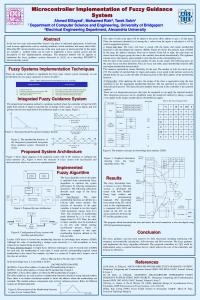
c) Fuzzy rule base: The fuzzy rule base is stored with the inputs and output membership functions in the knowledge base memory (KBM). Figure (6) shows the ...
during quiet stance with muscle onset defined as activity greater than 3 standard deviations. EMG amplitude was normalized and expressed as a percentage of ...
Detection and visualization of pospiviroids in potato,. Potato leafroll virus (PLRV) and aphids in support of transmission studies. Background. Viroids are highly ...
Detection and visualization of pospiviroids in potato, Potato leafroll virus (PLRV) and aphids in support of transmission studies Noémi Van Bogaert1,2, Kris De Jonghe2, Martine Maes2, and Guy Smagghe1 1Department
of Crop Protection, Faculty Bioscience Engineering, Ghent University, Coupure Links 653, 9000 Ghent, Belgium 2Plant Sciences Unit - Crop Protection, Institute for Agricultural and Fisheries Research (ILVO), Burgemeester Van Gansberghelaan 96, 9820 Merelbeke, Belgium
Background During virus replication in a plant cell, the virus Viroids are
highly stable, single-stranded RNA
coat opens and viroids can get encapsulated
plant pathogens that lack a protein coat. In potatoes (S. tuberosum, L.), Potato spindle tuber
Virion
viroid (PSTVd) and other pospiviroids cause severe malformations of the tubers and foliage, leading to important agricultural losses. Mixed
Viroid
infections of plant viruses and viroids might lead to “trans-encapsidation” of the viroid within the protein coat of the virus (e.g. Potato leafroll virus, PLRV). This system can potentially be vectored by aphids and ultimately (co-)infect new plants.
Aphids could transmit the virus with
Objectives
encapsidated viroid to new host plants
- How can we investigate transencapsidation (indirectly/directly)? - How common is transencapsidation (for different viruses/viroids)? - How can further transport to host plants (e.g. by aphids) occur?
Viruses and viroids in doubly inoculated potato leaves can be visualized microscopically by using specific antibodies
Indirect detection
Gold-labelled antibody
Start with potato material that is doubly infected (e.g. PLRV + PSTVd)
Specific antibody
Virus purification by ultracentrifugation
Target Detection of both pathogens by (q)RT-PCR
Treatment with endo-exonuclease
Direct detection
Detection of both pathogens by q(RT)-PCR
Via transmission electron microscopy (TEM) If both pathogens are still detected after nuclease treatment it can be assumed that the viroid was protected from degratation by being encapsulated within the protein coat of the virus
- Specific antibodies and Protein-A-Gold for the virus (e.g. PLRV) - Biotinylated