3429
Development 127, 3429-3440 (2000) Printed in Great Britain © The Company of Biologists Limited 2000 DEV3218
Left-right asymmetry in C. elegans intestine organogenesis involves a LIN-12/Notch signaling pathway Greg J. Hermann1, Ben Leung1,2 and James R. Priess1,2,3,4,* 1Division of Basic Sciences, Fred Hutchinson Cancer Research Center, Seattle, Washington 98109, USA 2Molecular and Cellular Biology Program and 3Department of Zoology, University of Washington, Seattle, Washington 4Howard Hughes Medical Institute, Seattle, Washington 98109, USA
98195, USA
*Author for correspondence (e-mail:
[email protected])
Accepted 31 May; published on WWW 20 July 2000
SUMMARY The C. elegans intestine is a simple tube consisting of a monolayer of epithelial cells. During embryogenesis, cells in the anterior of the intestinal primordium undergo reproducible movements that lead to an invariant, asymmetrical ‘twist’ in the intestine. We have analyzed the development of twist to determine how left-right and anterior-posterior asymmetries are generated within the intestinal primordium. The twist requires the LIN12/Notch-like signaling pathway of C. elegans. All cells within the intestinal primordium initially express LIN-12, a receptor related to Notch; however, only cells in the left half of the primordium contact external, nonintestinal cells that express LAG-2, a ligand related to Delta. LIN-12 and LAG-2 mediated interactions result in the left primordial cells expressing lower levels of LIN-12 than the right
primordial cells. We propose that this asymmetrical pattern of LIN-12 expression is the basis for asymmetry in later cell-cell interactions within the primordium that lead directly to intestinal twist. Like the interactions that initially establish LIN-12 asymmetry, the later interactions are mediated by LIN-12. The later interactions, however, involve a different ligand related to Delta, called APX-1. We show that the anterior-posterior asymmetry in intestinal twist involves the kinase LIT-1, which is part of a signaling pathway in early embryogenesis that generates anteriorposterior differences between sister cells.
INTRODUCTION
been designated arbitrarily as dextral handedness. Wild-type C. elegans are invariably dextral when cultured using standard laboratory conditions; however, culturing C. elegans at cold temperatures can result in some sinistral animals, with reversed left-right body axes (Wood et al., 1996). Sinistral animals also can be generated experimentally through micromanipulation of specific early embryonic blastomeres (Wood, 1991). The first apparent left-right asymmetry is observed during the 4-cell stage of embryogenesis when two blastomeres divide obliquely with respect to the left-right axis. If the left-right asymmetry of this division axis is reversed by micromanipulation, the resulting animal is sinistral. Changing the positions of the early blastomeres can alter the pattern of certain cell-cell interactions that determine some cell fates. The blastomeres that divide obliquely, and the descendants of these blastomeres, all express GLP-1, a receptor related to Notch (Evans et al., 1994). The division asymmetries cause only a subset of these blastomeres to contact a signaling cell; the resulting GLP-1 mediated interactions result in left blastomeres adopting different lineages from right blastomeres (Gendreau et al., 1994; Hutter and Schnabel, 1994, 1995). It is not understood whether, or how, these early interactions lead to specific organ asymmetries. The development of the C. elegans intestine provides a very
Although most animals have bilaterally symmetrical surface features, their internal organs often show pronounced left-right asymmetries in morphology and placement. For example, the tube of embryonic cells that forms the vertebrate heart loops to the right, and organs like the stomach and liver are positioned on the left and right, respectively. Although little is known at the molecular level about how left-right asymmetry originates during animal embryogenesis, or how this leads to subsequent asymmetries in organ morphology, recent studies have identified a few genes that show early left-right asymmetries in expression. In chick, frog, mouse and zebrafish embryos, an asymmetry in TGF-β signaling results in the asymmetric expression of transcription factors, such as Pitx2, within organ primordia (reviewed in Ramsdell and Yost, 1998; Capdevila et al., 2000). Through mechanisms that are not yet understood, Pitx2 and related factors may regulate morphogenetic events that result in organ asymmetry. The nematode C. elegans displays numerous left-right asymmetries in tissues and organs, the most prominent being the asymmetrically positioned, interwoven helices of the intestine and gonad in adults (reviewed in Wood, 1998). These and other asymmetries result in an overall body pattern that has
Key words: Left-right asymmetry, Anterior-posterior asymmetry, Intestinal organogenesis, Notch signaling, APX-1, GLP-1, LAG-1, LAG-2, LIN-12, LIT-1, POP-1, SPN-1
3430 G. J. Hermann, B. Leung and J. R. Priess simple model of organ morphogenesis, suggesting that a molecular explanation for asymmetry in the intestine should be possible. The fully formed intestine is an epithelial tube containing only 20 clonally derived cells (Sulston et al., 1983). During embryogenesis, cells within the intestinal primordium become polarized with respect to a central, anterior-posterior axis through the primordium (Leung et al., 1999). In crosssection, the early primordium displays radial symmetry; however, cells in the left half of the primordium intercalate with other left cells, and cells in the right half intercalate with other right cells. These cell movements result in a bilaterally symmetrical tube composed of a single row of left cells that abut a single row of right cells; the intestinal lumen forms at the interface between the left and right rows. Asymmetry in the intestine first becomes evident shortly after cell intercalation. Three of the anterior cells in the right row of the primordium move counterclockwise to the left, crossing the plane of bilateral symmetry, as the contralateral three left cells move simultaneously toward the right (see Fig. 1; Sulston et al., 1983; Leung et al., 1999). This movement initiates a left-handed ‘twist’ in the anterior of the intestine; by the time the larva hatches, the twist is about 180° (Sulston and Horvitz, 1977). The superhelical twist resulting from these cell movements has been proposed to displace the anterior end of the intestine to the left of the larval body, and the posterior end to the right (Sulston and Horvitz, 1977). This asymmetry may influence the subsequent morphogenesis of the gonad as it wraps around the intestine during larval development. The development of the intestinal twist must involve both a left-right, rotational asymmetry, as well as an anterior-posterior asymmetry that limits the twist to the anterior end. In this report, we describe a genetic and cellular analysis of how asymmetry is generated in the intestine during embryogenesis. We show that the Notch-like signaling pathway of C. elegans is required for the intestinal twist. This pathway involves the receptor LIN-12, the ligands LAG-2 and APX-1, and the nuclear protein LAG-1. We show that lin-12(+) function is required for intestinal twist, and that the LIN-12 protein is expressed asymmetrically in the right half of the intestinal primordium. The asymmetrical expression of LIN-12 requires lag-1(+) and lag-2(+) functions, and involves interactions between cells in the intestinal primordium and cells external to the primordium. Finally, we show that a separate polarity pathway, involving the genes pop-1 and lit-1, is required to specify the anterior-posterior boundary between cells that undergo twist and cells that do not. MATERIALS AND METHODS Strains and alleles N2 was used as the wild-type strain. Mutant alleles used are listed by chromosome: linkage group I (LGI): spn-1(it143), unc-40(e271), unc-40(e1430); LGIII: ceh-13(sw1), glp-1(e2141ts), glp-1(e2142ts), glp-1(q224ts), lin-12(n302sd), lin-12(n676n930ts), lin-12(n941), lin-12(n950sd), lin-39(n1760), lit-1(t1512ts), mab-5(e1239); LGIV: lag-1(q385), unc-5(e53); LGV: apx-1(zu347ts), lag-2(q387), lag2(q411), lag-2(q420ts); LGX: elt-2(ca15), unc-6(e78), unc6(ev400). Strains containing lag-2::GFP, JK2003 (qEx233) and JK2822 (qIs19) (Blelloch et al., 1999), were provided by Judith Kimble (University of Wisconsin, Madison, USA). The JR662 (wIs47) hs-end-1 strain was provided by Joel Rothman (UC, Santa
Barbara, USA) (Zhu et al., 1998). C. elegans strains were handled as described (Brenner, 1974). Microscopy We scored intestinal twist in 1.5-fold embryos by Nomarski microscopy. Only embryos that had a lateral presentation such that the rectum was present in the same focal plane as the intestine were analyzed. Al Candia and Stuart Kim (Stanford University) provided the affinity-purified rat anti-LIN-12 antiserum used in this study. Embryos were immunostained with the LIN-12 antiserum using the immunofluorescence protocol described in Leung et al. (1999). Immunostaining with mAb 1CB4 and mAb RL2 was performed as described (Lin et al., 1998; Leung et al., 1999). Laser ablations were performed as described (Leung et al., 1999). For immunofluorescence, embryos that developed to the E8 or E16 stage were transferred to 0.1% polylysine-coated slides and immunostained for LIN-12. JR662 hs-end-1 embryos were heatshocked as described (Zhu et al., 1998) and fixed and immunostained for LIN-12 after 2.5 hours. To quantify the number of LIN-12 expressing cells following heat shock, immunostained embryos were optically sectioned using a Delta Vision microscope. The resulting images were analyzed to determine the total number of nuclei and total number of LIN-12 positive cells in each embryo.
RESULTS Left-right asymmetry in the intestine The embryonic cell that produces the C. elegans intestine is called the E blastomere, and the various developmental stages of the intestinal primordium are named with reference to the number of E descendants present; E, E2, E4, E8, E16 and E20. The development of the intestine has been described in detail previously (Sulston et al., 1983; Leung et al., 1999). Once cell intercalation shapes the primordium into a bilaterally polarized tube, it is convenient to refer to specific left-right pairs of intestinal cells as intestinal rings (int ring I through int ring IX; Fig. 1A). Asymmetrical movements of cells in the int II, III and IV rings occur during stages E16 to E20. When viewed from the posterior end of the intestine, cells in the right half of the primordium are seen to move counterclockwise toward the left, as cells on the left move toward the right (Fig. 1A). This invariant pattern of cell movement can be summarized as a rotation of the int II, III and IV rings, and for convenience we refer to this rotation as intestinal twist. Since several of the mutants analyzed in the present study do not complete normal development and hatch, we selected the embryonic stage called the 1.5-fold (Fig. 1D) to score twist in all experiments; at this stage, twist is about halfway complete (90°). Twist can be scored in fixed embryos after immunostaining with an antibody that recognizes the intestinal cells (Fig. 1B), and in living embryos by following the positions of intestinal nuclei by light microscopy (Fig. 1D). A Notch-like pathway is required for intestinal twist An isolated E blastomere grown in culture produces a relatively symmetrical cyst of intestinal cells, suggesting that cell-cell interactions are likely to play a role in intestinal twist (Leung et al., 1999). Since a Notch-like signaling pathway mediates several cell-cell interactions in the early embryo (reviewed in Schnabel and Priess, 1997), we began by asking whether either of the two Notch-related receptors in C. elegans were required
Notch signaling and organ left-right asymmetry 3431
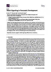
Fig. 1. Intestinal twist in wild-type and lin-12 embryos. (A) Diagram of the dorsal surface of a wild-type intestine showing the development of twist. Before twist, left (light green) and right (dark green) cells are organized with bilateral symmetry. Each int ring contains two cells, except int ring I, which contains four cells (not shown). The int rings rotate in the sequence III, II, then IV. The rotation initially is 90°, but in the newly hatched larvae the anterior intestine is twisted about 180°. (B,C) Dorsal views of embryos at the 1.5-fold stage immunostained with mAb 1CB4 to show intestinal cells (green) and DAPI to show nuclei (red); orientation as in A. (D,E) Lateral views of living embryos at the 1.5-fold stage. Black arrowheads indicate the anterior end of the intestinal primordium, and white arrowheads in B and D indicate the posterior boundary of twist. Embryos are 50 µm in length; in D and E anterior is to the left and dorsal is up. Bar, 5 µm.
for twist. These receptors are called GLP-1 and LIN-12 (Kimble and Simpson, 1997; Greenwald, 1998). Mutations that disrupt glp-1(+) functions after the 4-cell stage of embryogenesis appeared to have no effect on intestinal twist (Table 1). In contrast, mutations in the lin-12 gene resulted in prominent twist defects. All of the embryos from hermaphrodites homozygous for the null allele lin-12(n941) had defective twist, as did over 80% of the embryos from hermaphrodites homozygous for the conditional allele lin12(n676n930ts) (Table 1, Fig. 1C,E). In addition, we found that wild-type embryos had a fully penetrant defect in twist when lin-12(+) function was inhibited with double-stranded RNA (lin-12(RNAi); Table 1). We found that 19% (n=57) of the progeny of heterozygous lin-12(n941)/+ hermaphrodites had a twist defect, indicating that zygotic expression of lin-12(+), rather than maternal expression, is required for twist. In the course of these experiments we noticed that some of the homozygous lin-12(n941) and lin-12(RNAi) larvae and adults had aberrant positioning of the gonad relative to the intestine; although this defect was not analyzed in detail, it is consistent with the view that intestinal twist contributes to subsequent gonad morphogenesis (see Introduction). We found that twist requires the lag-1 and lag-2 genes (Table 1), which encode proteins related, respectively, to the Drosophila Notch pathway proteins Suppressor of Hairless, a transcription factor (Christensen et al., 1996), and Delta, a Notch ligand (Henderson et al., 1994; Tax et al., 1994). Since 92% of embryos homozygous for lag-1(q385) had a twist defect, we initially were surprised to find that only 43% of embryos homozygous for lag-2(q411), a null allele, had a twist defect. We discovered, however, that 100% of embryos homozygous for lag-2(q387) had a twist defect; the lag2(q387) mutation is a deletion that removes lag-2 as well as the neighboring gene apx-1, which encodes a second Delta-
related ligand (Henderson et al., 1994; Mello et al., 1994). APX-1 is capable of substituting for LAG-2 in certain cell-cell interactions (Fitzgerald and Greenwald, 1995; Gao and Kimble, 1995), suggesting that LAG-2 and APX-1 might have either partially redundant, or distinct, functions required for intestinal twist. Consistent with these hypotheses, we found that 85% of the self-progeny of apx-1(zu347ts) homozygous mothers cultured at the semipermissive temperature (23°C) had a defect in intestinal twist (Table 1). We found that 24% (n=50) of the self-progeny of apx-1(zu347ts)/+ heterozygous mothers cultured at the nonpermissive temperature (26°C) lacked twist, indicating that zygotic expression of apx-1(+) is required for twist. Therefore, a Notch-like pathway consisting of the receptor LIN-12, the effector protein LAG-1, and the ligands LAG-2 and APX-1, is required for intestinal twist. LIN-12 is expressed asymmetrically in the intestinal primordium We examined the expression pattern of LIN-12 before and during intestinal twist using an affinity-purified antiserum against the LIN-12 protein (A. Candia and S. Kim, unpublished). The patterns we describe are not observed in embryos from lin-12(n941) hermaphrodites or from lin12(RNAi) hermaphrodites, and are thus specific for LIN-12 (data not shown; K. Mickey and J. Priess, unpublished). LIN12 expression is first detected at very low, but above background, levels in each of the cells in the E4 intestinal primordium (arrows and arrowheads; Fig. 2A). By the E8 stage, LIN-12 appears most abundant in cells in the right half of the primordium (arrowheads; Figs 2B, 5E), and is present at lower or undetectable levels in cells in the left half (arrows; Figs 2B, 5E). The left-right asymmetry in LIN-12 expression persists to the E16 stage, when dynamic changes in LIN-12 expression occur along the anterior-posterior axis of the primordium (Figs
3432 G. J. Hermann, B. Leung and J. R. Priess Table 1. Intestinal twist Embryo type Wild type
glp-1 (e2142ts)1 glp-1 (e2141ts)2 lin-12(n941)3 lin-12(RNAi)4 lin-12(n676n930ts)5 lin-12(n302sd)6 lin-12(n950sd)6 lag-1(q385)7 lag-2(q411) 7 lag-2(q420ts)8 lag-2(q387)7 apx-1(zu347ts)9 10
MSap ablation unc-5 (e53)11 unc-6 (e78)11 unc-6 (ev400)11 unc-40 (e271)11 unc-40 (e1430)11 ceh-13 (sw1)12 lin-39(n1760) 12 mab-5 (e1239) 12
Temperature (°C) 15 20 23 26 26 26 20 20 15 20 26 20 20 20 20 15 23 20 15 20 23 20 20 20 20 20 20 20 20 20
% embryos with twist defect 0 0 0 0 0 0 100 100 92 85 87 0 0 92 43 54 55 100 0 23 85 36 0 5 10 8 11 6 0 0
n 21 23 29 33 26 17 30 21 25 27 15 23 19 24 21 24 20 24 27 26 27 11 23 20 20 25 27 18 24 21
The rotation of int rings II, III, and IV was examined in 1.5-fold embryos by Nomarski microscopy. Embryos were scored as having a twist defect when 2-3 rings did not rotate. 1glp-1(e2142ts) is defective for glp-1(+) functions at, and after, the 12-cell stage of embryogenesis (Priess et al., 1987; Kodoyianni et al., 1992). Embryos were raised at 26°C throughout embryogenesis. 2glp-1(e2141ts) is defective for glp-1(+) functions at, and after, the 4-cell stage of embryogenesis (Hutter and Schnabel, 1994; Mello et al. 1994; Moskowitz et al., 1994). Embryos were shifted to 26°C between the 8 and 24-cell stage (E to E2 stages of the intestinal primordium, respectively). 3lin-12(n941) embryos examined were the rare self-progeny of semisterile hermaphrodites homozygous for the null allele of lin-12(n941) (Greenwald et al., 1983). 4lin-12(RNAi) embryos were from parents injected with double stranded RNA generated using the eighth exon of lin-12 as a template. 5The lin-12(n676n930ts) allele is temperature-sensitive for only a subset of lin-12(+) functions (Sundaram and Greenwald, 1993). 6n302sd and n950sd are semi-dominant hypermorphic alleles of lin-12 (Greenwald et al., 1983). 7Homozygous lag-1 and lag-2 embryos were identified at the 1.5-fold stage by the absence of an excretory cell and rectum; the Lag phenotype (Lambie and Kimble, 1991). 8At 15°C, 21 embryos were Lag; of these, 9 did not have a twist defect. Of the 3 embryos at 15°C that were not Lag, one had a twist defect. At 23°C, all embryos scored were Lag. 9Intestinal twist was only scored in apx-1(zu347ts) embryos where APX-1/GLP-1 signaling at the 4-cell stage was not disrupted (Mickey et al., 1996). 10MSap was ablated using a laser microbeam. 11All are severe loss-of-function alleles (Hedgecock et al., 1990). 12sw1, n1760, and e1239 are, or appear to be, null alleles (Kenyon, 1986; Clark et al., 1993; Brunschwig et al., 1999); the posterior boundary of twist appeared normal in all mutants examined.
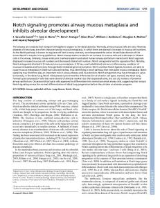
2C, 4A; see Fig. 2 legend). At the E16 stage, LIN-12 is prominent in punctate structures near the midline of the primordium (Figs 2C, 4A); these structures resemble in shape, position and timing of appearance the ‘apical vesicles’ described in an ultrastructural study of the primordium (Leung et al., 1999). Regulation of LIN-12 expression in the intestinal primordium A previous analysis of LIN-12 expression in nonintestinal, embryonic cells showed a requirement for GLP-1 mediated, cell-cell interactions (Moskowitz and Rothman, 1996). Briefly, LIN-12 expression was shown to occur only in cells that had prior expression of GLP-1, and that were in contact with ligand-expressing cells. In contrast, we found that LIN-12 was expressed in the E8 primordium in mutants lacking glp-1(+) function (Table 2; Fig. 5F). We asked whether cell-cell interactions were required for LIN-12 expression by using a laser microbeam to kill all of the blastomeres except for E. The E descendants were allowed to develop to the equivalent of the E8 or early E16 cell stage, then fixed and stained for LIN-12. In 8/8 cases, LIN-12 appeared to be expressed in all of the E descendants (Fig. 3A). Thus neither glp-1(+) function nor cellcell interactions after the birth of E appear to be required for cells in the intestinal primordium to express LIN-12. Several aspects of intestine-specific differentiation are controlled by a small group of ‘endoderm-determining’ transcription factors that are expressed very early in the E lineage. For example, the transcription factor END-1 is expressed in the E blastomere itself (Zhu et al., 1997), and ELT2 is expressed in the daughters of E (Fukushige et al., 1998). Ectopic, heat-shock-induced expression of an end-1 transgene is sufficient to cause a majority of embryonic cells to undergo at least partial intestine-specific differentiation (Zhu et al., 1998). To determine whether END-1 expression could cause ectopic expression of LIN-12, we fixed and immunostained embryos after heat-shock induction of the end-1 transgene. The pattern of cells expressing LIN-12 in these transgenic embryos (Fig. 3B,B′) appeared very different from the pattern in normal embryos at approximately equivalent stages (Fig. 2C). After heat shock, 41% of the cells in the transgenic embryos expressed LIN-12 (range, 18-65%; n=8 embryos), compared to only 18% of the cells in heat-shocked, wild-type controls (range, 14-21%, n=4 embryos). We looked for, but did not observe, a requirement for ELT-2 in LIN-12 expression; elt2(ca15) homozygous embryos expressed LIN-12 in the intestinal primordium, and appeared to have normal intestinal twist (data not shown). We conclude that LIN-12 expression in the E lineage is controlled by some of the endodermdetermining transcription factors, rather than by the GLP-1 activation pathway described previously. LAG-2-expressing cells induce LIN-12 asymmetry in the intestinal primordium Although the E blastomere appears to have an ‘autonomous’ ability to express LIN-12, we did not observe any evidence of LIN-12 asymmetry between the E descendants when neighboring blastomeres were killed (Fig. 3A). Thus cell-cell interactions appear to be required for LIN-12 asymmetry. Since lin-12(+) activity can alter the levels of LIN-12 expression in certain postembryonic cells (Wilkinson et al., 1994), we asked
Notch signaling and organ left-right asymmetry 3433 whether the LIN-12 signaling pathway was required for LIN-12 asymmetry. Although neither of two semidominant, activating mutations of lin-12 affected the expression of LIN-12 (lin-12(sd); Table 2) or intestinal twist (Table 1), mutations in either lag-1 or lag-2 affected LIN-12 asymmetry. 21% of the F2 self-progeny of lag-1(q385)/+ heterozygous hermaphrodites had approximately equal levels of LIN-12 expression in the left and right halves of the E8 and E16 primordia (Fig. 4B and Table 2). Similar results were observed in 27% and 22% of the F2 self-progeny of hermaphrodites heterozygous for lag-2(q387)/+ and for lag-2(q411)/+, respectively (Fig. 4C and Table 2). lag-2(q420ts) embryos showed symmetrical LIN-12 expression in the intestinal primordium at all temperatures (Table 2), consistent with our finding that this allele of lag-2 results in intestinal twist defects at all temperatures (Table 1). The lag-1 and lag-2 genes could function in cell-cell interactions within the intestinal primordium that lead to LIN12 asymmetry, or in interactions between nonintestinal cells and cells in the primordium. To distinguish between these possibilities, we examined the expression patterns of LAG-1
Fig. 2. LIN-12 expression. Embryos at various stages of intestinal development (E4, E8, E16) stained for LIN-12 (top row) and with DAPI (bottom row). Intestinal cells in the left and right halves of the primordium are indicated with white arrows and arrowheads, respectively; the anterior end of the primordium is indicated with black arrowheads for the E8 and E16 stages. During the E16 stage, the pattern of LIN-12 expression in the right half of the primordium changes markedly (also see Fig. 4A). Using the numbering system described in Leung et al. (1999), LIN-12 is detected in all of the right primordial cells except numbers 5 and 8 in the early E16 stage, then only in numbers 3 and 4 (arrowheads in C; note that the LIN-12 expressing cell at the posterior (bottom) of the embryo is not an intestinal cell), then only in number 4. At the time that the int rings begin to rotate, LIN-12 is not detected in the primordium.
and LAG-2 using an antiserum and a Green Fluorescent Protein (GFP) reporter, respectively (J. Kimble, unpublished; Blelloch et al., 1999). LAG-1 appeared to be expressed in the nuclei of all embryonic cells during the stages that the intestine forms, and was thus not informative (data not shown). We could not detect lag-2::GFP expression in any of the cells in the intestinal primordium; however, strong expression was observed in a row of 2-4 nonintestinal cells outside the left of the E4 (arrows, Fig. 5B) and E8 primordia (data not shown). A previous study reported that several embryonic cells express LAG-2, including the daughters and granddaughters of an embryonic blastomere named MSap (Moskowitz and Rothman, 1996). Through lineage analysis of the lag-2::GFP strain, we identified the cells adjacent to the intestinal primordium as the MSap descendants. To determine if the MSap descendants were required for LIN-12 asymmetry and intestinal twist, we used a laser microbeam to kill MSap. In each of eight operated embryos, LIN-12 was expressed symmetrically on both sides of the intestinal primordium (Fig. 4D and Table 2). We observed twist defects in 4/11 of the operated embryos, which is comparable to the frequency of twist defects observed in lag-2 mutants (Table 1). glp-1 mutant embryos, like wild-type embryos, lack or have very low levels of LIN-12 in the left half of the E8 primordium (see above). On the right half of the primordium, however, there are two additional cells that fail to express LIN-12 in 98% of glp-1(e2141ts) and glp-1(q224ts) embryos (asterisks, Fig. 5F; Table 3). We therefore examined LAG-2 expression in the glp-1 mutants. In addition to the normal pattern of LAG-2 expression by MSap descendants outside the left of the
Fig. 3. Control of LIN-12 expression. (A) LIN-12 staining of an E8 primordium derived from an ‘isolated’ E blastomere. All 8 of the E descendants showed LIN-12 expression; 7 are visible. Cells lacking LIN-12 expression are the laser-killed blastomeres, visible in the DAPI-stained image in (C). (B,B′) Two different focal planes of a single, heat-shocked hs-end-1 embryo stained for LIN-12. The embryo was allowed to develop to an age corresponding to approximately the E16 stage. The corresponding DAPI images are shown in (D,D′).
3434 G. J. Hermann, B. Leung and J. R. Priess
Fig. 4. Control of LIN-12 asymmetry. All images show E16 stage embryos prepared and labeled as in Fig. 2; genetic or cellular defects are listed above each panel.
Fig. 5. LAG-2 and LIN-12 asymmetry. (A-D) Nomarski micrographs of a wild-type embryo (A) and a glp-1 mutant embryo (C) containing a lag-2::GFP transgene at the E4 stage of intestinal development (the intestinal primordium is outlined). LAG-2 expression (GFP fluorescence) in the embryos in A and C is shown in B and D, respectively. The MSap daughters (two arrows in D) and granddaughters (four arrows in B) each express LAG-2 during the E4 stage. Note the ectopic, LAG-2 expressing cell (arrowhead) in D. (E-J) E8 stage embryos prepared as in Fig. 2; genotypes listed above each panel. Labeling is as in Fig. 2 except that the two anterior, right intestinal cells are indicated with asterisks in each panel for comparison.
Notch signaling and organ left-right asymmetry 3435
Fig. 6. Control of lag-2 expression. (A) Diagram showing the early divisions of the MS and E blastomeres. Dashes link sister cells. (B) Nomarski micrograph showing five of the first eight descendants of an ‘isolated’ MS blastomere; the daughters of MSap are labeled (arrows). (C) lag-2::GFP expression in the embryo shown in B. (D) Nomarski micrograph of a temperature-shifted lit-1 mutant embryo showing a subset of the first 16 descendants of the MS blastomere in addition to other cells; the granddaughters of MSap (arrows) and MSpp (arrowheads) are labeled. (E) lag-2::GFP expression in the embryo shown in D.
primordium (arrows, Fig. 5D), we observed an additional expressing cell outside the right of the E4 primordium in 14/14 glp-1(e2141ts) and 8/8 glp-1(q224ts) mutants (arrowhead; Fig. 5D). We identified this ectopic cell as ABpraap, and found that it was in contact with the E4 primordial cell that is the parent of the two, right E8 primordial cells that lack LIN-12 expression. lag-2(+) activity was required for the lack of LIN12 in the two right primordial cells, since these cells expressed Table 2. Asymmetric expression of LIN-12 in the intestine % embryos expressing LIN-12 Parental genotype Wild type glp-1 (e2142ts)1 glp-1 (e2141ts)1 glp-1 (q224ts)1 lin-12( n302sd) lin-12(n950sd) lag-1(q385)/+2 lag-2(q411)/+2 lag-2(q420ts) lag-2(q387 )/+2 apx-1 (zu347ts) MSap ablation spn-1(it143) 3
Temperature (°C) 15 20 26 26 26 26 20 20 20 20 15 23 20 15 23 20 20
Left half
Right half
0 0 0 0 0 0 0 0 0 0 0 0 0 0 0 0 17
100 100 100 100 100 100 100 100 79 73 2 0 78 100 100 0 83
Both halves 0 0 0 0 0 0 0 0 21 27 98 100 22 0 0 100 0
n 65 173 76 80 74 51 28 21 107 171 119 107 294 60 92 8 244
LIN-12 expression was scored in E8 and E16 intestinal primordia. For allele descriptions, see Table 1. 1Embryos were raised at 26°C throughout embryogenesis. 2Embryos scored were F2 progeny from single heterozygous parents. Since lag-1 and lag-2 cause L1-lethality, 17% of the F2 progeny are predicted to be homozygous for lag-1 or lag-2. 3Since spn-1(it143) is semi-embryonic lethal (Bergmann et al., 1998), only embryos with wild-type morphology were scored.
LIN-12 in glp-1(e2141ts); lag-2(q411) and glp-1(q224ts); lag2(q411) double mutants (Fig. 5G; Table 3). Thus, in both wildtype and in mutant embryos, contact with a LAG-2 expressing cell causes primordial cells to develop reduced levels of immunodetectable LIN-12 (see Discussion). Initiation of left-right asymmetry LAG-2 is expressed by MSap descendants outside the left of the intestinal primordium; however, the contralateral MSpp descendants, outside the right of the primordium, do not express LAG-2 (Fig. 6A). We found that the descendants of MSap, but not MSpp, expressed lag-2::GFP in lag-1(q385) mutants (5/5 embryos) and in glp-1(e2141ts) mutants (4/4 embryos), suggesting that the Notch-like signaling pathway is not required for the difference between MSap and MSpp. To investigate whether any cell-cell interactions were required for this difference, we used a laser microbeam to kill all of the early embryonic blastomeres except for MS, the precursor to both MSap and MSpp. In 5/5 experiments, lag-2::GFP was expressed only in the MSap daughters (arrows, Fig. 6C) and granddaughters (data not shown), and not in the MSpp descendants. Since MSap and MSpp are descendants of sister cells that are born in an anterior-posterior division, we considered the possibility that the LIT-1/POP-1 polarity pathway was involved in regulating LAG-2 expression. In early embryogenesis, all sister cells that are born in anterior-posteror divisions appear to differ in their levels of POP-1, a transcription factor that is related to vertebrate TCF/LEF1 and Drosophila Pangolin (Lin et al., 1995, 1998; Brunner et al., 1997). LIT-1 is a serine/threonine protein kinase that is required for POP-1 asymmetry (Meneghini et al., 1999; Rocheleau et al., 1999). Mutations in lit-1 cause anterior-posterior sisters to both adopt the anterior fate, and mutations in pop-1 cause the reciprocal transformation (Lin et al., 1995, 1998; Kaletta et al., 1997). We constructed a strain containing the temperature-sensitive allele lit-1(t1512ts) and the lag-2::GFP marker, and shifted embryos
3436 G. J. Hermann, B. Leung and J. R. Priess to the nonpermissive temperature (25oC) after the birth of the MS blastomere. The level of lag-2::GFP expression varied in each of four embryos examined, presumably from the incomplete penetrance of the lit-1(t1512ts) mutation (see Kaletta et al., 1997); however, all showed expression in the granddaughters of both MSap (arrows, Fig. 6E) and MSpp (arrowheads, Fig. 6E). We thus infer that LAG-2 expression in wild-type embryos is determined by an anterior-posterior cellfate decision that requires lit-1(+) activity. Although MSap and MSpp are descendants of sister cells that are born in an anterior-posterior division, this division is skewed slightly along the left-right axis of the embryo (Fig. 6A). This asymmetry results in MSap descendants contacting the left side of the intestinal primordium while MSpp descendants contact the right side. Mutations in several genes have been shown to disrupt the normally invariant division axes of the early embryonic cells (see Discussion). While most of these mutations result in grossly disorganized embryos, a mutation in the spn-1 gene can result in a few embryos that have a simple inversion of the first left-right asymmetries in blastomere divisions; 19% of viable spn-1(it143) embryos grow into adults that have a reversal of left-right asymmetry (Bergmann et al., 1998). We examined LIN-12 expression at the E8 and E16 stages of the intestinal primordium in embryos from spn-1(it143) homozygous hermaphrodites. Although the majority of embryos had the normal pattern of LIN-12 asymmetry in the intestinal primordium, 17% (n=244) had a left-right reversal of LIN-12 asymmetry (Fig. 4E; Table 2).
Fig. 7. LIT-1/POP-1 and twist boundary. (A,D) Nomarski micrographs of living embryos oriented as in Fig. 1D. Black and white arrowheads indicate the anterior end of the intestinal primordium and the posterior boundary of twist, respectively. The cells in int VI are the posterior sisters of cells in int IV (Sulston et al., 1983; Leung et al., 1999). (B, E) Comparably staged wild-type (B) and lit-1 mutant (E) embryos immunostained for POP-1; anterior-posterior sister cells in the E8 primordium are indicated by double-headed arrows. (C, F). DAPI images of embryos shown in B and E, respectively.
APX-1 functions after LIN-12 becomes asymmetric Our genetic analysis of twist indicates that the ligand APX-1, in addition to LAG-2, is essential for intestinal twist (Table 1). In contrast to the defects in LIN-12 asymmetry observed in lag2 mutants, LIN-12 appeared to be expressed with normal leftright asymmetry in the intestinal primordia of apx-1(zu347ts) mutant embryos cultured under conditions that cause these same embryos to show a highly penetrant twist defect (Tables 1, 2). This result suggested that apx-1(+) functions in twist after LIN-12 asymmetry is established at the E8 stage. Consistent with this hypothesis, we found that nearly all of the apx-1(zu347ts) mutant embryos that were shifted at the early E16 stage to the nonpermissive temperature had twist defects (Table 4). We have not yet been able to determine whether APX-1 is expressed asymmetrically in the intestinal primordium. The currently available APX-1 antiserum (Mickey et al., 1996) stains at or near the apical surfaces of cells in the E16 primordium (our unpublished results). The immunostaining, however, does not appear to be specific because it persists in apx-1 mutant embryos, as well as in wild-type embryos after dsRNA inhibition of apx-1 (our unpublished results). Cell movements in intestinal twist Because twist involves the circumferential migrations of cells in the anterior of the intestine, and the intestine is surrounded by a basement membrane when twist occurs (our unpublished observations), we addressed the possibility that the UNC-6 guidance system was required for twist. UNC-6, a lamininrelated basement membrane component, acts as a guidance signal for circumferential migration by a wide variety of cell types in C. elegans including axons, mesodermal cells and
Notch signaling and organ left-right asymmetry 3437 Table 3. LIN-12 expression in glp-1 mutants Embryo type Wild type glp-1 (e2141ts) glp-1 (e2141ts); lag-2(q411) glp-1 (q224ts) glp-1 (q224ts); lag-2(q411)
% embryos lacking LIN-12 expression in anterior, right E descendants* 6 98 0 98 0
n 34 53 29 44 12
Embryos were raised at 26°C throughout embryogenesis and scored for LIN-12 expression in the E8 primordium. The homozygous mutant progeny from glp-1; lag-2/+ parents were identified by the expression of LIN-12 on both halves of the intestinal primordium. *These cells (see asterisks in Fig. 5F) are daughters of the E descendant called Ear.
gonadal cells (Hedgecock et al., 1990; Ishii et al., 1992). UNC5 and UNC-40 are cell-surface receptors that guide cell migrations away from or towards UNC-6 cues, respectively (Hedgecock et al., 1990; Leung-Hagesteijn et al., 1992; Chan et al., 1996). Almost all of the embryos produced by unc-5, unc-6 or unc-40 mutants appeared to have normal twist, indicating that these genes do not play a major role in the cell movements associated with twist (Table 1). Some unc-6 and unc-40 mutant embryos, however, had subtle and variable defects in the movements and positions of anterior intestinal cells (Table 1 and data not shown). Control of the posterior boundary of intestinal twist The twist in the wild-type embryonic intestine extends to, but does not include, int ring V (Fig. 1A). Cells in int ring V are unique in that two nonintestinal cells, the germ cell precursors, invariably insert processes into their ventral surfaces (Sulston et al., 1983; our unpublished observations). Attachment of the germ cell precursors, however, does not appear to prevent twist in int ring V; we found that twist did not extend into int ring V in all experiments where either the parent (n=6) or grandparent (n=16) of the germ cell precursors was killed with a laser microbeam. Although in most experiments the laser-killed cell remained in contact with some region of the intestine, in two cases the grandparent was extruded from the embryo during gastrulation and so did not contact the intestine. We also examined the possibility that the C. elegans homeotic complex genes, ceh-13, lin-39 and mab-5, could have a role in specifying anterior-posterior differences between intestinal cells that determined the posterior boundary of twist. The posterior boundary appeared normal in strains with mutations in each of these genes (Table 1). Since POP-1, the downstream effector of the LIT-1/POP-1 polarity, is expressed with anterior-posterior asymmetry in at least some cells in the intestinal primordium (Lin et al., 1998), we asked whether lit-1(+) function was required to stop twist at int ring V, and whether lit-1(+) was required for POP-1 asymmetry. We found that twist extended beyond int ring V, in 100% (n=21) of lit-1(t1512ts) embryos shifted to the nonpermissive temperature at the E8 stage (Fig. 7D), and in 5% (n=22) of the embryos shifted at the E16 stage. Twist occurred in cells up to, and including, int ring VI. These temperatureshift experiments did not appear to affect the pattern of LIN12 expression in the E8 or E16 primordia (data not shown), but
had a marked effect on the expression of POP-1. In 98% of wild-type control embryos (n=62; Fig. 7B), each anteriorposterior pair of sister cells in the E8 primordium showed higher levels of POP-1 immunostaining in the anterior sister than in the posterior. In contrast, all pairs of sisters had approximately equivalent levels of POP-1 in lit-1(t1512ts) embryos (n=32; Fig. 7E) that were shifted to the nonpermissive temperature at about the E4 stage. Thus lit-1 appears to function to regulate POP-1 asymmetry in the intestinal primordium, as it does in the early embryo, and lit-1(+) function is required to define the posterior boundary of intestinal twist. DISCUSSION Development of the C. elegans intestine provides a simple model system for a genetic and molecular analysis of organogenesis (Leung et al., 1999). In this report, we have addressed how asymmetry is generated in the intestinal primordium by analyzing the asymmetrical ‘twist’ that develops at the anterior of the intestinal tube. Our results show that the LIN-12 signaling pathway of C. elegans, which is used repeatedly during development in cell-fate decisions, also has a role in controlling morphogenetic differences between otherwise identical intestinal cells. Anterior-posterior asymmetry in the intestine appears to be controlled by a cellpolarity pathway involving the lit-1 gene. Initial events in left-right asymmetry We have shown that lin-12(+) activity is essential for cells in the intestinal primordium to undergo the asymmetric, patterned movements we call intestinal twist. All of the cells in the E4 primordium express LIN-12, apparently in response to endoderm-determining transcription factors like END-1. A nonintestinal cell, MSap, plays an important role in intestinal twist, since about half of the embryos completely lack twist when MSap is killed. MSap descendants express LAG-2 and contact the left side of the primordium. Since loss of lag-2(+) activity causes a twist defect comparable to that caused by killing MSap, we propose that interactions between the LAG2-expressing cells outside the primordium and LIN-12expressing cells in the primordium initiate the events that lead to twist. Table 4. Temperature-shift analysis of apx-1 (zu347ts) Stage of primordium at shift E2 E4 E8 Early E16 Late E16
% embryos with twist defect 100 96 90 72 4
n 12 25 40 25 27
Embryos were shifted from 15°C to 26°C at the indicated stage and scored for the rotation of int rings II, III, and IV at the 1.5-fold stage. Embryos were scored as having a twist defect when 2-3 rings did not rotate. Early E16 is prior to, and late E16 is after, migration of intestinal nuclei (Leung et al., 1999).
3438 G. J. Hermann, B. Leung and J. R. Priess The lit-1 gene, and presumably other genes in the LIT1/POP-1 polarity pathway (see Rocheleau et al., 1997; Thorpe et al., 1997), appears to create an anterior-posterior difference between the sister cells MSa and MSp that leads to an MSa daughter (MSap) expressing LAG-2, while the corresponding MSp daughter (MSpp) does not. We propose that this anteriorposterior difference is transduced into a left-right difference by the slight skewing of the division axis of the MS blastomere. Cells surrounding MS appear to influence the pattern of MS division, since this pattern is not normal when the surrounding cells are killed (unpublished observations) or when their positions are inverted (see Introduction; Wood, 1991). The surrounding cells could provide specific signals that alter the MS division orientation, such as the effect of the C. elegans Wnt-like pathway on the division of the early blastomere ABar (Rocheleau et al., 1997; Thorpe et al., 1997), or could simply provide steric constraints on division. The development of left-right asymmetry in intestinal organogenesis has some similarity to the mechanism that establishes left-right asymmetry in the lineages of certain early blastomeres in C. elegans (see Introduction). Both events involve a Notch-related signaling pathway, although LIN-12, rather than GLP-1, is used as the receptor for intestinal asymmetry. Early lineage asymmetry results from skewed division axes that position receptor-expressing cells asymmetrically with respect to a signaling cell. In contrast, intestinal asymmetry appears to result from skewed division axes that position a signaling cell asymmetrically with respect to receptor-expressing, intestinal cells. In this case, division asymmetry changes what is fundamentally a LIT-1 and POP-1 mediated, anterior-posterior difference between MS daughters into a left-right difference. Left-right asymmetry in the primordium Although LIN-12 expression appears at low, uniform levels throughout the E4 primordium, by the E8 stage LIN-12 appears at much higher levels in the right half than in the left. The asymmetry appears to result both from an increase in the level of LIN-12 in the right half of the primordium, and a decrease in the low levels in the left half. We have shown that this LIN12 asymmetry requires MSap, and the wild-type functions of the genes lag-2 and lag-1; defects in any of these components lead to uniform, relatively high levels of LIN-12 throughout the E8 primordium. We propose, therefore, that a LAG-2 mediated activation of LIN-12 in the left half of the E4 primordium leads to low levels, or lack of, LIN-12 in the left half of the E8 primordium, and this hypothesis is supported by our analysis of the effect of ectopic LAG-2 on LIN-12 expression in glp-1 mutants. After exposure to ligand, receptors in the Notch family undergo proteolytic cleavage events that are essential for signal transduction (reviewed in Annaert and De Strooper, 1999; Artavanis-Tsakonas et al., 1999; Chan and Jan, 1999). Nevertheless, it is unusual for ligand exposure to cause a visible decrease in level of receptor. For example, GLP-1 levels on the surfaces of early embryonic blastomeres are not noticeably different between blastomeres that either do, or do not, interact with ligand-expressing cells (Evans et al., 1994). Moreover, LIN-12 expression appears to increase, rather than decrease, after exposure to ligand in the interaction between the AC and VU cells during postembryonic development of C.
elegans (see below; Wilkinson et al., 1994). Similarly, Drosophila embryonic cells that are exposed to ligand can increase their relative levels of Notch (Huppert et al., 1997). The proteolytic cleavage of LIN-12 that normally results from ligand exposure is, by itself, unlikely to account for the low levels of LIN-12 in the left half of the E8 primordium, since the transcription factor LAG-1, the downstream effector of the activated receptor, is required for these low levels. We propose that LIN-12 asymmetry in the E8 primordium serves to pattern a second, subsequent, LIN-12 mediated interaction that leads to twist asymmetry. Our temperature-shift experiments demonstrate that the ligand APX-1 is essential for intestinal twist during the early E16 stage of the intestinal primordium, after LIN-12 asymmetry has been established through the earlier, LAG-2 mediated, interaction. Since LIN12, but not GLP-1, is required for twist, we infer that APX-1 must be interacting with LIN-12 at the E16 stage. In addition, the observation that lag-1 mutations cause a complete loss of twist, while lag-2 mutations cause a variable and partial loss, supports the hypothesis that there are two distinct LIN-12 mediated interactions. We consider it likely that apx-1(+) function is required within the cells of the intestinal primordium for twist. The anterior primordial cells, which are the cells that undergo twist, express LIN-12 but do not contact any nonintestinal cells that express LIN-12, suggesting that APX-1 interacts with LIN-12 on the intestinal cells. Furthermore, the surfaces of the intestinal cells in the E16 primordium are separated from the surface membranes of all surrounding, nonintestinal cells by two extracellular basement membranes (our unpublished results). Thus we consider it likely that APX-1 and LIN-12 interactions occur between cells within the E16 primordium, rather than between primordium cells and surrounding cells. In principal, the asymmetric, high levels of LIN-12 in the right half of the E8 and E16 primordia could provide a basis for reproducible, asymmetric cell-cell interactions, irrespective of whether APX-1 is expressed in all, or only a subset of, the primordial cells. If APX-1 is expressed asymmetrically, one interesting possibility is that lin-12(+) activity determines the expression pattern of APX-1. A well-characterized example of cell interactions affecting LAG-2 and LIN-12 expression in C. elegans is the AC/VU cell-fate decision during postembryonic development (Wilkinson et al., 1994). An interaction between two initially equivalent cells, each expressing both LIN-12 and LAG-2, leads to one cell adopting the AC fate and the other adopting the VU fate. A feedback mechanism appears to increase the expression of LAG-2 in whichever cell initially has slightly less lin-12(+) activity. Since cells in the left half of the E8 and E16 primordia have low levels of LIN-12, a similar feedback mechanism could increase the levels of APX-1 in those cells. This general model would not explain how LIN-12 asymmetry is generated between the E4 and E8 stages, since the LAG-2 mediated interaction leads to a decrease, rather than an increase, in the levels of LIN-12. We have shown that lag-2 mutants have a twist defect that is only partially penetrant. Interestingly, rotation of the various int rings appears to be variable in the abnormal intestines of lag-2 mutants; often one or two of the three int rings appear to rotate (our unpublished observations). Since lag-2 mutants have equivalent levels of LIN-12 in the right and left halves of the E8 primordium, one possibility is that the feedback
Notch signaling and organ left-right asymmetry 3439 mechanism described above could lead to variable asymmetry in APX-1 and LIN-12 mediated interactions between primordial cells that initially have equivalent levels of both APX-1 and LIN-12. In future studies it will be important to develop methods for detecting APX-1 expression, and LIN-12 activation, in the primordium to analyze this second LIN-12 mediated interaction directly. Anterior-posterior asymmetry in the primordium The Homeotic Complex (HOM-C) genes, and the anteriorposterior differences in the expression patterns of these genes, appear to be highly conserved in most animals, including C. elegans. Anterior-posterior differences observed in tissues or organs often depend on HOM-C function, and the C. elegans HOM-C genes are required during postembryonic development for numerous, position-specific differences between epithelial cells (reviewed in Krumlauf, 1994; Capecchi, 1997; Kenyon et al., 1997; Beck et al., 2000; GrapinBotton and Melton, 2000). However, the only anteriorposterior asymmetries that have been characterized thus far in the intestine (our present study; Schroeder and McGhee, 1998) are not determined by HOM-C genes, but rather by a distinct polarity pathway involving LIT-1 and POP-1. Our results paired with those of Lin et al. (1998) demonstrate that sister cells at each anterior-posterior division of the intestinal lineage show POP-1 asymmetry, and that lit-1(+) activity is required for this asymmetry. In the analysis by Schroeder and McGhee (1998) of a transgene that is expressed in the anterior of the intestine, expression was found to be regulated by pop-1(+) activity. In the present report, we have shown that lit-1(+) activity is required to limit twist to the anterior of the intestine, and that lit-1(+) function is required at or after the E8 stage. Thus the LIT-1/POP-1 pathway appears to function late in embryogenesis, and appears to be a major source of anteriorposterior asymmetry in the intestine. Asymmetric left-right morphogenesis Once the anterior-posterior boundary of twist is specified, what is the basis for the circumferentially oriented movements of intestinal cells that result in twist? Since the intestinal primordium is covered with an extracellular matrix at the time of rotation, it is possible that cells recognize, and respond to, guidance cues in the matrix. The circumferential migration of several other cell types in C. elegans has been shown to be controlled by the well-characterized UNC-6 guidance system (Hedgecock and Norris, 1997). We found, however, that mutants defective in this pathway had only minor, if any, defects in twist. A second possibility is that intestinal cells move in response to changes in the adhesive properties of other intestinal cells. Prior to the development of twist, there is a period of cell intercalation in the primordium. The behavior of cells during intercalation suggests that there may be left-right differences in cell adhesivity (Leung et al., 1999): cells in the right half of the primordium intercalate with other right cells, but never with left cells, and vice versa. The development of twist requires that some of the left cells exchange ipsilateral contacts with other left cells for new contacts with right cells, and vice versa. For example, the right int IV cell establishes new contacts with the left int V cell, while reducing ipsilateral contacts with the right int V cell (Fig. 1A). In theory, twist could result by creating heterotypic interactions between left
and right cells that were stronger than homotypic interactions. Studies of Drosophila support the general hypothesis that the Notch pathway might play a role in controlling adhesive differences between otherwise equivalent cells. Notch appears to have a role in the epithelial cell movements that result in dorsal closure of the embryo (Zecchini et al., 1999), and in the axonal outgrowth of some neurons (Giniger et al., 1993). It will be interesting to determine if other developmental events that require Notch function involve changes in adhesivity, for example in the control of ommatidial rotation in the Drosophila eye. We thank Stuart Kim, Al Candia, and Judith Kimble for allowing us to use their antisera against LIN-12 and LAG-1 prior to publication. We thank members of the Priess laboratory for helpful discussion and Katie Mickey for sharing unpublished observations. For strains and antibodies we thank Dominique Bergmann, Tetsunari Fukushige, Dali Gao, Judith Kimble, Jim McGhee, Joel Rothman, Jim Thomas, Bill Wood and Jiangwen Zhu. Some nematode strains used in this work were provided by the Caenorhabditis Genetics Center, which is funded by the NIH. G. Hermann is supported by a postdoctoral fellowship from the Damon Runyon-Walter Winchell Foundation (DRG1561); B. Leung is supported by a training grant from the National Cancer Institute (CA09657-09); J. Priess is supported by the Howard Hughes Medical Institute.
REFERENCES Annaert, W. and De Strooper, B. (1999). Presenilins: molecular switches between proteolysis and signal transduction. Trends Neurosci. 22, 439-443. Artavanis-Tsakonas, S., Rand, M. D. and Lake, R. J. (1999). Notch signaling: cell fate control and signal integration in development. Science 284, 770-776. Beck, F., Tata, F., Chawengsaksophak, K. (2000). Homeobox genes and gut development. BioEssays 22, 431-441. Bergmann, D. C., Crew, J. R., Kramer, J. M. and Wood, W. B. (1998). Cuticle chirality and body handedness in Caenorhabditis elegans. Dev. Genet. 23, 164-174. Blelloch, R., Anna-Arriola, S. S., Gao, D., Li, Y., Hodgkin, J. and Kimble, J. (1999). The gon-1 gene is required for gonadal morphogenesis in Caenorhabditis elegans. Dev. Biol. 216, 382-393. Brenner, S. (1974). The genetics of Caenorhabditis elegans. Genetics 77, 7194. Brunner, E., Peter, O., Schweizer, L. and Basler, K. (1997). pangolin encodes a Lef-1 homologue that acts downstream of Armadillo to transduce the Wingless signal in Drosophila. Nature 385, 829-833. Brunschwig, K., Wittmann, C., Schnabel, R., Burglin, T. R., Tobler, H. and Muller, F. (1999). Anterior organization of the Caenorhabditis elegans embryo by the labial-like Hox gene ceh-13. Development 126, 1537-1546. Capdevila, J., Vogan, K. J., Tabin, C. J. and Belmonte, J. C. I. (2000). Mechanisms of left-right determination in vertebrates. Cell 101, 9-21. Capecchi, M. R. (1997). Hox genes and mammalian development. Cold Spring Harb. Symp. Quant. Biol. 62, 273-281. Chan, S. S., Zheng, H., Su, M. W., Wilk, R., Killeen, M. T., Hedgecock, E. M. and Culotti, J. G. (1996). UNC-40, a C. elegans homolog of DCC (Deleted in Colorectal Cancer), is required in motile cells responding to UNC-6 netrin cues. Cell 87, 187-195. Chan, Y. M. and Jan, Y. N. (1999). Presenilins, processing of beta-amyloid precursor protein, and notch signaling. Neuron 23, 201-204. Christensen, S., Kodoyianni, V., Bosenberg, M., Friedman, L. and Kimble, J. (1996). lag-1, a gene required for lin-12 and glp-1 signaling in Caenorhabditis elegans, is homologous to human CBF1 and Drosophila Su(H). Development 122, 1373-1383. Clark, S. G., Chisholm, A. D. and Horvitz, H. R. (1993). Control of cell fates in the central body region of C. elegans by the homeobox gene lin-39. Cell 74, 43-55. Evans, T. C., Crittenden, S. L., Kodoyianni, V. and Kimble, J. (1994). Translational control of maternal glp-1 mRNA establishes an asymmetry in the C. elegans embryo. Cell 77, 183-194.
3440 G. J. Hermann, B. Leung and J. R. Priess Fitzgerald, K. and Greenwald, I. (1995). Interchangeability of Caenorhabditis elegans DSL proteins and intrinsic signalling activity of their extracellular domains in vivo. Development 121, 4275-4282. Fukushige, T., Hawkins, M. G. and McGhee, J. D. (1998). The GATA-factor elt-2 is essential for formation of the Caenorhabditis elegans intestine. Dev. Biol. 198, 286-302. Gao, D. and Kimble, J. (1995). APX-1 can substitute for its homolog LAG2 to direct cell interactions throughout Caenorhabditis elegans development. Proc. Natl. Acad. Sci. USA 92, 9839-9842. Gendreau, S. B., Moskowitz, I. P., Terns, R. M. and Rothman, J. H. (1994). The potential to differentiate epidermis is unequally distributed in the AB lineage during early embryonic development in C. elegans. Dev Biol 166, 770-781. Giniger, E., Jan, L. Y. and Jan, Y. N. (1993). Specifying the path of the intersegmental nerve of the Drosophila embryo: a role for Delta and Notch. Development 117, 431-440. Grapin-Botton, A. and Melton, D. A. (2000). Endoderm development from pattern to organogenesis. Trends Genet. 16, 124-130. Greenwald, I. (1998). LIN-12/Notch signaling: lessons from worms and flies. Genes. Dev. 12, 1751-1762. Greenwald, I. S., Sternberg, P. W. and Horvitz, H. R. (1983). The lin-12 locus specifies cell fates in Caenorhabditis elegans. Cell 34, 435-444. Hedgecock, E. M., Culotti, J. G. and Hall, D. H. (1990). The unc-5, unc-6, and unc-40 genes guide circumferential migrations of pioneer axons and mesodermal cells on the epidermis in C. elegans. Neuron 4, 61-85. Hedgecock, E. M. and Norris, C. R. (1997). Netrins evoke mixed reactions in motile cells. Trends Genet. 13, 251-253. Henderson, S. T., Gao, D., Lambie, E. J. and Kimble, J. (1994). lag-2 may encode a signaling ligand for the GLP-1 and LIN-12 receptors of C. elegans. Development 120, 2913-2924. Huppert, S. S., Jacobsen, T. L. and Muskavitch, M. A. T. (1997). Feedback regulation is central in Delta-Notch signalling required for Drosophila wing vein morphogenesis. Development 124, 3283-3291. Hutter, H. and Schnabel, R. (1994). glp-1 and inductions establishing embryonic axes in C. elegans. Development 120, 2051-2064. Hutter, H. and Schnabel, R. (1995). Establishment of left-right asymmetry in the Caenorhabditis elegans embryo: a multistep process involving a series of inductive events. Development 121, 3417-3424. Ishii, N., Wadsworth, W. G., Stern, B. D., Culotti, J. G. and Hedgecock, E. M. (1992). UNC-6, a laminin-related protein, guides cell and pioneer axon migrations in C. elegans. Neuron 9, 873-881. Kaletta, T., Schnabel, H. and Schnabel, R. (1997). Binary specifications of the embryonic lineage in Caenorhabditis elegans. Nature 390, 294-298. Kenyon, C. (1986). A gene involved in the development of the posterior body region of C. elegans. Cell 46, 477-487. Kenyon, C. J., Austin, J., Costa, M., Cowing, D. W., Harris, J. M., Honigberg, L., Hunter, C. P., Maloof, J. N., Muller-Immergluck, M. M., Salser, S. J. et al. (1997). The dance of the Hox genes: patterning the anteroposterior body axis of Caenorhabditis elegans. Cold Spring Harb. Symp. Quant. Biol. 62, 293-305. Kimble, J. and Simpson, P. (1997). The LIN-12/Notch signaling pathway and its regulation. Ann. Rev. Cell Dev. Biol. 13, 333-3361. Kodoyianni, V., Maine, E. M. and Kimble, J. (1992). Molecular basis of loss-of-function mutations in the glp-1 gene of Caenorhabditis elegans. Mol. Biol. Cell 3, 1199-1213. Krumlauf, R. (1994). Hox genes in vertebrate development. Cell 78, 191-201. Lambie, E. J. and Kimble, J. (1991). Two homologous regulatory genes, lin-12 and glp-1, have overlapping functions. Development 112, 231240. Leung, B., Hermann, G. J. and Priess, J. R. (1999). Organogenesis of the Caenorhabditis elegans intestine. Dev. Biol. 216, 114-134. Leung-Hagesteijn, C., Spence, A. M., Stern, B. D., Zhou, Y., Su, M. W., Hedgecock, E. M. and Culotti, J. G. (1992). UNC-5, a transmembrane protein with immunoglobulin and thrombospondin type 1 domains, guides cell and pioneer axon migrations in C. elegans. Cell 71, 289-299. Lin, R., Hill, R. J. and Priess, J. R. (1998). POP-1 and anterior-posterior fate decisions in C. elegans embryos. Cell 92, 229-239. Lin, R., Thompson, S. and Priess, J. R. (1995). pop-1 encodes an HMG box protein required for the specification of a mesoderm precursor in early C. elegans embryos. Cell 83, 599-609. Mello, C. C., Draper, B. W. and Priess, J. R. (1994). The maternal genes
apx-1 and glp-1 and establishment of dorsal-ventral polarity in the early C. elegans embryo. Cell 77, 95-106. Meneghini, M. D., Ishitani, T., Carter, J. C., Hisamoto, N., NinomiyaTsuji, J., Thorpe, C. J., Hamill, D. R., Matsumoto, K. and Bowerman, B. (1999). MAP kinase and Wnt pathways converge to downregulate an HMG-domain repressor in Caenorhabditis elegans. Nature 399, 793-797. Mickey, K. M., Mello, C. C., Montgomery, M. K., Fire, A. and Priess, J. R. (1996). An inductive interaction in 4-cell stage C. elegans embryos involves APX-1 expression in the signaling cell. Development 122, 17911798. Moskowitz, I. P., Gendreau, S. B. and Rothman, J. H. (1994). Combinatorial specification of blastomere identity by glp-1-dependent cellular interactions in the nematode Caenorhabditis elegans. Development 120, 3325-3338. Moskowitz, I. P. G. and Rothman, J. H. (1996). lin-12 and glp-1 are required zygotically for early embryonic cellular interactions and are regulated by maternal GLP-1 signaling in Caenorhabditis elegans. Development 122, 4105-4117. Priess, J. R., Schnabel, H. and Schnabel, R. (1987). The glp-1 locus and cellular interactions in early C. elegans embryos. Cell 51, 601-611. Ramsdell, A. F. and Yost, H. J. (1998). Molecular mechanisms of vertebrate left-right development. Trends Genet. 14, 459-465. Rocheleau, C. E., Downs, W. D., Lin, R., Wittmann, C., Bei, Y., Cha, Y., Ali, M., Priess, J. R. and Mello, C. C. (1997). Wnt signaling and an APCrelated gene specify endoderm in early C. elegans embryos. Cell 90, 707716. Rocheleau, C. E., Yasuda, J., Shin, T. H., Lin, R., Sawa, H., Okano, H., Priess, J. R., Davis, R. J. and Mello, C. C. (1999). WRM-1 activates the LIT-1 protein kinase to transduce anterior/posterior polarity signals in C. elegans. Cell 97, 717-726. Schnabel, R. and Priess, J. R. (1997). Specification of cell fates in the early embryo. In Caenorhabditis elegans II (ed. D. L. Riddle, T. Blumenthal, B. J. Meyer and J. R. Priess), pp. 361-382. Cold Spring Harbor Laboratory Press. Schroeder, D. F. and McGhee, J. D. (1998). Anterior-posterior patterning within the Caenorhabditis elegans endoderm. Development 125, 4877-4887. Suderam, M. and Greenwald, I. (1993). Genetic and phenotypic studies of hypomorphic lin-12 mutants in Caenorhabditis elegans. Genetics 135, 755763. Sulston, J. E. and Horvitz, H. R. (1977). Post-embryonic cell lineages of the nematode, Caenorhabditis elegans. Dev. Biol. 56, 110-156. Sulston, J. E., Schierenberg, E., White, J. G. and Thomson, J. N. (1983). The embryonic cell lineage of the nematode Caenorhabditis elegans. Dev. Biol. 100, 64-119. Tax, F. E., Yeargers, J. J. and Thomas, J. H. (1994). Sequence of C. elegans lag-2 reveals a cell-signaling domain shared with Delta and Serrate of Drosophila. Nature 368, 150-154. Thorpe, C. J., Schlesinger, A. J., Carter, C. and Bowerman, B. (1997). Wnt signaling polarizes an early C. elegans blastomere to distinguish endoderm from mesoderm. Cell 90, 695-707. Wilkinson, H. A., Fitzgerald, K. and Greenwald, I. (1994). Reciprocal changes in expression of the receptor lin-12 and its ligand lag-2 prior to commitment in a C. elegans cell fate decision. Cell 79, 1187-1198. Wood, W. B. (1991). Evidence from reversal of handedness in C. elegans embryos for early cell interactions determining cell fates. Nature 349, 536538. Wood, W. B. (1998). Handed asymmetry in nematodes. Semin. Cell Dev. Biol. 9, 53-60. Wood, W. B., Bergmann, D. and Florance, A. (1996). Maternal effect of low temperature on handedness determination in C. elegans embryos. Dev. Genet. 19, 222-230. Zecchini, V., Brennan, K. and Martinez-Arias, A. (1999). An activity of Notch regulates JNK signalling and affects dorsal closure in Drosophila. Curr. Biol. 9, 460-469. Zhu, J., Fukushige, T., McGhee, J. D. and Rothman, J. H. (1998). Reprogramming of early embryonic blastomeres into endodermal progenitors by a Caenorhabditis elegans GATA factor. Genes Dev. 12, 38093814. Zhu, J., Hill, R. J., Heid, P. J., Fukuyama, M., Sugimoto, A., Priess, J. R. and Rothman, J. H. (1997). end-1 encodes an apparent GATA factor that specifies the endoderm precursor in Caenorhabditis elegans embryos. Genes Dev. 11, 2883-2896.