1943
Development 127, 1943-1951 (2000) Printed in Great Britain © The Company of Biologists Limited 2000 DEV1507
Specification of neuropeptide Y phenotype in visual cortical neurons by leukemia inhibitory factor Petra Wahle*, Thorsten Gorba, Marcus J. Wirth and Kirstin Obst-Pernberg AG Entwicklungsneurobiologie, Ruhr-Universität, ND 7/31, 44780 Bochum, Germany *Author for correspondence (e-mail:
[email protected])
Accepted 7 February; published on WWW 6 April 2000
SUMMARY Building the complex mammalian neocortex requires appropriate numbers of neurochemically specified neurons. It is not clear how the highly diverse cortical interneurons acquire their distinctive phenotypes. The lack of genetic determination implicates environmental factors in this selection and specification process. We analysed, in organotypic visual cortex cultures, the specification of neurons expressing neuropeptide Y (NPY), a potent anticonvulsant. Endogenous brain-derived neurotrophic factor and neurotrophin 4/5 play no role in early NPY phenotype specification. Rather, the decision to express NPY is made during a period of molecular plasticity during which differentiating neurons with the potential to express
NPY compete for the cytokine leukemia inhibitory factor which is produced in the cortex, but is negatively regulated by thalamic afferences. The neurons that fail in this competition are parvalbuminergic basket and chandelier neurons, which express NPY transiently, but will not acquire a permanent NPY expression. They switch into a facultative NPY expression mode, and remain responsive to the neurotrophins which modulate NPY expression later in development.
INTRODUCTION
In the adult rat cortex interneurons constitute three families characterized by the expression of parvalbumin, or calretinin, or somatostatin (Gonchar and Burkhalter, 1997). The parvalbuminergic neurons are fast-spiking basket and chandelier cells which do not express neuropeptides or calbindin (Demeulemeester et al., 1989, 1991; Gonchar and Burkhalter, 1997). Calretinin neurons are mostly bipolar cells, while the somatostatin neurons constitute a variety of types which colocalize neuropeptide Y (NPY) and calbindin. In developing rat however, the diversity appeared much higher. Cauli et al. (1997) using single-cell RT-PCR followed by Southern blots reported that in postnatal day-16 to -22 neocortex, many parvalbuminergic neurons coexpressed NPY, somatostatin and calbindin. During this time, parvalbuminergic neurons acquire their fast spiking properties (Massengill et al., 1997) and begin to downregulate their transient calbindin expression (Alcantara et al., 1996). In adult cortical interneurons, calbindin and peptidergic phenotypes (Gonchar and Burkhalter, 1997) are associated with non-fast spiking physiological properties (Kawaguchi and Kubota, 1997). Most likely, the segregation of neurochemical properties observed in the adult has not yet been completed by the age analyzed by Cauli et al. (1997) with a very sensitive technique. With regard to the enormous phenotypic plasticity it must be assumed that sets of neurons of any given phenotype become selected from a larger number of neurons with the genetic potential to express this phenotype. This implies that differentiating neurons must somehow decide which phenotype to adopt.
The interneurons in the mammalian cortex are highly diverse and may constitute up to 100 distinct cell types characterized by morphology and axon termination, neurochemical and molecular profile, firing properties, and responsiveness to transmitters (DeFelipe, 1993; Parra et al., 1998; Stevens, 1998). To complicate matters, during corticogenesis interneurons not only arise from the telencephalic ventricular zone, but invade the neocortex from the lateral ganglionic eminence, and they display a notoriously nonradial dispersal behavior (Anderson et al., 1997; Tan et al., 1998). No wonder then that the neurochemically defined interneuron types (Demeulemeester et al., 1988, 1989; DeFelipe, 1993; Gonchar and Burkhalter, 1997) are not related by genetic lineage, but apparently become specified locally by environmental factors (Mione et al., 1994; Götz and Bolz, 1994; Götz et al., 1995). Once arrived at their final destination many differentiating interneurons, and also pyramidal neurons, transiently express neuropeptides, calcium-binding proteins or receptors, and subsequently undergo phenotypic shifts which are influenced by afferent innervation, trophic factors and sensory experience (Shaw et al., 1986; Parnavelas and Cavanagh, 1988; Wahle, 1994; Alcantara et al., 1996; Obst and Wahle, 1995, 1997; Obst et al., 1998). These shifts appear essential for the establishment of the adult neurochemical architecture because they shape the laminar- and areal-specific sets of distinct interneuron types likely required for proper function of the mature cortex.
Key words: Neuropeptide Y, Visual cortex, Leukemia inhibitory factor, Neurotrophins, Rat
1944 P. Wahle and others We studied neurons expressing NPY mRNA in rat visual cortex. In the adult, NPY delineates a subset of non-fast spiking interneurons of small basket and Martinotti cell morphology. Recent studies have emphazised the important cellular and behavioral functions of NPY. It is implicated in feeding behavior, anxiety, the preference for alcohol, and most importantly, NPY is a major endogenous antiepileptic peptide and modulator of excitatory synaptic transmission (Wahlestedt et al., 1993; Erickson et al., 1996a,b; Klapstein and Colmers, 1997; Baraban et al., 1997; Woldbye et al., 1997; Ehlers et al., 1998; Thiele et al., 1998, Tecott and Heberlein, 1998; Vezzani et al., 1999). It is likely then that NPY expression is under tight regulatory control. However, NPY expression is highly dynamic and is easily promoted by neuronal excitation and epileptiform activity (Gall et al., 1990; Carnahan and Nawa, 1995, for review; Nawa et al., 1995; Schwarzer et al., 1996; Marty and Onteniente, 1999). NPY expression is also promoted by brain-derived neurotrophic factor (BDNF), neurotrophin 4/5 (NT-4/5), and the cytokine leukemia inhibitory factor (LIF) and the neurons express tyrosine receptor kinase trkB and the LIF receptor β (Carnahan and Nawa, 1995; Marty et al., 1997; Wirth et al., 1998a,b; Marty and Onteniente, 1999; Gorba and Wahle, 1999). In these conditions (high levels of neuronal activity, exposure to exogenous trophic factors) NPY mRNA is upregulated to high levels in many more neurons than normal, including parvalbuminergic cortical interneurons and pyramidal neurons (M. Engelhardt, G. diCristo, N. Berardi, L. Maffei, P. W., unpublished) or hippocampal granule and pyramidal cells (Gall et al., 1990). Together, this suggests the existence of constitutive and facultative NPY expressers. Clearly, the ability of the nervous system to upregulate an anticonvulsant like NPY in a usedependent way is highly adaptive. Yet, the mechanisms by which developing neurons learn to express NPY in a permanent, constitutive versus a transient, facultative mode are unknown. MATERIALS AND METHODS Cultures Organotypic cortex monocultures and thalamocortical cocultures were prepared from pigmented Long-Evans rats at the day of birth and kept in vitro for up to 70 days (DIV; Obst and Wahle, 1995, 1997). Cocultures regenerate the axonal connections within the first 2 weeks in vitro (Bolz et al., 1990; Obst and Wahle, 1997). Control cocultures were implanted with lipophilic dyes, and axonal connections in fiber bridges between the explants were seen in all these cocultures. MCM was harvested from 10-30 DIV monocultures every other day. It was either directly applied to cocultures, or preincubated for 1 hour with the anti-LIF antibody before being applied to cocultures. BDNF, NT4/5, neurotrophin-3, nerve growth factor and LIF (all from Peprotech) were applied at 20 ng/ml medium every third day for the time periods indicated in the legends. The neutralizing anti-LIF antibody (R&D Systems) was applied at 4 µg/ml medium every second day. Its neutralizing efficiency was proved in a control experiment: we reported (Wirth et al., 1998b) that exogenous LIF stimulates NPY mRNA expression in monocultures grown in the presence of 10 mM MgSO4 to block spontaneous bioelectric activity. When anti-LIF antibody was applied to activity-blocked monocultures supplemented with exogenous LIF, the antibody completely blocked the effect of LIF (2 µg/ml and 4 µg/ml anti-LIF antibody were equally effective, the neutralization in the present study was done with a 4 µg/ml
concentration). TrkB-IgGs and TrkC-IgGs were kindly provided by Dr Yancopoulos (Regeneron Pharmaceuticals). They were applied at a concentration of 20 µM, which was shown to effectively neutralize endogenous neurotrophins in tissue cultures (McAllister et al., 1996; Routbort et al., 1997). Biolistic transfection Thalamocortical cocultures were transfected at 5 DIV using the Helios Gene Gun (Biorad) and 1.6 µm gold particles coated with expression plasmid cDNAs encoding either p-EGFP-N1 under cytomegalovirus promoter (Clontech; 1 µg/mg gold) or p-EGFP-N1 (1 µg/mg gold) plus pCAGGS-LIF under beta-actin promoter (2 µg/mg gold; generously provided by Dr Austin Smith, Center of Genome Research, Edinburgh, UK; for vector construction including the LIF vector see Miyazaki et al., 1989; Niwa et al., 1991). Both plasmids were prepared endotoxin-free (Endofree, Qiagen), precipitated onto gold particles according to the manufacturer’s recommendation (Biorad), and 0.2 mg gold was loaded per cartridge. The gene gun was mounted into a support, with the distance from muzzle to tissue set at 1 cm, pressure was 160 psi (1.1×10⁶ Pa). EGFP expression was checked at 7 DIV, and only well expressing cultures were kept. Expression was observed for about 2 further weeks suggesting that also LIF was expressed through the presumed period of molecular plasticity. At 75 DIV, OTC were checked for visible fiber bridges between the two explants and only cultures passing this examination went into NPY in situ hybridization. In situ hybridization and quantitative analysis NPY mRNA-expressing cells were labeled by in situ hybridization in paraformaldehyde-fixed, freefloating cultures. Digoxigenin-labeled riboprobes complementary to exon 2 and/or the signal peptide of NPY were transcribed (Roche; following the manufacturer’s protocol) and used under established conditions and controls (Obst and Wahle, 1997). Both probes yielded identical results. The coculture and the monoculture study were each performed with riboprobes from one transcription to yield probe constancy. For analysis (Obst and Wahle, 1997; Wirth et al., 1998) labeled neurons were plotted (Eutectics Neuron Reconstruction System, Raleigh, NC, USA), and subsequently total neuron number per culture was determined, after counterstaining with thionin, to calculate the percentage of NPY mRNA-expressing neurons. Non-parametric statistics (MannWhitney U-Test) was performed followed by corrections for multiple testing according to Holm (1979). Fluorescent double-labeling with mouse monoclonal antibodies against parvalbumin (SWANT) followed by biotinylated secondary and Texas Red-conjugated streptavidin was performed as described by Gorba and Wahle (1999) in 40 DIV and 60 DIV cortex monocultures and thalamocortical cocultures. The percentage of cortical parvalbuminergic neurons coexpressing NPY was determined (about 150 neurons were analyzed for every age and experimental condition). PCR Five monocultured cortices and five cortices from thalamocortical cocultures per age, at the six ages indicated in Fig. 3, were pooled, mRNA was isolated (mRNA Direct kit, Dynal), and single-stranded cDNA was reverse transcribed with AMV reverse transcriptase (20 U/µl, Stratagene) at 42°C for 45 minutes. PCR was performed in a total volume of 50 µl with GoldStar DNA Polymerase (Eurogentec). PCR conditions were kept in the linear range determined for each product (30 cycles for BDNF, 36 cycles for NT-4/5 and LIF). The two sets of cDNA libraries were probed for BDNF (bases 154-393 specific for 3′ exon V; Maisonpierre et al., 1991), NT-4/5 (bases 309-605; Berkemeier et al., 1991), and LIF (bases 179-513; Yamamori et al., 1989). Relative band intensities were densitometrically determined with the Eagle Eye System (Stratagene). Intensities were normalized to glucose-6-phosphate dehydrogenase expression (bases
NPY phenotype specification in visual cortex 1945
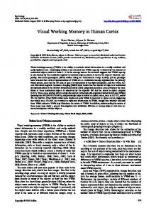
Fig. 1. NPY mRNA expression in representative monocultures (VC) and thalamocortical cocultures (VC-Th). (A) VC at 60 DIV. (B) VC-Th at 70 DIV. (C) VC-Th at 70 DIV, MCM from 3-20 DIV. (D) VC-Th at 70 DIV, MCM from 60-70 DIV. (E) VC-Th at 70 DIV, NT-4/5 from 60-70 DIV. (F) VCTh at 70 DIV, LIF from 3-20 DIV. (G) VC-Th at 70 DIV, LIF from 60-70 DIV. (H) VC-Th at 70 DIV, MCM plus anti-LIF from 3-20 DIV. The labeling intensity per neuron reflects the amount of mRNA (Emson, 1993). (I,J) High degree of overlap between NPY mRNA and parvalbumin immunofluorescence in 60 DIV monocultures. Scale bar in A-H, 300 µm; I,J, 50 µm.
2112-2272; Ho et al., 1988), and were expressed relative to intensities measured at postnatal day 0 (P0), which had been set to 1. Values from at least three PCRs were used to construct the graphs with standard deviation from mean.
RESULTS Transient NPY expression and phenotype restriction Many developing neurons in vivo express NPY transiently and stop the expression later in development. This process has been termed phenotype restriction (Obst and Wahle, 1995, 1997). It occurs during the second postnatal month in rat, concurrent with the decline phase of the sensitive period of cortical plasticity (Fagiolini et al., 1994), and shapes the areal- and laminar-specific NPY mRNA expression pattern of the mature visual cortex. Surprisingly, the phenotype restriction fails to occur in visual cortex raised as organotypic monoculture (Figs 1A, 2, bar 1). Apparently, all neurons fated to express NPY become committed to do so under this condition (Fig. 1A). The phenotype restriction is therefore not a cell-autonomous
process, but rather the consequence of a decision which NPY neurons make. The decision likely occurs during an early period of molecular plasticity, and it depends on environmental factors. A major factor is the afferent innervation, since it downregulates NPY mRNA expression. The phenotype restriction occurs only in thalamocortical (Fig. 1B, note also a decline in intensity of labeled neurons; Fig. 2, bar 2) and corticocortical, but not in efferently connected corticotectal cocultures, during the second month in vitro (between 30-60 DIV) with a time course precisely as in vivo (Obst and Wahle, 1995). Afferents thus influence the neurochemical architecture of their target region. The phenotype restriction is prevented by diffusible factor(s) present in monoculture-conditioned medium (MCM). MCM was applied to thalamocortical cocultures from 3-20 DIV (which overlaps in time with the in vitro regrowth of afferents and neuronal maturation) followed by normal medium until 60 DIV. Despite their temporally limited presence, the MCM factor(s) are able to override the phenotype-restricting capacity of the afferents and to confer a permanent NPY expression (Fig. 1C, note the high levels of expression and the much
1946 P. Wahle and others NPY mRNA expressing neurons [%]
10
26
30
16
15
10
9
12
7
9
*
*
8
9
9 8 7
**
6 5
**
4 3 2 1 0 1
2
3
4
5
6
7
Fig. 2. Percentages of NPY mRNA-expressing neurons in monocultures (VC) and thalamocortical cocultures (VC-Th). Mean values with standard deviation are given, the number of cultures analyzed is given above every bar. Bar 1, VC 60-90 DIV; 2, VC-Th 60-90 DIV; 3, VC-Th 60-70 DIV, MCM from 3-20 DIV; 4, VC-Th 70 DIV, MCM from 60-70 DIV; 5, VC-Th 60-70 DIV, NT-4/5 from 3-20 DIV; 6, VC-Th 70 DIV, NT-4/5 from 60-70 DIV; 7, VC-Th 60 DIV, LIF from 3-20DIV; 8, VC-Th 70 DIV, LIF from 60-70 DIV; 9, VC-Th 60 DIV, MCM plus anti-LIF from 3-20 DIV. **P