sm70n2227-2025
25/5/06
16:02
Página 227
SCIENTIA MARINA 70 (2) June 2006, 227-234, Barcelona (Spain) ISSN: 0214-8358
Observations on the ontogenetic and intraspecific changes in the radula of Polycera aurantiomarginata García and Bobo, 1984 (Gastropoda: Opistobranchia) from Southern Spain INES MARTÍNEZ-PITA, JOSE M. GUERRA-GARCÍA, ANA I. SÁNCHEZ-ESPAÑA and FRANCISCO J. GARCÍA Laboratorio de Biología Marina, Departamento de Fisiología y Zoología, Facultad de Biología, Universidad de Sevilla, Avda. Reina Mercedes 6, 41012 Sevilla, Spain. E-mail:
[email protected]
SUMMARY: Polycera aurantiomarginata García and Bobo 1984 has a stable population in the intertidal area of El Portil beach (Huelva, SW Spain). This fact allowed specimens of different sizes to be collected from March 2001 to December 2003. In this paper, the ontogenetic variations of the radula of P. aurantiomarginata are studied. The radulae of 141 specimens were examined, 138 from El Portil and 3 from La Herradura (Granada, SE Spain). Specimens of 1.5-2 mm in length lack the typical radula described for P. aurantiomarginata. They have the so called pre-radula whose teeth are different in size and shape from the typical radula of the adults. In the specimens of 3 and 4 mm the pre-radula coexists with the characteristic radula, which is the single structure present in the specimens larger than 4 mm. The following features of the radula are included in this study: radular length, number of teeth rows and length of the outer lateral teeth. According to the three measured variables, the affinities among specimens without a pre-radula were established through cluster analysis, which defined three different groups (4-10 mm, 11-22 mm and 23-48 mm). Correlations between specimen length and radula length, number of rows and mean length of outer lateral teeth were significant. Feeding strategies could be related to the different morphology of the radula established by the Cluster analysis. Keywords: Polycera aurantiomarginata, Opisthobranchia, Gastropoda, radula, ontogenetic variation. RESUMEN: CAMBIOS ONTOGENÉTICOS E INTRAESPECÍFICOS OBSERVADOS EN LA RÁDULA DE POLYCERA AURANTIOMARGINATA GARCÍA BOBO, 1984 (GASTROPODA OPISTHOBRANCHIA). – Polycera aurantiomarginata García y Bobo, 1984 muestra una población estable y en la localidad de El Portil (Huelva, SW de España), lo que ha permitido la recolección, desde marzo de 2001 hasta diciembre de 2003, de un alto número de ejemplares de todos los tamaños. En el presente estudio se ha extraído la rádula de 141 animales, 138 recogidos en la zona intermareal de El Portil y 3 en La Herradura (Granada). Los tamaños de los animales han oscilado entre 1.5 mm y 48 mm. Se ha podido observar en los individuos de entre 1.5 y 2 mm la existencia de una pre-rádula cuyos dientes son morfológicamente diferentes a los de la rádula de los individuos mayores; sin embargo, en los ejemplares de 3 y 4 mm esta pre-rádula coexiste con la rádula típica, siendo esta estructura la única presente en individuos de longitud igual o mayor a 4 mm. A cada una de las rádulas extraídas, tanto con pre-rádula o sin ella, se le ha medido la longitud total de la cinta, la longitud del diente lateral externo y el número de filas de dientes. Considerando los tres parámetros medidos, las afinidades entre los ejemplares sin pre-rádula se establecieron a partir de análisis de Cluster, que definieron tres grupos distintos (4-10 mm, 11-22 mm y 23-48 mm). Las correlaciones existentes entre la longitud de los individuos y la longitud de la rádula, el número de filas y la longitud media de los dientes fueron significativas. Las diferencias morfológicas reconocidas en los grupos considerados podrían estar relacionadas con distintas estrategias alimentarias. AND
Palabras clave: Polycera aurantiomarginata, Opisthobranchia, Gastropoda, rádula, variación ontogenética.
sm70n2227-2025
25/5/06
16:02
Página 228
228 • I. MARTÍNEZ-PITA et al.
INTRODUCTION The morphology of the radular teeth, including aspects such as the number of teeth rows and teeth per half-row, has often been used to differentiate species of opisthobranchs. Nevertheless, the intraspecific and ontogenetic radular variations in the opisthobranchs have been proved in some species. Bleakney (1989) revealed a morphological variation of the radula of Placida dendritica (Alder and Hancock, 1843), comparing populations from the Pacific and Atlantic. Bertsch (1976, 1978a, 1978b) studied the radulae of Discodoris evelinae Marcus, 1955 and several genera of Chromodorididae, concluding that there is a morphological variation between the newly formed teeth, situated in the newer rows, and the other teeth, situated in older rows. Furthermore, the number of teeth rows increases with the length of the animal. Pruvot-Fol (1926) described the presence of a “pre-radula” to distinguish the first teeth rows from the remaining rows, during the juvenile stage of the policerid nudibranch Polycera quadrilineata (Müller, 1776). Focusing on the family Polyceratidae, Ferreira (1977) reported different radular patterns between juvenile and adults in species of the genus Triopha Bergh, 1880. Recently, Ocaña et al. (2004) provided data on the ontogenetic variation in number and morphology of radular teeth in several species of genus Tambja Burn, 1962, comparing juvenile and adult stages. This paper examines intraspecific radular variations of Polycera aurantiomarginata García and Bobo, 1984 from southern Spain, and provides data on the variation in number and morphology of teeth rows in specimens at different stages, between 1.5 and 48 mm in length.
IBERIAN PENINSULA
EL PORTIL LA HERRADURA
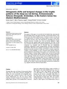
FIG. 1. – Map of Spain showing the sampling locations.
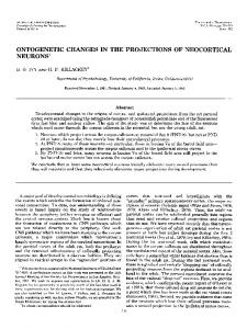
the juveniles were also photographed using SEM. The length of the radula, the number of rows and the length of the outer lateral tooth were measured with a micrometer ocular. To determine the size of the radular teeth we considered the length of the outer lateral tooth, from the apical cusp to the base (Fig. 2). The affinities among specimens according to the three measured variables (radula length, number of rows and the mean values of tooth length) were established through Cluster analysis using the UPGMA method (Unweighted Pair Group Method using Arithmetic averages) (Sneath and Sokal, 1973), based on Euclidean distance. For these analyses the four specimens with a pre-radula were not included. Once the groups of specimens were established through the Cluster analysis, the possible differences of the three
MATERIAL AND METHODS The present study was conducted using specimens from El Portil beach (Huelva, SW Spain) (37º12´40´´N, 7º7´50´´W) and La Herradura (Granada, SE Spain) (36º44´N, 3º43´W) (Fig. 1). Three animals were collected in La Herradura and 138 of them in El Portil from March 2001 to December 2003. The specimens were carried alive to the laboratory, where they were measured and fixed in 4% formalin. The radulae of all the specimens were studied by using optic microscopy and those of SCI. MAR., 70(2), June 2006, 227-234. ISSN: 0214-8358
FIG. 2. – Outer lateral tooth. The bar shows the measured length to determine the size of the teeth.
sm70n2227-2025
25/5/06
16:02
Página 229
ONTOGENETIC AND INTRASPECIFIC RADULAR CHANGES OF P. AURANTIOMARGINATA • 229
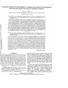
FIG. 3. – SEM photographs of the radula and pre- radula in different specimens of P.aurantiomarginata. A. Older portion of the radula in an individual 1.5 mm long. B. Detail of a bifid tooth in the radula of an individual 1.5 mm long. C. Radula and pre-radula of a specimen 4 mm in length. D. Detail of the pre-radula portion from a juvenile 4 mm in length. E-F. Adult radula.
measured variables between groups were tested using the non-parametric Kruskal-Wallis test, after testing the data for normality using the KolmogorovSmirnov test and Levene’s test for homogeneity of variances. Multivariate analyses (cluster) were carried out using the PRIMER package (Clarke and Gorley, 2001) and for univariate analyses (mean comparison through Kruskal Wallis) the BMDP was used (Dixon, 1983).
RESULTS Morphological variation of the radular teeth The radula of P. aurantiomarginata is defined as having the formula 8-15 x 4.2.0.2.4 (Figs. 3E, F). The four marginal teeth are quadrangular, without cusps and the size decreases outwards (Fig. 3F; Fig. 4C). The two internal teeth are hamate, as the inner SCI. MAR., 70(2), June 2006, 227-234. ISSN: 0214-8358
sm70n2227-2025
25/5/06
16:02
Página 230
230 • I. MARTÍNEZ-PITA et al.
FIG. 4. – Ontogenetic variations in the radula and pre-radula. A, B and C, variation of the marginal teeth in animals smaller than 3 mm (A), 3-4 mm in length (B) and bigger than 4 mm (C). D, E, F and G, variation in the outer lateral tooth in animals smaller than 3 mm (D), 3-4 mm in length in the pre-radula portion (E) and radula portion (F) and individuals bigger than 4 mm (G). H, I and J, variation of the inner lateral tooth in animals smaller than 3 mm (H), 3-4 mm in length (I) and individuals bigger than 4 mm (J). The value of the scale bars are 10mm in H, 20mm in A, B, D, E and I, 100mm in F and 200mm in C, G and J.
tooth is smaller and thinner than the second one; they have an apical cusp and a spur approximately in the middle of the tooth (Figs. 3E, F; Figs. 4G, J). There is no rachidian tooth. This radular pattern, considered as the adult type of the species, appears in almost all the specimens studied. However, some morphological differences are seen in specimens smaller than 4 mm in length. Specimens of 3-4 mm in length have the radula divided into two portions, with morphological variations of the teeth. The anterior teeth rows, which SCI. MAR., 70(2), June 2006, 227-234. ISSN: 0214-8358
are the oldest, have the marginal teeth provided by an enlarged basal part and a filamentous cusp (Fig. 3D; Fig. 4B); the lateral teeth have the adult morphology, although the spurs are very small (Figs. 4E, I). The newer rows have the adult pattern described before (Fig. 3C; Figs. 4F, J). In this situation, the anterior portion of the radula is considered as a preradula, according to Pruvot-Fol (1926). Specimens smaller than 3 mm lack the adult radular pattern (Fig. 3A), or it only appears in the last rows of the ribbon. The marginal teeth are filamentous, lacking
sm70n2227-2025
25/5/06
16:02
Página 231
ONTOGENETIC AND INTRASPECIFIC RADULAR CHANGES OF P. AURANTIOMARGINATA • 231 180
3.5
1.5 mm 2 mm 3 mm 4 mm 4 mm´ 4 mm´´
160
Y=0.69+0.04X
3.0
Teeth length (µm)
Radula length (mm)
140 2.5 2.0 1.5 1.0
120 100 80 60 40
0.5
20 0
0.0 0
10
20
30
40
50
60
0
5
10
15
20
25
30
35
Row
Animal length (mm)
FIG. 5. – Relation between the animal’s length and the length of the radula.
the basal enlarged portion; some marginal teeth have the tip bifid (Figs. 3A, B; Fig. 4A). The outer lateral teeth lack the spur and the cusp is provided by a longitudinal furrow (Fig. 4D). The inner lateral teeth have the adult morphology, but the spurs are very small (Fig. 4H).
FIG. 7. – Size of the outer lateral tooth in each row of the radula ribbon in specimens up to 4 mm in length. The arrow indicates the beginning of the radula part. Individuals 4 mm in length without a pre-radula are included.
P