On-eye Performance of Custom Wavefrontguided Soft Contact Lenses in a Habitual Soft Lens-wearing Keratoconic Patient Jason D. Marsack, PhD; Katrina E. Parker, OD; Yafei Niu, MS; Konrad Pesudovs, BS Optom, PhD; Raymond A. Applegate, OD, PhD ABSTRACT PURPOSE: To assess visual, optical, and fitting characteristics for wavefront-guided soft contact lenses produced for one habitual soft lens-wearing moderate keratoconic eye. METHODS: A process for production and evaluation of custom wavefront-guided soft contact lenses was developed. Wavefront aberrations were quantified with the COAS-HD wavefront sensor (Wavefront Sciences); soft contact lenses containing both high and low order aberrations were designed with custom software and produced using an ophthalmic lens lathe. Photopic high and low contrast logMAR visual acuity were recorded with the lens in place over an artificial 5-mm pupil and residual 2nd to 10th order root-mean-square (RMS) aberrations were analyzed over a 5-mm pupil. Comparisons were made to the eye’s habitual toric soft contact lens using t tests. RESULTS: Photopic high contrast values for habitual and final custom contact lenses for a 5-mm pupil were 0.07⫾0.06 and ⫺0.08⫾0.05, respectively. Photopic low contrast values were 0.73⫾0.06 and 0.62⫾0.07, respectively. Habitual and final custom correction low order RMS over a 5-mm pupil were 2.08 and 0.34 µm, and high order RMS levels were 0.77 and 0.39 µm, respectively. CONCLUSIONS: The final custom contact lens showed 1.5 lines of improvement for photopic high contrast (P=.03) and 1 line for photopic low contrast (P=.11) over a 5-mm pupil compared to habitual correction. Low and high order aberrations were reduced by 84% and 50% over a 5-mm pupil, respectively. Further improvements in performance of custom lenses may be achieved with further wavefront iterations. [J Refract Surg. 2007;23:960-964.]
K
eratoconus is a corneal disease that leads to thinning, weakening, and alteration of the stroma.1,2 Due to the fact that the stroma makes up the bulk of the thickness of the cornea, deformations in the stroma are responsible for alterations of the shape of the cornea as a whole. Because the cornea acts as an optical instrument (the most powerful such instrument in the ocular system), the morphological changes associated with keratoconus are accompanied by the induction of optical defects or aberrations. Previous studies in the literature have detailed such aberrations that are present in keratoconic eyes.3,4 Data have demonstrated that, on average, best corrected visual performance in non-scarred eyes with keratoconus (regardless of the correction method used) is reduced compared to normal levels.5,6 Further, recent work has linked uncorrected, residual ocular aberrations in keratoconic patients wearing rigid gas permeable (RGP) contact lenses to reduced visual performance through the use of optical quality metrics.7 This finding suggests that although RGP lenses significantly reduce optical aberrations induced by keratoconus, methods should be sought to further target residual optical aberrations. From Visual Optics Institute, College of Optometry, University of Houston, Houston, Tex (Marsack, Parker, Niu, Applegate); and the NH&MRC Centre for Clinical Eye Research, Department of Ophthalmology, Flinders Medical Centre and Flinders University, Bedford Park, South Australia, Australia (Pesudovs). Supported by grants NEI P30 EY07551 (University of Houston, College of Optometry), NEI T32 EY07024 (Marsack), and NEI ROI EY08520 (Applegate), an American Optometric Foundation soft contact lens research grant (Marsack), and a National Keratoconus Foundation research grant (Marsack). Drs Marsack, Pesudovs, and Applegate have a patent under review with the US Patent and Trademark Office in the area of custom contact lens design. The remaining authors have no proprietary interest in the materials presented herein. Presented at the 8th International Congress of Wavefront Sensing & Optimized Refractive Corrections; February 22-25, 2007; Santa Fe, New Mexico. The authors thank Wavefront Sciences (Albuquerque, NM), DAC International (Carpinteria, Calif), and Metro Optics (Austin, Tex) for their assistance. Correspondence: Jason D. Marsack, PhD, Visual Optics Institute, College of Optometry, 505 J Davis Armistead Bldg, University of Houston, Houston, TX 77204. Tel: 713.743.1791; Fax: 713.742.2053; E-mail:
[email protected]
960
journalofrefractivesurgery.com
Custom Wavefront-guided Soft Contact Lenses/Marsack et al
One such method under investigation comes in the form of a custom wavefront-guided soft contact lens. This idea has been the subject of numerous reports that have covered relevant topics such as the impact on visual performance of lens rotation and displacement,8-10 design principles,11 the feasibility of implementing custom lenses,12-14 the optical impact of correcting higher order aberrations,15,16 and how to represent aberration structures.17-20 These theoretical studies have been applied to practice in recent demonstrations of wavefrontguided corrections.21,22 The purpose of this study was to investigate the process implemented at the Visual Optics Institute, College of Optometry, University of Houston for the design, manufacture, and evaluation of wavefrontguided soft contact lenses for one habitual soft contact lens-wearing, moderate keratoconic patient. PATIENTS AND METHODS Appropriate University Institutional Review Board and informed consent approval was obtained prior to initiating data collection. One patient (SC1) with clinically diagnosed keratoconus was enrolled in this initial proof-of-principle custom wavefrontguided soft contact lens study. The right eye of this patient with moderate keratoconus (determined from max simK reading of 47.10 D using the Keratron corneal topographer [Optikon, Rome, Italy]) was studied. Moderate keratoconus severity was identified in a similar manner to the Collaborative Longitudinal Evaluation of Keratoconus (CLEK) baseline study,5 where the distinction between mild, moderate, and severe keratoconus was between steep keratometric powers of 45.00 and 52.00 D (the CLEK study used keratometry to obtain these measurements). Visual performance testing was conducted on the habitual soft contact lens (SofLens 66; Bausch & Lomb, Rochester, NY) and a series of four iteratively produced custom lenses. No attempt was made to further optimize the habitual lens for the purposes of the experiment reported here. Both photopic high contrast and photopic low contrast visual acuity measurements were collected. Photopic high contrast was recorded because it is the clinical standard for assessing visual performance. Photopic low contrast was recorded because it has been shown that low contrast visual acuity is more sensitive to optical degradation than high contrast visual acuity.23 The Michelson contrast level was 4% for the low contrast charts and 87% for the high contrast charts. Visual acuity on each chart was determined using previously published procedures.24-26 Visual acuity measurements were recorded and reported for the final custom wavefront-guided soft contact lens Journal of Refractive Surgery Volume 23 November 2007
and the habitual soft contact lens for a dilated pupil viewing the target through a 5-mm artificial pupil. A series of four custom wavefront-guided soft contact lenses was designed and produced for the right eye of the patient. The initial lens designed and tested (L0) was a fitting lens with a toric posterior surface and a spherical, prism-ballasted anterior surface. After insertion of any given lens (Lx) in the series, the patient was given 10 to 20 minutes to allow the lens to equilibrate to the eye. Rotation and translation of a custom lens (Lx) on the eye were determined by quantifying the position of the two alignment marks on the anterior surface of the eye relative to the pupil center with a custom-built digital slit lamp. The macro properties (eg, base curve, prism, and edge radius) of L0 were conserved, where possible, in successive lens designs. Changes were made to the macro properties of the lens only when clinical evaluation of the lens deemed modification necessary to improve the lens fit or facilitate stability. Residual eye ⫹ lens optical aberration for each lens was quantified using a 10th order Zernike polynomial with a COAS-HD wavefront sensor (Wavefront Sciences, Albuquerque, NM). The Zernike terms targeted for correction in this study varied by lens. L1 contained full 2nd to 3rd order correction and L2 and L3 contained full 2nd to 4th order correction. Modes in higher orders were left uncorrected. Data reported in the literature predict a full 4th order correction may provide a patient who has 47.10 D max steep keratoconus with high contrast logarithm of the minimum angle of resolution (logMAR) visual acuity of 0.0.7 In this series of lenses, the aberration patch described by wavefront aberration data was quantified over a maximum non-dilated pupil size (which varied slightly depending on the iteration) and was then scaled to describe a 6.5-mm diameter pupil. Scaling of the wavefront patch was meant to artificially increase the size of the wavefront zone of the lens beyond the measured pupil. Although it was known that this would induce errors in the wavefront correction, it was hypothesized that these errors would be corrected through the iterative prescription of future custom lenses and that the errors would become smaller with each iteration. The aberration structure present in the 6.5-mm diameter wavefront patch of each lens (Lx), where x⬎0, was determined by adding the aberration structure previously measured through Lx-1 on the eye to the wavefront patch present in Lx-1. To account for rotation of the lens on the eye, the rotational offset quantified through Lx-1 using the procedure described above was applied before addition to the aberration structure of Lx-1. In this series of custom 961
Custom Wavefront-guided Soft Contact Lenses/Marsack et al
TABLE 1
TABLE 2
Low Contrast and High Contrast Visual Acuity Data Recorded With the Habitual Soft Contact Lens and the Final Custom Wavefront-guided Soft Contact Lens (L3) Over a 5-mm Pupil
Low Order Root-Mean-Square (RMS) and High Order RMS Levels for the Habitual Soft Contact Lens and Final Custom Wavefront-guided Soft Contact Lens (L3) Over a 5-mm Pupil
Lens Habitual contact lens L3 P value
High Contrast (logMAR, 87%)
Low Contrast (logMAR, 4%)
0.07⫾0.06
0.73⫾0.06
⫺0.08⫾0.05
0.62⫾0.07
.03
.11
lenses, translational offsets are not incorporated into the design and placement of the wavefront patches. After visual performance testing was complete with each custom contact lens, the lens was removed and optically profiled in the ClearWave contact lens aberrometer (Wavefront Sciences). This specialized Shack-Hartmann wavefront sensor is designed to quantify and report the Zernike aberration structure of a contact lens. The patient completed the study after four custom contact lenses were tested, regardless of visual performance or the level of aberration correction achieved. Comparisons between the habitual soft contact lens and the final custom wavefront-guided soft contact lens were done using t tests. RESULTS Entrance photopic high contrast visual acuity (undilated) for the physiological pupil size is ⫺0.04 logMAR. This level of entrance visual acuity is better than average entrance visual acuity for patients with keratoconus (regardless of correction method or scarring) as reported by the CLEK study, where only 33.9% of patients had high contrast entrance visual acuity of 0.0 or better in at least one eye.5 Table 1 reports both the low contrast and high contrast logMAR visual acuity data recorded with the habitual soft contact lens and final custom wavefront-guided soft contact lens for a dilated pupil of patient SC1 viewing through a 5-mm artificial pupil. Only the visual acuity with the final custom contact lens is reported because visual performance data for the 5-mm pupil were not collected on the interim custom wavefront-guided soft contact lenses. The impact of uncorrected low and high order aberrations on visual performance can be seen in this 5-mm pupil dataset. 962
Average Low Order RMS (µm)
Average High Order RMS (µm)
Habitual contact lens
2.076
0.770
L3
0.336
0.386
Lens
Note. Low order aberration was reduced by 84% and high order aberration was reduced by 50% when lens L3 is compared to the habitual lens.
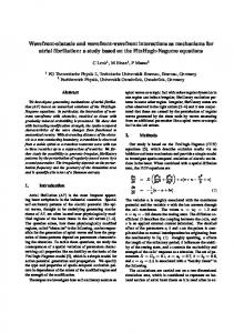
Table 2 reports the on-eye low order root-meansquare (RMS) and high order RMS for the habitual contact lens correction and final custom contact lens correction studied over a 5-mm pupil. These data are reported at the same pupil size used for quantification of visual performance measures in Table 1. Both low order aberrations and high order aberrations are reduced for the final custom contact lens compared to the habitual contact lens. Low order aberrations are reduced from 2.076 to 0.336 µm, or by 84%. The residual low order aberration present during final custom wavefront-guided soft contact lens wear of 0.336 µm has a spherical equivalent manifest refraction of ⫺0.373 D over a 5-mm pupil. Importantly, high order aberrations (the aberrations that cannot be effectively treated by the habitual soft contact lens correction) are reduced from 0.770 to 0.386 µm, or by 50%. However, this level of higher order aberration remains outside the normal range for a sample matched for age and pupil size of 0.174⫾0.062 µm.27 The Figure represents the optical results for this experiment by showing the uncorrected higher order wavefront error of the eye (0.999 µm) in panel (a), the higher order wavefront error carved into custom contact lens L3 (0.809 µm) in panel (b), and the residual higher order aberration when the study eye is wearing the lens (0.386 µm) in panel (c). The Figure demonstrates that a custom contact lens designed and manufactured based on the measured wavefront error of a keratoconic eye can partially compensate for the higher order aberrations. DISCUSSION The goal of this study was to design, manufacture, and evaluate basic custom wavefront-guided soft contact lenses for one habitual soft contact lens-wearing, journalofrefractivesurgery.com
Custom Wavefront-guided Soft Contact Lenses/Marsack et al
Figure. The optical results of correction of higher order aberration are shown. The uncorrected higher order wavefront error (0.999 µm) in panel (a) is negated by the higher order wavefront error in custom contact lens L3 (0.809 µm) in panel (b), resulting in a reduced residual higher order aberration (0.386 µm) in panel (c).
moderate keratoconic patient. Although this patient may not be considered as typical for keratoconus due to the habitual mode of correction, we considered this as a logical starting point for evaluation of custom wavefront-guided soft contact lenses based on the patient’s familiarity with soft contact lens wear and ability to achieve excellent habitual logMAR visual acuity with a traditional soft lens correction. Table 1 shows that high contrast logMAR visual acuity statistically significantly improved from the habitual soft contact lens value of 0.07⫾0.06 to the final custom contact lens value of ⫺0.08⫾0.05 over a 5-mm pupil (P=.03). Low contrast values for the same correction modalities improved from 0.70⫾0.06 to 0.62⫾0.07 µm over a 5-mm pupil, but did not reach statistical significance (P⫽.11). Table 2 demonstrates low order RMS over a 5-mm pupil zone decreased from 2.076 µm to 0.336 µm for the habitual contact lens correction and custom contact lens correction, respectively. High order RMS levels decreased from 0.770 µm to 0.386 µm for the habitual contact lens correction and custom contact lens correction, respectively. A reduction in both low order (84%) and high order (50%) RMS error was demonstrated for the custom contact lens compared to the habitual contact lens at a 5-mm pupil size. It seems plausible that the patient would be relatively unhappy with the habitual correction considering the elevated level of residual aberration this correction leaves in place. However, under physiological conditions, where the pupil only reached approximately 4 mm, the patient achieved ⫺0.04 logMAR visual acuity with residual low and high order aberrations of 1.27 and 0.44 µm, respectively. The importance of pupil size is demonstrated, resulting in a decrease in low and high order aberraJournal of Refractive Surgery Volume 23 November 2007
tions for the habitual contact lens correction of 39% and 42%, respectively, from the 5- to 4-mm pupil. The considerable reduction in aberration for the smaller pupil size may help explain why the patient is satisfied with the habitual soft contact lens visual acuity. The goals of this study were achieved by demonstrating the ability to design, manufacture, and test custom wavefront-guided soft contact lenses on a keratoconic eye. The study also demonstrated the ability of such lenses to improve visual performance and reduce both high and low order optical aberrations. REFERENCES 1. Krachmer JH, Feder RS, Belin MW. Keratoconus and related noninflammatory corneal thinning disorders. Surv Ophthalmol. 1984;28:293-322. 2. Rabinowitz YS. Keratoconus. Surv Ophthalmol. 1998;42:297-319. 3. Pantanelli S, Yoon G, Jeong TM, MacRae S. Aberration characterization of abnormal eyes using the large dynamic range Shack-Hartmann wavefront sensor. Invest Ophthalmol Vis Sci. 2004;45:E-Abstract 2848. 4. Maeda N, Fujikado T, Kuroda T, Mihashi T, Hirohara Y, Nishida K, Watanabe H, Tano Y. Wavefront aberrations measured with Hartmann-Shack sensor in patients with keratoconus. Ophthalmology. 2002;109:1996-2003. 5. Zadnik K, Barr JT, Edrington TB, Everett DF, Jameson M, McMahon TT, Shin JA, Sterling JL, Wagner H, Gordon MO. Baseline findings in the Collaborative Longitudinal Evaluation of Keratoconus (CLEK) Study. Invest Ophthalmol Vis Sci. 1998;39:25372546. 6. Elliott DB, Yang KC, Whitaker D. Visual acuity changes throughout adulthood in normal, healthy eyes: seeing beyond 6/6. Optom Vis Sci. 1995;72:186-191. 7. Marsack JD, Parker KE, Pesudovs K, Donnelly WD III, Applegate RA. Uncorrected wavefront error and visual performance during RGP wear in keratoconus. Optom Vis Sci. 2007;84:463-470. 8. Guirao A, Williams DR, Cox IG. Effect of rotation and translation on the expected benefit of an ideal method to correct the eye’s higher-order aberrations. J Opt Soc Am A Opt Image Sci Vis. 2001;18:1003-1015.
963
Custom Wavefront-guided Soft Contact Lenses/Marsack et al
9. Guirao A, Cox IG, Williams DR. Method for optimizing the correction of the eye’s higher-order aberrations in the presence of decentrations. J Opt Soc Am A Opt Image Sci Vis. 2002;19:126-128. 10. de Brabander J, Chateau N, Marin G, Lopez-Gil N, Van Der Worp E, Benito A. Simulated optical performance of custom wavefront soft contact lenses for keratoconus. Optom Vis Sci. 2003;80:637-643. 11. Thibos LN, Cheng X, Bradley A. Design principles and limitations of wave-front guided contact lenses. Eye Contact Lens. 2003;29:S167-S170. 12. Marsack J, Milner T, Rylander G, Leach N, Roorda A. Applying wavefront sensors and corneal topography to keratoconus. Biomed Sci Instrum. 2002;38:471-476.
optical aberrations and the corneal surface of the eye. Invest Ophthalmol Vis Sci. 2005;46:1915-1926. 19. Smolek MK, Klyce SD. Goodness-of-prediction of Zernike polynomial fitting to corneal surfaces. J Cataract Refract Surg. 2005;31:2350-2355. 20. Pantanelli SM, Yoon GY. Can the Zernike polynomials reliably represent the aberration in normal and abnormal eyes? Invest Ophthalmol Vis Sci. 2006;47:E-Abstract 1204. 21. Yoon G, Jeong TM, Cox IG, Williams DR. Vision improvement by correcting higher-order aberrations with phase plates in normal eyes. J Refract Surg. 2004;20:S523-S527.
13. Kollbaum P. Seeing into the future with contact lenses. Contact Lens Spectrum. 2003;2:25-29.
22. Sabesan R, Carvalho L, Jeong T, Yoon GY, Somasundaram R, Cox IG. Correcting higher order aberrations using customized soft contact lenses in keratoconic eyes. Invest Ophthalmol Vis Sci. 2006;47:E-Abstract 1205.
14. Cox IG, Lagana M. Feasibility of wavefront customized contact lenses. In: Krueger R, Applegate R, MacRae S, eds. Wavefront Customized Visual Correction: The Quest for Super Vision. 2nd ed. Thorofare, NJ: SLACK Incorporated; 2004:279-284.
23. Pesudovs K, Marsack JD, Donnelly WJ III, Thibos LN, Applegate RA. Measuring visual acuity—mesopic or photopic conditions, and high or low contrast letters? J Refract Surg. 2004;20:S508S514.
15. Guirao A, Porter J, Williams DR, Cox IG. Calculated impact of higher-order monochromatic aberrations on retinal image quality in a population of human eyes. J Opt Soc Am A Opt Image Sci Vis. 2002;19:620-628.
24. Applegate RA, Sarver EJ, Khemsara V. Are all aberrations equal? J Refract Surg. 2002;18:S556-S562.
16. Marsack JD, Pesudovs K, Sarver EJ, Applegate RA. The impact of Zernike-fit error on simulated high- and low-contrast acuity in keratoconus: implications for using Zernike-based corrections. J Opt Soc Am A Opt Image Vis Sci. 2006;23:769-776. 17. Klyce SD, Karon MD, Smolek MK. Advantages and disadvantages of the Zernike expansion for representing wave aberration of the normal and aberrated eye. J Refract Surg. 2004;20:S537-S541. 18. Carvalho LA. Accuracy of Zernike polynomials in characterizing
964
25. Applegate RA, Ballentine C, Gross H, Sarver EJ, Sarver CA. Visual acuity as a function of Zernike mode and level of root mean square error. Optom Vis Sci. 2003;80:97-105. 26. Applegate RA, Marsack JD, Ramos R, Sarver EJ. Interaction between aberrations to improve or reduce visual performance. J Cataract Refract Surg. 2003;29:1487-1495. 27. Applegate RA, Donnelly WJ III, Marsack JD, Koenig DE, Pesudovs K. Three-dimensional relationship between high-order root-mean-square wavefront error, pupil diameter, and aging. J Opt Soc Am A Opt Image Sci Vis. 2007;24:578-587.
journalofrefractivesurgery.com