sensors used, it is not possible to record the heart rate simultaneously with fMEG by ... on a large sensor array, the maternal and fetal magnetocardiographic ...
Proceedings of the 14th Biennial BIOMAG Conference, Boston, Massachusetts, USA, August 2004.
1
On-line Fetal Heart Rate Monitoring Using SQUID Sensor Arrays D. Guti´errez1 , A. Nehorai1 , D. McKenzie2 , H. Eswaran3 , C. L. Lowery3 , and H. Preissl3,4 1
University of Illinois at Chicago, USA. 2 VSM MedTech Ltd., Coquitlam, BC, Canada. 3 University of Arkansas at Little Rock, USA. 4 University of T¨ubingen, Germany.
ABSTRACT Objective: Fetal magnetoencephalography (fMEG) is the only complete non-invasive method to record fetal brain signals in the utero. When using this method in a clinical setting, it is necessary to monitor the fetal heart rate during the recordings. Based on the high sensitivity of the magnetic sensors used, it is not possible to record the heart rate simultaneously with fMEG by conventional methods like Doppler ultrasound. However, based on a large sensor array, the maternal and fetal magnetocardiographic signals (mMCG and fMCG) are recorded simultaneously with the fMEG with adequate temporal and spatial resolution. Method: Recordings were performed on a 151 channel SQUID array covering the whole maternal abdomen (SARA system). We use a multichannel adaptive filter to cancel the maternal signal and dynamically select those channels with high content of fetal signal. Our technique can be implemented in real time using a recursive steepest descent algorithm. From the extracted fMCG signal, we obtain an on-line estimate of the heart rate with an R-wave detector that uses a smoothed first derivative magnitude of the signal. Results: We show through real data that, with this technique, we obtain real-time estimates of the fetal heart rate with a small standard deviation. Furthermore, our proposed criterion for channel selection can identify the channels with low or high content of fetal signal. Conclusion: We developed a fast and reliable fMCG extraction method, which has on-line capability. Currently, initial studies are conducted with an experimental setup, allowing the on-line access to the magnetic signals of all 151 channels. Our method will help to establish fetal magnetometry in a clinical setting, where continuous control of vital signals of the fetus is necessary. KEYWORDS Fetal magnetoencephalography, magnetocardiography, adaptive filtering, signal cancellation. INTRODUCTION Fetal magnetoenchepalography (fMEG) is a completely passive and non-invasive technique for the study of fetal brain function in utero [Lenge, 2001]. fMEG is measured in the presence of environmental noise and various near-field biological signals: maternal magnetocardiogram (mMCG), fetal magnetocardiogram (fMCG) uterine smooth muscle (magnetomyogram), and motion artifacts [Wakai, 2002]. In clinical studies, it is necessary to monitor the fetal heart rate during the recordings. However, traditional techniques such as fetal electrocardiogram (fECG), uterine pressure sensors, or Doppler ultrasound, do not provide with complete information of the cardiac function. fMCG has multiple advantages for the analysis of xk m ˜ 1k m ˜ 2k ... m ˜ Lk the morphology and temporal parameters of the fetal heart signals [Lewis, 2003]. It fills the diagnostic gap R−wave detector left by the difficulties involved in recording fECG due to the insulating effect of the vernix caseosa and the existence of preferred conduction pathways between the fetal heart and maternal abdomen. Furthermore, fMCG is xk [−Nw :Nw ] m ˜ 1k [−Nw :Nw ] Steepest descent detected as early as the 16th week of gestation, and measurements can be obtained simultaneously with fMEG −1/L using a sufficiently large sensor array and adequate sample rate. However, the detection of the fMCG signal depends m ˜ 2k [−Nw :Nw ] Steepest descent on the ability to separate the fetal heart component from the composite signal. In general, the mMCG and fMCG −1/L signals are the dominant components of the ensemble, . . . . . . . . . with an average magnitude as large as 10 pT at the fetal thorax location. Closer to the maternal heart, the mMCG can be as large as 100pT [Vrba, 2003]. Therefore, algom ˜ Lk [−Nw :Nw ] Steepest descent rithms for heart rate monitoring based on MCG have to overcome not only this problem, but need to have a low computational complexity and low processing delay, and −1/L must be able to detect rapid and dramatic changes in the heartbeats in a beat-to-beat manner. fˆk [−Nw :Nw ] fˆk In this paper, we present an algorithm based in a multichannel filter to adaptively separate the fMCG sigFigure 1: Diagram of the multichannel adaptive filter. nal from the mMCG, as well as dynamically select those channels with highest content of fetal signal and monitor the fetal heart activity on-line. METHODS We define our measurement model as x(n) = f (n)+m(n), where x(n) is the measurement of a single channel, f (n) is the fetal signal component, m(n) is the maternal signal component plus noise, and n = 1, 2, . . . , N time samples. Our goal is then to cancel m(n) from x(n). This problem can
Proceedings of the 14th Biennial BIOMAG Conference, Boston, Massachusetts, USA, August 2004.
2
EXAMPLE
Heart Rate (BPM)
Estimated signal (fT)
Input signal (fT)
be seen from the point of view of adaptive signal processing as a case of signal cancellation [Hayes, 1996] in which the process f (n) is to be estimated from a corrupted observation x(n). Consider that a group of L channels can be used as reference, i.e. they contain mostly maternal signal. Refer to each of the reference measurements as m ˜ i (n), where i = 1, 2, . . . , L. Although m ˜ i (n) will be correlated with m(n), the two processes will not be equal. Therefore, it is not possible to estimate f (n) simply by subtracting m ˜ i (n) from x(n). Instead, we use an adaptive filter to estimate m(n) and then subtract it from x(n), i.e. fˆ(n) = x(n) − m(n), ˆ where fˆ(n) and m(n) ˆ are the estimates of the fetal and maternal signals, respectively, at the nth time sample. If we assume that we can temporarily store NT time samples of our on-line measurements in a buffer, then we can define ˜ ik = [m x k = [x(n0 ), x(n0 + 1), x(n0 + 2), . . . , x(n0 + NT − 1)]T , and m ˜ i (n0 ), m ˜ i (n0 + 1), m ˜ i (n0 + 2), . . . , m ˜ i (n0 + NT − 1)]T , where n0 is an arbitrary starting time P sample such that n0 + NT − 1 ≤ N at the kth buffer stored. Now, the estimate of the maternal signal at the kth buffer ˆ k = (1/L) i wik m ˜ ik , where wik is the adaptive coefficient corresponding to the ith reference signal. We use the steepest descent is given by m algorithm to compute the adaptive coefficients recursively while keeping the mean square error of fˆ k minimized. According to this algorithm, our 2 2 ˜ ik , and σ adaptive coefficients are given by wi,k+1 = (1 + µˆ σm σx2m ˆm ˆx2m ˜ )wik + µˆ ˜ , where µ is the step size, σ ˜ is the sample variance of m ˜ is the sample ˜ ik . covariance between x k and m 4 Accurate on-line removal of the maternal signal requires all calculations to be done in 2 synchrony with the peak produced by the maternal heart beat. The detection of this peak is achieved through an R-wave detector that uses a smoothed first derivative of the sig0 ˜ 01k |}, where Θ is the threshnal [K¨ohler, 2002], i.e. Θ = 0.4 max{[0.25, 0.5, 0.25]T ∗ |m −2 54 54.5 55 55.5 56 56.5 57 57.5 58 ˜ 01k is the first derivative of m ˜ 1k . Note that only the first of the old of the detector, and m 4 reference channels is passed through the R-wave detector as we consider that the delay between the reference signals is negligible. Once we have detected the peak of the maternal 2 signal, all computations are done for windows of 2Nw + 1 time samples from both x k and 0 ˜ ik : Nw samples before and Nw after the peak, where Nw is the window size. m ˆ ˆ k. Finally, the estimate of the fetal signal at the kth iteration is obtained as f k = x k − m −2 54 54.5 55 55.5 56 56.5 57 57.5 58 A complete diagram of our adaptive filter is shown in Fig. 1. Further details of our method can be found at http:°cyclone.ece.uic.edu\˜david\onlinefHRM. 150 100
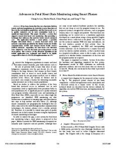
We applied our adaptive filter to real magnetic data from a 151 channel SQUID array 50 0 10 20 30 40 50 covering the whole maternal abdomen (CTF SARA system). Our data was sampled at Time (s) 312.5 Hz and then low-pass filtered at a cut-off frequency of 40 Hz. The 7 upper rim channels were used as our reference channels m ˜ i (n), i.e. L = 7. We considered a buffer Figure 2: From top to bottom: Input signal (x k ), outˆ size NT = 300 and a window size Nw = 80. These values were chosen accordingly to the put signal (f k ), and estimate of the fetal heart rate (mean shown in dotted line). Nyquist rate. For the steepest descent algorithm, we chose a step size µ = 0.1. Our preliminary results showed that |wik | → µ for those channels with high content of fetal signal. Using this criterion, we chose the signal from one of these channels to estimate the heart rate. This estimate was obtained at each window through an R-wave to detect the peaks of the estimated fetal signal fˆ k . Such R-wave detector was implemented in the same way as explained in the previous section. A snapshot of x k and fˆ k for the selected channel, as well as the estimated fetal heart rate, are shown in Fig. 2. CONCLUDING REMARKS We have presented a reliable and fast method with on-line capabilities to extract the fMCG signal. Our method is based in an adaptive filter to cancel the maternal signal component. This technique also allowed us to identify those channels with poor, regular, or high content of fetal signal by keeping track of the values of the adaptive coefficients. This capability may be also used to track on-line changes of the fetus position and allow adaptive channel selection. Finally, using this procedure for a specific channel, we can obtain an accurate estimate of the fetal heart rate. Our method will help to establish fetal magnetometry in a clinical setting, where continuous control of vital signals of the fetus is necessary. ACKNOWLEDGMENT This work was supported by NIN/NINDS Grant 2 R01 NS36277-04A1 and NSF Grant CCR-0105334.
REFERENCES Hayes MH. Statistical digital signal processing and modeling. 1st. ed. New York. John Wiley & Sons, Inc. 1996. K¨ohler BU, Hennig C, Orglmeister R. The principles of software QRS detection. IEEE Eng. Med. Bio. 2002;21:42-57. Lenge JM, Chen M, Wakai RT. Improved neuromagnetic detection of fetal and neonatal auditory evoked response. Clin. Neurophysiol. 2001;112:785-92. Lewis MJ. Review of electromagnetic source investigations of the fetal heart. Medical Engineering & Physics 2003;25:801-10. Vrba J. Multichannel SQUID biomagnetic systems. In: H. Weinstock editor. Applications of superconductivity. Dordrecht. Kluwer Academic Publishers. 2000. pp. 61-138. Wakai RT, Lutter WJ. Matched-filter template generation via spatial filtering: application to fetal biomagnetic recordings. IEEE Trans. Biomed. Eng. 2002;49:1214-17.