J Pharm Pharmaceut Sci (www.cspsCanada.org) 16(2) 217 - 230, 2013
Optimisation and Validation of a High Throughput Screening Compatible Assay to Identify Inhibitors of the Plasma Membrane Calcium ATPase Pump - a Novel Therapeutic Target for Contraception and Malaria Tamer M A Mohamed 1,2, Simon A Zakeri1, Florence Baudoin1, Markus Wolf3, Delvac Oceandy1, Elizabeth J Cartwright1, Sheraz Gul3, Ludwig Neyses1 1 Institute of Cardiovascular Sciences, Manchester Academic Health Sciences Centre, University of Manchester, Manchester M13 9PT, UK. 2 Dept. of Biochemistry, Faculty of Pharmacy, Zagazig University, Zagazig, Egypt. 3 European ScreeningPort GmbH, 22525 Hamburg, Germany.
Received, March 13, 2013; Revised, April 25, 2013; Accepted, May 8th, 2013; Published, May 10th, 2013.
ABSTRACT – Purpose. ATPases, which constitute a major category of ion transporters in the human body, have a variety of significant biological and pathological roles. However, the lack of high throughput assays for ATPases has significantly limited drug discovery in this area. We have recently found that the genetic deletion of the ATP dependent calcium pump PMCA4 (plasma membrane calcium/calmodulin dependent ATPase, isoform 4) results in infertility in male mice due to a selective defect in sperm motility. In addition, recent discoveries in humans have indicated that a single nucleotide polymorphism (SNP) in the PMCA4 gene determines the susceptibility towards malaria plasmodium infection. Therefore, there is an urgent need to develop specific PMCA4 inhibitors. In the current study, we aim to optimise and validate a high throughput screening compatible assay using recombinantly expressed PMCA4 and the HTRF® Transcreener® ADP (TR-FRET) assay to screen a drug library. Methods and Results. PMCA4 membrane microsomes were prepared from HEK293 cells overexpressing PMCA4. Western blot quantification revealed nearly nine-fold increased expression of PMCA4 compared to LacZ (control virus)-infected cells. Maximal PMCA4 microsomal activity was achieved in the TR-FRET assay with 15ng/μl microsomal concentration, 30-minute pre-incubation with compounds at 37°C, and calcium buffering with 1mM EGTA providing 1μM free-calcium. Finally a dose-response curve for carboxyeosin (a non-specific PMCA inhibitor) under optimised conditions showed significant PMCA4 inhibition. Upon confirmation that the assay was suitable for high-throughput screening, we have screened the ChemBioNet small molecule library (~21,000 compounds) against the PMCA4 assay to identify those that are its apparent inhibitors. This screening yielded 1,494 primary hits. Conclusions. We have optimised the HTRF® Transcreener® ADP assay for high-throughput screening to identify PMCA4 inhibitors. The output of the screening campaign has provided preliminary chemical starting points that could be further developed to specific PMCA4 inhibitors for non-hormonal contraception or anti-malaria therapy. This article is open to POST-PUBLICATION REVIEW. Registered readers (see “For Readers”) may comment by clicking on ABSTRACT on the issue’s contents page. _______________________________________________________________________________________ neurones and muscle cells it maintains cell resting potential by extruding three sodium ions from the cell in exchange for two potassium ions. The activity of this enzyme energizes such diverse functions as the maintenance of the membrane potential and the renal and intestinal handling of solutes [1-3]. The gastric H+/K+-ATPase exploits a very similar enzymatic mechanism to catalyze the exchange of intracellular protons for ________________________________________
INTRODUCTION P-type ATPases are a group of protein which utilise the energy generated from adenosine triphosphate (ATP) hydrolysis predominantly to facilitate the movement of cations, Na+, K+, H+ or Ca2+ against the concentration gradient. In the human body, examples include the sodiumpotassium pump (Na+/K+-ATPase), the proton pump (H+/K+ ATPase), sarco-endoplasmic reticulum calcium ATPase (SERCA), and the plasma membrane calcium/calmodulin dependent ATPase (PMCA). The Na+/K+-ATPase pump, which is ubiquitously expressed, facilitates cell ionic gradients, ionic transport, and in both
Corresponding Author: Ludwig Neyses, Institute of Cardiovascular Sciences, Manchester Academic Health Sciences Centre, University of Manchester. Room 1.302 Stopford Bldg, Oxford Road, Manchester,, United Kingdom; Email.
[email protected]
217
J Pharm Pharmaceut Sci (www.cspsCanada.org) 16(2) 217 - 230, 2013
extracellular potassium ions, thus generating the enormous proton gradients associated with gastric acid secretion [4]. SERCA is ubiquitously expressed, and is known to facilitate uptake of calcium into the sarco/endoplasmic reticulum from the cytosol. This is important in cardiac muscle relaxation, in which the sarcoplasmic reticulum is essentially refuelled with calcium for subsequent contraction to take place [5-9]. Finally, the PMCA family, are a group of pumps that extrude calcium from the cytosol into the extracellular space, control calcium homeostasis, and in some cases regulate intracellular signalling pathways. There are four main isoforms: PMCA1, 2, 3 and 4, each one being encoded by a different gene. Every cell contains at least one PMCA isoform, with PMCA activity being driven by the hydrolysis of ATP [10, 11]. Recent Genome Wide Association Studies (GWAS) and studies of genetically modified mice have revealed potentially important roles for PMCA isoforms in human disease. A number of the human GWAS have identified single nucleotide polymorphisms (SNPs) in the PMCA1 gene as the single strongest association with blood pressure (BP) variance in humans [12-15]. PMCA2 which is expressed in the cilia of the inner ear, Purkinje cells and the mammary gland, has been implicated in deafness [16-18]. PMCA3 is expressed in brain tissue, pancreatic islet cells, and skeletal muscle, but less is known of the pathophysiological significance [19-21]. However, a recent study in humans has related a single mutation in PMCA3 with ataxia [22]. PMCA4 is by far the most extensively studied isoform. Our own studies in mice lacking PMCA4 [23] revealed that the males were infertile due to a defect in sperm motility, but were otherwise healthy under physiological conditions and had a normal lifespan, as did the females. These findings were confirmed by Okunade et al. who generated a similar PMCA4 knockout mouse model [24]. In addition, a GWAS has shown the PMCA4 gene to be associated with resistance to infection by malaria plasmodium in humans [25]. These findings have collectively implicated PMCAs in a variety of diseases and have led us to optimise and validate a generic PMCA screening compatible assay to be performed for drug discovery purposes. In the current study we used PMCA4 as a model to optimise and validate the HTRF® Transcreener® ADP (TR-FRET) assay for high throughput screening (HTS) of ATPasetargeting compounds. This assay can also be applied to study other ATPases.
METHODS Microsome preparation HEK293 cells overexpressing PMCA4 or βgalctosidase (LacZ) were used to prepare PMCA4 and LacZ microsomes as previously described [26]. Briefly, HEK293 cells were cultured and maintained in 175cm2 flasks, until cells were at 60-70% confluence. Cells were then infected by 25 Multiplicity Of Infection (MOI) with either recombinant PMCA4 or LacZ adenovirus, for 48 hours. Cells were washed three times with PBS and then harvested in 5ml harvest solution (1x PBS, 0.26% 2mg/ml aprotinin, 0.11% 2mg/ml leupeptin, 0.1% 0.1M PMSF). Harvested cells were centrifuged at 1000g for 10 minutes at 4ºC. 3ml hypotonic solution (10mM Tris-HCl, pH7.5, 1mM MgCl2, 0.5mM EGTA, 2mM DTT, 0.2% 2mg/ml aprotinin, and 0.05% 2mg/ml leupeptin) was added to the cell pellet for 10 minutes on ice. Swelled cells were homogenised in a Dounce homogenizer before addition of 3 ml homogenate solution (10mM Tris-HCl, pH7.5, 2mM DTT, 0.38M sucrose, 0.3M KCl, 0.2% 2mg/ml aprotinin, and 0.05% 2mg/ml leupeptin) and homogenization was completed to seal the vesicles. The cell homogenate was centrifuged at 1500g for 20 minutes to remove the cellular debris. 60µl 0.25mM EDTA and 1.08 ml of 2.5M KCl were added to the supernatant and centrifuged at 100,000g for 40 minutes at 4ºC. The resulting pellet was re-suspended in 0.4ml final solution (10mM Tris-HCl, pH7.5, 1mM DTT, 0.19M sucrose, 0.15M KCl, 0.2% 2mg/ml aprotinin, 0.05% 2mg/ml leupeptin and 0.02mM CaCl2). Western blotting Microsomes overexpressing PMCA4 or βgalactosidase (LacZ) where lysed with RIPA buffer (1x PBS, 1% Igepal, 0.5% sodium deoxycholate, 0.1% sodium dodecyl sulphate [SDS], 20µM phenylmethylsulfonyl fluoride [PMSF], 500ng/ml leupeptin, 1.0µg/ml aprotinin, and 500ng/ml pepstatin). Protein concentrations were determined using a BCA protein assay kit (Pierce). 30µg of protein extracts were then separated by SDS-polyacrylamide gel electrophoresis and transferred on to a nitrocellulose membrane. Mouse monoclonal antiPMCA4 antibody [JA9] (Abcam) was used for the Western blot analysis and tubulin expression (Abcam) was used as a loading control. The levels of protein expression were quantified using ImageJ software (NIH).
218
J Pharm Pharmaceut Sci (www.cspsCanada.org) 16(2) 217 - 230, 2013
Homogeneous Time Resolved Fluorescence (HTRF®) Transcreener® ADP assay The HTRF® Transcreener® ADP assay (Cisbio) was used as per manufacturer’s instructions with the following modifications. The assay was performed in low volume 384 well black microtitre plates in two steps, namely the enzymatic reaction step followed by a detection step. During the enzymatic reaction, 1µg of PMCA4 microsomes were incubated at 37ºC in a total volume of 10µl of the optimised reaction mixture containing 50mM Hepes-Tris pH 7.8, 160mM KCl, 2mM MgCl2 , 5mM NaN3, 1mM CaCl2, 1 mM EGTA, 1 unit calmodulin, 100µM ATP. For HTRF® Transcreener® ADP assay detection of ATP hydrolysis, 5µl of Eu3+-cryptate antibody and 5µl ADP-d2 solutions were added and the plate incubated at room temperature for 60 minutes. The HTRF signal from each well was assessed using the FLUOstar Omega Multidetection Microplate Reader, with excitation filter at 337nm and using emission filters at 680nm and 620nm for ADP-d2 and Eu3+-cryptate antibody respectively, the data analysis was performed using Omega software version 1.30 (BMG Labtech). For all wells, F was calculated as the ratio between the 680nm reading and 620nm reading. The data were presented as ATPase activity (%) = (1- (dF/dF max)) x 100. dF = ((FSample – FBlank) / (FBlank)) x 100 and dFmax = dF of inactive microsomes which fully inhibited with 25mM EGTA. The TR-FRET ratio = signal665nm/ signal615nm = emmissiond2 acceptor dye/emissionEu3+cryptate.
RESULTS HTRF® Transcreener® ADP (TR-FRET) assay The HTRF® Transcreener® ADP (TR-FRET) assay (Cisbio) relies upon the hydrolysis of ATP to ADP. The detection method is based on an antibody specific to ADP which is labelled with Eu3+-cryptate, which is also able to bind to either (d2 acceptor dye)-coupled-ADP (to produce a TR-FRET signal) or ADP released from the enzymatic ATPase reaction (no TR-FRET signal). The resulting TR-FRET signal is inversely proportional to the concentration of the nonlabelled ADP in the sample. As PMCA4 is an ATPase pump that has the ability to convert ATP to ADP with the release of inorganic phosphate (Pi) in a stoichiometric ratio, the released ADP due to ATP hydrolysis by PMCA4 would compete with ADP-d2 and reduce the TR-FRET signal (Figure. 1A). To ascertain that the HTRF assay is suitable to measure the ATPase activity, a standard curve was performed for different ATP:ADP ratios (Table 1). This dose response curve showed a constant increase in the 1(dF/dFmax) values in response to increased ADP/ATP ratios until a plateau phase was reached (Figure. 1B). It was important to use different ADP/ATP ratios for the dose response curve to mimic the ATPase reaction (as ADP is produced, ATP is depleted). Generation of PMCA4 overexpressing microsomes PMCA4 microsomes were generated from the membranes of HEK293 cells infected with a PMCA4 adenovirus construct. Western blot analysis revealed ~10 times higher PMCA4 protein expression in the PMCA4-infected cells compared to the LacZ-infected cells (P80%, with a maximum around 90%) under all conditions with no inter-variability, suggesting oversaturation of PMCA4 microsomes with free calcium. Even when no calcium was added, the amount of calcium in the final solution where the microsomes were resuspended (20µM CaCl2) was sufficient to activate PMCA4 (Figure. 2C). By buffering calcium in the presence of 1mM EGTA, there was a complete inhibition of PMCA4 activity in all CaCl2 concentrations, except in the presence of 1mM CaCl2 where almost 100% ATPase activity in the PMCA4 microsomes was observed (Figure. 2D). This suggests that 1mM EGTA does not fully chelate the calcium and sufficient calcium is available to activate PMCA4. Using an online software tool [27] based on the Fabiato formula [28], we calculated that 1mM EGTA in the presence of 1mM CaCl2 would provide 4μM free calcium under these reaction conditions; which appear to be the optimal for PMCA4 activity to be maximized. At higher concentrations of EGTA (25mM), essentially all free calcium would be expected to be chelated and thus fully inhibit PMCA4 activity (Figure. 2E), which was confirmed by the fact that ≤30% ATPase activity was achieved across all CaCl2 concentrations. Finally, the inclusion of EDTA at 25mM indicated that ≤20% ATPase activity remained across all CaCl2 concentrations (Figure. 2F). Therefore, 4μM free calcium as achieved in the presence of 1mM EGTA and 1mM CaCl2 solution was verified as the optimum concentration to maximize PMCA4 activity and used in all the following experiments.
Automating the HTRF® Transcreener® ADP (TR-FRET) assay for high-throughput screening The feasibility of automating the assay using a robotic liquid handling system was assessed. The activity of the PMCA4 microsomes was determined in 384 well microtitre plates that were preloaded with 100nl DMSO in each well to determine if any edge effects and/or false positives were detected. Column 24 in each microtitre plate served as a negative control in which 1µM carboxyeosin was used to inhibit PMCA4. The signal was stable up to 4 hours with no significant variation between wells indicating low chances of producing false positives. In
Optimisation of the HTRF® Transcreener® ADP (TR-FRET) assay to screen for PMCA4 inhibitors In order to determine the optimal conditions for the HTRF® Transcreener® ADP assay to screen for PMCA4 inhibitors, three further experiments were performed (Figures. 3A-C). First, PMCA4
220
J Pharm Pharmaceut Sci (www.cspsCanada.org) 16(2) 217 - 230, 2013
addition, these results did not show edge effect that is usually apparent in assays that involve incubation at 37°C (Figure. 4A-C). Z' analysis was carried out to measure for the consistency of the readings across the positive and the negative control replicates in the plate (Z' = 1 - ((3 x (Standard Deviation of the Maximum Signal Control) + 3 x (Standard Deviation of the Minimum Signal Control))/((Mean of the Maximum Signal Control) – (Mean of the Minimum Signal Control))) [32]. The Z' analysis showed stability over different timing points which indicated the stability and consistency of the TR-FRET signal over time (Figure. 4D).
stoichiometrically to ATP hydrolysis. These assays depend on the hydrolysis of ATP to ADP and inorganic phosphate under the action of PMCA ATPase activity with the resulting inorganic phosphate forming a complex with the malachite green dye to produce a green colour which can be detected [36]. The colorimetric nature of these assays represents an obstacle for use in high or medium throughput screenings as coloured compounds might interfere with the results and produce false positive results. Moreover, compounds that either contain a free phosphate group or one that resembles it would be expected to yield false positive results. Therefore, our proposed TR-FRET assay is a robust and reliable ATPase assay that offers the ability to detect ADP release by ATPases using specific antibodies, thus more accurately and reliably measuring the rate of ATP hydrolysis. Furthermore, the flexibility of the fluorescent readings allows both inhibition and stimulation to be detected, enabling the assay to detect both inhibitors and stimulators of ATPase targets. In addition, our PMCA4 overexpression system using adenovirus resulted in ~10 fold overexpression of the PMCA4 protein which minimized the interference of the ecto-ATPases already expressed in the microsomes which was confirmed when comparing the PMCA4 microsomes activity against LacZ microsomes. Using the current assay, it was possible to screen the ChemBioNet small molecule library for PMCA4-inhibitors in a single day, which contrasts with the enzyme-coupled assays that would not only take longer but would require suitable counter assays to identify those compounds that are acting upon the enzymecoupled system. The TR-FRET assay also allows the rapid identification of compounds that are either quenchers/autofluorescent which often appear as false positives in fluorescence based screening assays; these can be identified by comparing the signal at 620nm from the Eu3+cryptate channel with the DMSO controls. We chose a generous cut-off of 130% for classifying compounds as quenching and autofluorescent respectively; which overcomes the need to perform additional counter assays [37]. This systematic approach led to fewer falsepositive hits and therefore a more reliable screening process.
Screening of ChemBioNet drug library to identify PMCA4 inhibitors To demonstrate that this assay can be used efficiently in a high throughput screening campaign, the PMCA4 assay was evaluated against the ChemBioNet small molecule library (~21,000 compounds) at a concentration of 20µM. The TR-FRET assay allows the identification of optically interfering compounds by comparing the 620nm signal from the Eu3+cryptate channel in the presence of compounds with that of the DMSO control populations. Compounds with intensity in Eu3+-cryptate channel 130% of DMSO controls were classified as optical interfering compounds (quenching and auto-fluorescent respectively). This screening campaign revealed 1,494 apparent PMCA4 inhibitors when using a >50% inhibition cut-off after excluding the optical interfering compounds (Figure. 5). DISCUSSION The present study describes a HTRF® Transcreener® ADP (TR-FRET) assay that was successfully utilised in a high throughput screening campaign to identify PMCA4 inhibitors. In addition this assay can be modified for other ATPases and it also offers the ability to identify ATPase stimulators. Although there are other ATPase assays that have been optimised for high throughput screening, these rely largely upon undesirable methods to detect ATPase activity [33-35]. Examples include use of radioactive scintillation proximity assay [33] or colorimetric assays [35], as well as coupled-enzyme systems [34] that detect phosphate production linked
221
J Pharm Pharmaceut Sci (www.cspsCanada.org) 16(2) 217 - 230, 2013
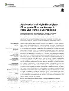
Figure 1. Homogeneous time resolved fluorescence assay (HTRF) assay principle and standard curve: (A) Schematic diagram showing the assay principle for the HTRF® Transcreener® ADP assay (Cisbio). The detection method is based on an antibody specific to ADP which is labelled with Eu3+-cryptate, which is also able to bind to either (d2 acceptor dye)-coupled-ADP (to produce TR-FRET) or ADP released from the enzymatic ATPase reaction (no TR-FRET . The resulting TR-FRET signal is inversely proportional to the concentration of the non-labelled ADP in the sample. As PMCA4 is an ATPase pump that has the ability to convert ATP to ADP with the release of inorganic phosphate in a stoichiometric ratio, the released ADP due to ATP hydrolysis by PMCA4 would compete with ADP-d2 and reduce the TR-FRET signal. (B) Dose response for different ADP/ATP ratio which mimic an ATPase reaction (as ADP is produced, ATP is depleted).
222
J Pharm Pharmaceut Sci (www.cspsCanada.org) 16(2) 217 - 230, 2013
Figure 2. PMCA4 expression in microsomal preparations and optimisation of calcium buffering: (A) Western blot analysis showing PMCA4 expression in PMCA4 microsomal membrane preparations against LacZ control microsomal preparations; α-tubulin was used as a loading control. (B) Quantification of PMCA4 overexpression in PMCA4 and LacZ control microsomal preparations (n=6, *p