devoid of significant side effects (Riggs and Melton, 1992). 'Musculo-Skeletal .... electric fields at 300 pAmps/cm2 for 34 h to show an enhance- ment of DNA ...
JOURNAL OF BONE AND MINERAL RESEARCH Volume 8, Supplement 2, 1993 Mary Ann Liebert, Inc., Publishers
Optimization of Electric Field Parameters for the Control of Bone Remodeling: Exploitation of an Indigenous Mechanism for the Prevention of Osteopenia CLINTON T. RUBIN,’ HENRY J. DONAHUE,’ JANET E. RUBIN,2 and KENNETH J. McLEOD’
ABSTRACT The discovery of piezoelectric potentials in loaded bone was instrumental in developing a plausible mechanism by which functional activity could intrinsically influence the tissue’s cellular environment and thus affect skeletal mass and morphology. Using an in vivo model of osteopenia, we have demonstrated that the bone resorption that normally parallels disuse can be prevented or even reversed by the exogenous induction of electric fields. Importantly, the manner of the response (i.e., formation, turnover, resorption) is exceedingly sensitive to subtle changes in electric field parameters. Fields below 10 pV/cm, when induced at frequencies between 50 and 150 Hz for 1 Mday, were sufficient to maintain bone mass even in the absence of function. Reducing the frequency to 15 Hz made the field extremely osteogenic. Indeed, this frequency-specific sinusoidal field initiated more new bone formation than a more complex pulsed electromagnetic field (PEMF), though inducing only 0.1% of the electrical energy of the PEMF. The frequencies and field intensities most effective in the exogenous stimulation of bone formation are similar to those produced by normal functional activity. This lends strong support to the hypothesis that endogenouselectric fields serve as a critical regulatory factor in both bone modeling and remodeling processes. Delineation of the field parameters most effective in retaining or promoting bone mass will accelerate the development of electricity as a unique and site-specific prophylaxis for osteopenia. Because fields of these frequencies and intensities are indigenous to bone tissue, it further suggests that such exogenous treament can promote bone quantity and quality with minimal risk or consequence.
INTRODUCTION
F
OR A GIVEN LOADING INCIDENT, the
potential for fracture is strongly dependent on the quantity of an individual’s bone mass. Any measure that can retard or prevent the loss of bone will provide a substantial reduction in the probability for fracture (Raisz, 1988). However, the principle of “the more the better” is not necessarily a panacea. Some agents (e.g., fluorides) may increase apparent bone density yet concurrently compromise its effective strength by incorporation of a structurally incompetent mineral (Kleerekoper and Balena, 1991; Riggs et a]., 1990). This emphasizes that the quality of the bone tissue is also critical to the skeleton’s capacity to resist monotonic or fatigue failure. Indeed, prophylaxes that preserve normal bone density by inhibition (e.g.,
bisphosphonates) (Giannini et al., 1993; Parftt, 1991) or intermption (e.g., calcitonin) (Chambers et al., 1984; Chestnut, 1984) of normal remodeling processes may predispose the skeleton to failure by destroying the tissue’s ability to repair microdamage or revitalize necrotic bone. Even estrogen, which holds a broadly acknowledged benefit to both the skeleton (Ettinger et al., 1985; Mitlak and Nussbaum, 1993) and the cardiac system (Hillneret al., 1986), is often avoided or abandoned because of undesirable side effects, including carcinogenicity(Henderson, 1989; Voight et al., 1991). With an ever escalating assortment of prophylactic measures targeted to inhibit bone loss or preserve bone quality, it is frustrating that no preventive measure has been universally acclaimed, shown uniformly effective, or acknowledged as devoid of significant side effects (Riggs and Melton, 1992).
‘Musculo-Skeletal Research Laboratory, Departmerit of Orthopaedics, State University of New York, Stony Brook, New York *Department of Medicine, Veterans Administration Medical Center, Emory University, Atlanta, Georgia.
s573
RUBIN ET AL.
s574 To develop efficacious, exogenous prophylaxes for osteopenia, it is first critical to understand the endogenous factors responsible for the normal control of bone modeling (e.g., formation) and remodeling (e.g., maintenance or resorption or both). At one level, osteopenia can be considered an inevitable consequence of the aging process. However, when this bone loss becomes symptomatic (i.e., when osteopenia compounds to osteoporosis), it occurs at relatively few, but consistent, sites in the skeleton. That this disease shows a preference for sites critical to the structural integrity of the axial and appendicular skeleton (e.g., hip, spine) implies that the skeleton’s structural effectiveness is locally, not systemically, controlled. Clearly, a prophylaxis that can strategically target selective sites in the skeleton would be ideal. Unquestionably, one of the most potent local mediators of bone mass and morphology is that associated with physical stimuli, in particular, mechanical loading. The structural prowess of the skeleton is due in large part to the bone’s capacity to recognize some aspect of its functional environment as a stimulus for the resorption or formation of tissue. This adaptive phenomenon has been recognized for a century as Wolff‘s law, reflecting Julius Wolff s 1892 treatise that mathematical laws might explain the structural objectives of skeletal adaptation. Unfortunately, although the skeleton’s ability to respond to its mechanical milieu is now widely accepted, identification of a reasonable mechanism through which load can be transformed to a signal relevant to the bone cell population has proven elusive. In the work presented here, a normal by-product of functional loading, the electric field, is considered as a critical, indigenous regulatory factor in the achievement and maintenance of skeletal morphology. Further, a case is made for exogenous electric field stimulation of the skeleton as a unique, focal treatment of osteopenia.
BACKGROUND AND HISTORY In 1954, Yasuda observed that electrical currents are generated when dry bone is loaded. The discovery of load-induced piezoelectric potentials in bone suggested a means by which stress or strain could instrinsically alter the bone’s cellular environment and thus influence proliferation and function (Bassett and Becker, 1962). This hypothesis became even more attractive when it was demonstrated that, in wet bone, two sources of electrical current coexisted: the piezoelectric currents arising from the deformation of collagen and the relatively large electrokinetic currents (streaming potentials) produced by the strain-induced interaction of charged constituents of extracellular fluids flowing past the mineral phase of the matrix (Hastings and Mahmud, 1988). The clinical application of electric fields for treatment of delayed fracture unions was based on the premise that a normal by-product of skeletal loading, the electric potential, had been eliminated by the fracture, and that the exogenous reintroduction of such intrinsic regulatory signals would stimulate the reparative process (see Bassett, 1989, for a review). Although the use of electric fields remains controversial in orthopedics, the precedent for their application has been bolstered by double-blind clinical trials demonstrating their efficacy (Barker et al., 1984; Mammi et al., 1993; Sharrard, 1990). But what constitutes the optimal electric field character-
istics for use in the clinic? And even if exogenously induced electric fields are capable of accelerating fracture repair, do functionally induced fields play a role in the normal processes of bone modeling and remodeling? Measurement of the electric potentials generated by strain suggests that the average field intensities in bone are quite small, on the order of 1 pV/pe (1 microvolt per microstrain) (Otter et al., 1990). Considering that the skeleton is seldom subject to strains exceeding 3000 microstrain (Rubin and Laynon, 1984a), the regulatory influence of functionally engendered endogenous fields must occur at thresholds below 3000 pV/cm. Further, since the great majority of bone tissue is rarely subject to strains greater than 500 p e (Gross et al., 1992) yet bone mass is retained, one could argue that fields well below 500 pV/cm successfully represent some regulatory role (McLeod and Rubin, 1993). Numerous in vitro preparations have been developed to establish the ability of extremely low intensity, exogenously induced electric fields to modulate bone cell activity. Working with bone explants, primary osteoblasts, and osteosarcoma cells, various groups have studied electric field effects on in vitro systems using capacitive coupling, pulsed electromagnetic induction, and direct electrode coupling techniques (see Brighton and McCluskey, 1986, for a review). These varied induction techniques resulted in a wide range of field frequencies and intensities being used, making it difficult to identify any common cellular response to any generic field parameter. For example, Noda et al. (l987), with rat osteosarcoma cells (ROS 1712.8). used direct electrodes to induce 60 Hz sinusoidal electric fields at 300 pAmps/cm2 for 34 h to show an enhancement of DNA synthesis (as assayed by 3H-thymidine uptake). Cain et al. (1987) used magnetic fields to induce 2 msec pulses (repeated at 15 Hz) of 0.6 mVlcm intensity into a preparation of confluent calvarial cells for 1 h to study the PTH-stimulated adenylate cyclase response. Ozawa et al. (1989), using a mouse osteoblast-like cell line (MC3T3-E1), have shown an enhancement of DNA synthesis using capacitively coupled 10 Hz pulses of 3 msec inducing peak fields of 32 V/cm. In contrast to these extremely high fields, Fitzsimmons et al. (1989) demonstrated a mitogenic effect of electric fields using capacitively coupled sinusoidal fields of 8-24 Hz at average induced field intensities of lo-’ V/cm. Even this brief review represents convincing evidence of the capacity of electric fields to modulate bone cell processes. However, because of the diversity and complexity of the signals and preparations used, it is difficult to identify those specific aspects of the field that have caused the response, much less what their physiologic relevance may be. Because bone tissue has the ability to both resorb and form, it is dangerous to extrapolate pertubations of cellular activity to an end p r o c e s e i.e. will the signal stimulate resorptive or formative activity? Finally, in contrast to stimulating the healing of delayed union, the ultimate objective of developing a prophylaxis for osteopenia would be to retain bone mass, and, therefore, the goal of electric field exposure should be to retain basal levels of activity. Clearly, this is difficult to assay in vitro. In vivo investigations, although removed somewhat from identifying specific cellular mechanisms responsible for the adaptive process, are valuable for their ability to demonstrate the true modelinghemodeling product of electric field stimulation. Although the predominant in vivo research effort of electric field
ELECTRIC FIELD MODULATION OF BONE REMODELING effects has been directed toward the enhancement of fracture healing, studies directed toward the prevention or treatment of osteoporosis have not been ignored. These studies provide an initial indication of the differential sensitivity of bone tissue to field parameters, such as frequency and exposure duration. McElhaney et al. (1968), using a rat model of disuse osteoporosis, demonstrated that capactively coupled 30 Hz sinusoidal fields were effective in preventing bone loss, whereas 3 Hz fields (at the same driving voltage) were not, suggesting that bone tissue may have distinct frequency response characteristics. Subsequent experiments by Kenner et al. (1973, using a rabbit model of disuse osteoporosis, showed that directly coupled 5 Hz fields could significantly reduce bone loss if these fields were pulsed. Bassett et al. (1979) further emphasized the importance of specific frequency components in the regulation of remodeling by showing that 70 Hz pulsed (325 psec) magnetic fields were equally effective in slowing osteoporosis as 10 Hz trains of pulses (twenty 325 psec pulses, separated by 200 psec). Cruess et al. (1983) demonstrated that these 70 Hz pulsed fields produced a dramatic reduction in collagenase activity in exposed bones as compared to bones subjected to disuse but with no field stimulation. Regarding dose duration, Martin and Gutman (1978) used the rat disuse model to show that the efficacy of a capacitively coupled 30 Hz field was not proportional to exposure duration, as a 2 Nday exposure was as effective as an 8 Wday exposure. In contrast to this work, Brighton et al. studied the efficacy of 60 kHz capacitively coupled electric fields to inhibit both sciatic. denervation osteoporosis ( 1985) and castration-induced osteopenia ( 1989) and demonstrated that only a 24 h constant-duty cycle was able to stimulate a beneficial result. Unfortunately, although this group has demonstrated some dose-response data, it is difficult to extrapolate their 60 kHz results to a mechanism relevant to the normal physiology of the tissue. As with the in vitro protocols, due to the complex nature of the waveforms and the varied animals models and induction modalities used to test the efficacy of the signals, we are able only to conclude that electricity has the potential to modulate bone remodeling processes. Identification and optimization of those specific field parameters controlling this response have proven both difficult and elusive. In summary, work at the cell and tissue level supports the hypothesis that electrical currents can regulate the activity of bone cells and, more generally, bone remodeling processes. Unfortunately, no systematic approach has been undertaken to evaluate the general range of electric field frequency and amplitudes that may affect bone remodeling, much less those associated with the spectrum of normal functional activity (and thereby reflective of an intrinsic regulatory signal). Because functionally induced skeletal strain is directly linked to the electrical currents that arise from the bone tissue, characterization of those exogenous currents dominating remodeling activity could become essential in identifying physiologic mechanisms of signal transduction (e.g., piezoelectric vs streaming processes). In turn, defining the characteristics of the optimal electrical signal could lead to identification of those specific mechanical parameters that are most influential to the noimal growth and maintenance of bone mass (e.g., strain energy density vs peak principal strain). Finally, although the potential benefits of electromagnetic fields are made apparent by these studies, the potential risks of exposure must be emphasized. Public aware-
s575
ness of the consequences of electromagnetic fields has escalated, fueled by recent epidemiologic evidence that long-term exposure to 60 Hz magnetic fields can be correlated with significant increases in childhood malignancies (Guenel et al., 1993; Savitz et al., 1988; Tomenius, 1986). Therefore, as treatment regimens for osteopenia will undoubtedly require chronic exposure to fields, identification of the minimal energies necessary to achieve these goals will be essential to ensure minimal risk. To isolate and identify those parameters of the electric field that are osteoregulatory, we have focused on the in vivo avian ulna model of disuse osteopenia as a bone preparation sensitive enough to discriminate beneficial from ineffective. In brief, the left ulna diaphysis of adult male turkeys is functionally isolated by removing a 3-4 mm section of the proximal and distal epiphysis and covering the metaphyseal ends with Delrin caps. This procedure leaves a 110 mm diaphyseal section of mechanically deprived mature lamellar bone, in which the musculature, as well as nutrient and nervous supplies, remain surgically undisturbed. The contralateral ulna is left surgically undisturbed and serves as an intraanimal control. Differences between the left (experimental) and right (control) ulnae in these birds are interpreted to reflect changes that occurred over the 8 week experimental period. In this model, 8 weeks of functional isolation alone will consistently result in a 10%-15% loss of bone (Rubin and Lanyon, 1987). This bone loss is generated by circumferential endosteal resorption and intracortical porosis, as evident from the microradiographs made from the central section of the ulna midshaft, approximately 50 mm from the site of either proximal or distal surgical intervention. When used for studying bone’s sensitivity to mechanical stimuli, this preparation has been used to identify the dependence of bone modeling and remodeling on number of load cycles (Rubin and Lanyon, 1984a), strain magnitude (Rubin and Lanyon, 1985), and dynamic vs static loads (Lanyon and Rubin, 1985). The model also has been used to study the impact on bone modeling and remodeling of nutrition (Lanyon et al., 1986), age (Rubin et al., 1992), growth (Bain and Rubin, 1990b). and endocrinopathy (Bain and Rubin, 1990a).
POWER-SPECIFIC EFFICACY OF ELECTRIC FIELDS TO PREVENT OSTEOPENIA Although many investigators have demonstrated that exogenous electromagnetic fields are capable of inhibiting osteopenia (Bassett, 1989; Brighton and McCluskey, 1986; Brighton et al., 1989; Cruess et al., 1983; Skeny et al., 1991), there is sparse evidence of a correlation between any specific physical parameter of the field (e.g., frequency, intensity, duration) and its relative efficacy in preventing loss of bone. We approached this limitation in a series of experiments (Rubin et al., 1989), and identified the power of the electric field as a parameter strongly correlated to the skeletal response. Pulsed electromagnetic fields (PEMFs), a complex electromagnetic waveform comprised of a repeated finite pulse train and similar to those used in the clinic to treat delayed fracture union, were induced in the turkey ulna preparation using Helmholtz coil pairs strapped to the wing of the animal. By varying the magnetic field rise time in these coils,
S576
RUBIN ET AL. the power of the induced electric field was altered, yet peak magnetic flux was held constant. In turkey ulnae isolated from function for 8 weeks, exposure only to inactive coils resulted in an 11% bone loss as compared to their intact contralateral ulnae. This was not significantly different from the bone loss caused by disuse alone. However, exposure of the ulna preparation to a PEMF for 1 Wday established a distinct dose-response to induced electrical power, with a maximum osteogenic effect between 0.001 and 0.04 (Tesla/s)* . s. In contrast to the 11% loss of bone caused by disuse, these specific signals were sufficient to stimulate an increase in bone area (9.5% and 12.8%, respectively, as compared to their intact, contralateral control ulnas), yielding a net 20% benefit of stimulation (Fig. 1). Importantly, not only did these signals stimulate new bone formation at the periosteal and endosteal surfaces, but also intracortical porosis was inhibited. Pulse power levels above or below this range were less effective in generating bone formation, and in some cases, field exposure was incapable of even inhibiting bone loss. Because peak magnetic flux was held constant in each of these groups, the doseresponse observed emphasizes that it was the induced electric field that served as the controlling agent. These data are strong evidence that PEMFs similar to those used in the clinic can prevent the bone loss normally associated with disuse and that the mediating factor in this response are the components of the electric field. With appropriate control of the induced electric field, new bone formation actually can be stimulated when using a fraction of the clinical exposure times. This work, therefore, identified a dose-response relationship of an electric field parameter to bone modeling and remodeling activity and suggested that optimal signal characteristics of the electric field may exist.
SELECTIVITY OF BONE ADAPTATION TO THE FREQUENCY OF THE ELECTRIC FIELD From the PEMF work it was clear that the bone tissue response to induced electric fields is dependent on specific characteristics of the field. However, because magnetic pulse rise time was varied in this study, causing both magnitude (energy) and temporal (frequency) changes in the induced electric fields, it was only possible to demonstrate the relative
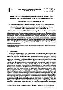
FIG. 1. Microradiographs of transverse sections of the ulna midshaft following 8 week regimens of disuse plus electric field treatment. After functional isolation without induction of an electric field (top), the ulna lost approximately 10% of its bone mass through endosteal resorption and intracortical porosis (refer to triangle symbols in Fig. 2). A functionally isolated ulna
subject to 1 Wday of a PEMF inducing 0.002 T2s-' maintained bone mass and minimized intracortical porosis (middle). This bone was difficult to distinguish from a microradiograph of the intact contralateral ulna from the same animal. By exposing the functionally isolated ulna preparation to 1 Wday of a PEMF engendering an average power proportional to 0.04 T's-', bone mass was increased substantially (bottom). This modeling activity represents a net increase of 33% as compared with the nonstimulated disuse. Although there is substantial periosteal and endosteal new bone formation (less mineralized woven bone on surfaces), there is very little remodeling activity within the cortex. Importantly, the woven bone seen at 8 weeks will remodel to lamellar bone by 16 weeks (Rubin and Bain, 1991). Despite the disuse, osteoporosis has been prevented and bone mass increased. (Modified from Rubin et al., 1989.)
ELECTRIC FIELD MODULATION OF BONE REMODELING role of induced power in the bone modeling arid remodeling response. To distinguish the independent roles of frequency vs energy characteristics of the electric field, an analytic evaluation of each PEMF used in the empirical study was performed ( M c k o d and Rubin, 1990). Using Fourier analysis techniques, it was possible to define the distribution of the PEMF's electrical energy as a function of frequency. The ability of any given PEMF to modulate bone modeling and remodeling was then correlated to the similarities and distinctions of' the frequency domain of the other PEMFs used in the study. In other words, modulating the magnetic field rise time of the PEMF in turn altered the spectral distribution of induced electric field energy. The spectral analysis of the PEMFs indicated that for induced electric fields spanning the frequency range of I Hz (one cycle/sec) to 250 kHz (250,000 cycles/sec), the singular component of the field that correlated with the PEMF's ability to prevent bone loss was that energy confined to frequencies below 120 Hz.If only the energy induced at frequencies below 75 Hz was considered, the fieldlosteogenesis correlation improved. Remarkably, even though signals with at least some energy committed to frequencies below 75 Hz were the most osteogenic, this component of the frequency band contained less than one one thousandth of the total power induced by the PEMF waveform. One immediate implication of this analytic study was that induced fields several orders of magnitude below those typically used in the clinic may, if the waveform were confined to the frequency range to which the bone tissue is most sensitive, be capable of preventing bone loss. To determine that both bone modeling and remodeling is indeed selectively responsive to frequency-specific, low-level electric fields, it is essential to abandon the complex PEMF waveform and concentrate on the ability of simple sinusoids to modulate modeling and remodeling.
s577
improvement over the 11% bone loss caused by disuse alone (Fig. 2). Although not osteogenic, both the 150 Hz and 75 Hz signals were successful in retarding bone loss (difference from intact controls: -2.6% and +5.4%, respectively). The 15 Hz sinusoid, however, was more effective in initiating new bone formation (20.4% increase in bone mass) than the PEMF. Even though the 15 Hz signal required only 0.1% of the PEMF's total power, it stimulated a 30% benefit of stimulation. Interestingly, the region of maximal sensitivity appears quite narrow, with the osteogenic response decreasing dramatically at frequencies below 10Hz. Because 15 Hz represented the most osteogenic frequency tested, experiments to determine the role of field magnitude were performed at this frequency as well (McLeod and Rubin, 1988). The dependence on magnetic flux density was evaluated over the range of 2.5 to 2500 pT. Over this range, the modeling and remodeling response of the turkey ulna preparation demon-
25 20
2U z
15
10
W
0
z a
I
5
W
0
T
/
2 W
n
T -5 -10 -15
OPTIMIZING THE OSTEOGENIC COMPONENTS OF THE ELECTRIC FIELD
1
5
0
€ .m1
.001
.01
.1
1
RELATIVE POWER
To test the hypothesis that bone tissue is preferentially sensitive to fields contained within an extremely low frequency band, the ulna preparation was used to determine the efficacy of exogenously induced, frequency-specific (sinusoid) electric fields to prevent disuse osteopenia. Specifically, the osteogenic capacity of a PEMF with a fundamental (lowest) frequency component at 75 Hz and harmonics (second, third, and so on frequency components) at and above 150 Hz W i f i compared to three sinusoidal waveforms tuned to 150 Hz,75 Hz,or 15 Hz (McLeod and Rubin, 1992). The total energy within each of the three sinusoidal waveforms was identical and similar to that contained only within the 75 Hz fundamental of the PEMF waveform. Within each of the four experimental groups, the functionally isolated ulna of five animals was exposed for 1 h/day over an 8 week period, and the modeling and remodeling response was compared to ulnae subject to inactive coils. Quantifying the modeling and remodeling response elicited by these waveforms again demonstrated the benefit of PEMF treatment even in the presence of disuse, stimulating an increase in bone area of 13.6%. This new bone formation reflects a 24%
FIG. 2. Summary of changes in the cross-sectional area (mean ? SE) of the functionally isolated ulna following exposure to a series of distinct electric fields as compared to the intact contralateral control ulnas. Area changes are plotted as a function of relative electrical power induced by the coil. Relative power, calculated as the square of induced field intensity times the induced field pulsed width, is a simplification of the many parameters that are affected as the characteristics of the induced field are changed. This graph represents a first step in defining the osteoregulatory components of the field. The five data points on the extreme right of the graph ( 0 ) represent the data from our first study, which used relatively high power, complex pulsed electric fields (Rubin et al., 1989). A PEMF with energy concentrated in the low-frequency domain (m) is shown to be as osteoinductive as PEMFs requiring two orders of magnitude more power. The osteogenic capacity of the 15 Hz sinusoidal electric field ( +), consuming one ten thousandth of the total power used in the original pulsed fields, was statistically more potent than any other signal. Also shown are the area changes caused by disuse treated with an inactive coil (A). (Modified from McLeod and Rubin, 1992.)
RUBIN ET AL.
S578 strated a distinct dose dependency. Increasing peak flux density from 2.5 p T to 25 p T modulated the remodeling process from that of resorptive activity to a state in which the level of bone turnover was minimal; i.e., osteopenia was prevented. Increasing the 15 Hz field to 250 p T stimulated substantial new bone on both the periosteal and endosteal surfaces. Increases in flux density beyond 250 pT, up to 2500 p T at 15 Hz, did not stimulate any additional effect. Preliminary calculations of the electric fields that would be induced in the ulna preparation under these magnetic flux conditions indicate that at 15 Hz, field intensities on the order of 1-10 pV/cm are capable of stimulating new bone formation even in the absence of function. In summary, these data suggest that maximum electric field efficacy will be achieved if the signal is confined to a frequency window between 10 Hz and 100 Hz. The sensitivity of the bone cell-matrix composite is such that fields as low as 1 pV/cm will influence bone mass. The manner of the response is strongly dependent on the frequency in which the field is applied. Unfortunately, this selectivity appears to be a physiologic paradox. Although most functional strain energy is contained in frequencies below 10 Hz (Antonssen and Mann, 1985) bone tissue is relatively insensitive to fields induced in this range.
PHYSIOLOGIC RELEVANCE OF 15 TO 40 Hz ELECTRIC FIELDS Although bone tissue may be most responsive to electric fields induced in the 10-100 Hz band, justifying their physiologic relevance in terms of a mediating role in normal modeling and remodeling processes becomes plausible only if such frequency components exist during normal functional activity. As even vigorous sprinting will not exceed 3 Hz (three cycles per sec), a selective sensitivity of bone to fields beyond this boundary could create a biologic anomaly. However, as demonstrated, the field intensities required to initiate an osteogenic response at these higher frequencies are extremely small. Therefore, it is not necessarily critical to engender enormous strains in this frequency range of 10-100 Hz, only some strain energy. By analyzing the spectral distribution of functionally induced strain generated within the long bones of three distinct species (horse, dog, and wild turkey), we were able to determine if these higher frequency strains did exist during functional activity (Rubin et al., 1990). Strains measured from rosette strain gauges attached to the subperiosteal bone surface of the tibia of dog and turkey, as well as the metacarpal of the horse, were analyzed using Fourier analysis techniques. Not surprisingly, during locomotion, the predominant strain energy is contained within the stride rate frequency component and its higher order harmonics up through 10 Hz. That is, if running speed is such that the left foot hits the ground every 0.5 sec, most of the strain energy will be found at 2 Hz (the fundamental frequency), with decaying strain energies at its harmonics of 4, 6, and 8 Hz. In addition to this predominant energy distribution, higher-frequency strain components consistently appear in the spectra in the vicinity of 15 Hz to 40 Hz, the magnitudes of which are on the order of 50/0-10% of the fundamental. Interestingly, these oscillations are present within the strain energy spectra regardless of animal or activity and implicate the dynamics of muscle contraction as the source of
this energy band. Even during rest, a dominant 25 Hz strain energy component was identified (Fig. 3). Given the persistent existence of low-magnitude, higherfrequency strain energy, an important role for these more subdued activities in the maintenance of bone mass becomes possible. As these relatively low-amplitude, high-frequency strains will in turn cause electrokinetic potentials specific to those frequencies, an endogenous link to the osteoregulatory capacity of the 10-100 Hz exogenous fields is made. Even though the amplitude of these potentials would be relatively small when compared to those caused by the fundamental and harmonics of locomotion, if the bone tissue and its cellular constituency are more selectively attuned to these higher frequencies, this frequency band could conceivably play the dominant role in osteoregulation.
TRANSDUCTION OF THE ELECTRIC FIELD TO A CELLULAR RESPONSE We have shown that at the level of the organ, both bone modeling and remodeling are extremely sensitive to very low
I6O
1
300
200
100
400
500
Milliseconds
0
5
10
15 Frequency
20
25
30
(Hz)
Fig. 3. Strain recorded from the cranial surface of the thoroughbred racehorse cannon bone (third metacarpal) while the animal was standing. The raw strain from a longitudinally oriented gauge is shown at the top, with the spectral distribution of this strain signal shown at the bottom. Low-frequency swaying of the animal is evident below 5 Hz, and physiologic tremor is seen by the 5 and 10 Hz peaks in the spectra. The dominant component of this standing spectra is a 26 Hz peak, which is most likely attributed to force variations arising from muscle dynamics. If bone tissue favors strain energy in this frequency range, this component of the functional strain milieu may represent a critical component of the regulatory signal for the maintenance of bone mass. (From Rubin et al., 1990.)
* s579
ELECTRIC FIELD MODULATION OF BONE REMODELING
frequency-specific electric fields. Bone resorption can be prevented by the exogenous induction of fields, and when concen- ** 500 trated within an appropriate frequency range, new bone formation can be initiated. At least one osteogenic frequency range is that between 10 Hz and 100 Hz, a bandwidth that corresponds to functionally induced oscillations in bone. Fields as small as 1 pV/cm, when induced at 15 Hz for 1 Nday, are sufficient to stimulate bone formation even in the absence of function. Although these in vivo experiments demonstrate that osteogenesis is selectively sensitive to electric fields of specific characteristics, they do not address the specific cellular mechanisms involved in the perception of and response to the fields. However, recent work from our laboratory shows that these osteogenic electric fields can affect bone cell activity directly. Both biochemical and biophysical measurements of osteoblastlike cells (ROS 17/2.8) demonstrate that exogenous electric 0 30 60 90 fields between 10 and 100 Hz at field intensities as low as 5 pV/cm will alter generic indicators of osteoblast activity, such Time, seconds as alkaline phosphatase activity (McLeod et al., 1993). Importantly, the ability of these fields to modulate osteoblast activity is strongly dependent on the density and shape of the cells, suggesting that fields may have a differential effect on bone lining cells vs osteocytes, osteoblasts vs osteoclasts, growing vs mature bone, or woven vs lamellar bone. These in vitro studies also indicate that the exposure time characteristics are important, since these osteoblast-like cells adapt slowly to the applied fields and have a delayed recovery following removal of the fields (McLeod et al., 1991). By recording changes in intracellular calcium concentration before, during, and after electric field exposure, the time course and sensitivity of the cellular response to this stimulus were quantified. ROS 17/2.8 cells were scrape loaded with the photoprotein aequorin (which luminesces in the presence of calcium), plated \* 40 6 onto glass coverslips at 200 x lo3 cells/cm2, and incubated for Q6 FREOUENCY IHd 8-12 h. The cells were then placed in a dark, temperaturecontrolled chamber, and a current source was used to induce FIG. 4. A. Spontaneous intracellular calcium transients (spiklow-frequency ( 6 4 0 0 Hz) electric fields of 10-1o00 pV/cm ing) as measured from an ensemble of ROS 17/23 cells in into the media via platinum electrodes. The mean light levels monolayer culture, preloaded with the luminescent photoprotein recorded were converted to intracellular calcium levels, and aequorin. Individual spikes represent transient rises in the higher-frequency components were analyzed by subtracting the intracellular calcium concentration of individual cells, possibly system noise power spectra from the total light spectra obtained associated with cell division. Spiking rates range from 1 to 10 in the presence of cells. This analysis of the luniinescent signal per min depending on cell density and the uniformity of aequorin permitted the determination of both mean intracellular calcium loading. B. The calcium spiking activity of cell ensembles can be quantified by performing a spectral power analysis of the levels and fluctuations in the intracellular calcium concentration temporal spiking pattern. Determination of the spectrum for the as a function of time. basal spiking activity demonstrates a lorentzian spectrum with a Mean basal calcium levels in the cells (averaged over 9 min) cutoff frequency near 0.25 Hz, consistent with an average spike were approximately 190 nM (SE 25 nM). Moreover, the intra- width of 4 sec. Low-frequency electric field exposure results in cellular calcium levels demonstrated distinct fluctuations or a substantial decrease in the magnitude of the spectra with no spiking (Fig. 4A). which were found to have a lnrentzian power change in the cutoff frequency, indicating a decreased rate of spectrum (i.e., above a characteristic frequency, the fluctuations spiking with no change in the average spike width. On removal decreased with the square of the frequency). Sinusoidal electric of the field, frequency of the spiking is reestablished over fields of 10-1o00 pV/cm at frequencies between 6 and 600 Hz approximately the same period originally required to suppress it. did not significantly alter mean basal calcium levels. However, (B from McLeod et al., 1991.) electric field exposure dramatically reduced the magnitude of the calcium fluctuations (Fig. 4B). The greatesl attenuation of activity, whereas at lo00 pV/cm, this suppression was achieved this spiking was achieved via a 20 Hz field exposure, where within 3 min. Following removal of the field, calcium spiking calcium fluctuations were reduced by 90% with fields as low as was reestablished within a period similar to that required to 10 pV/cm. The time course of this attenuation was strongly suppress it. dependent on the magnitude of the applied field. At 60 Hz, a 15 Given the ability of fields to suppress calcium spiking, one pV/cm field required close to 1 h to suppress 90%of the spiking could argue that the relatively quiescent level of remodeling
'
RUBIN ET AL.
s580 observed in mature bone is at least partially a product of an ever present, functionally induced, low-level electric field. The persistent calcium spiking that appeared in the absence of the field implies that in conditions such as immobilization, the field normally responsible for the suppression of cellular spiking is delinquent. The role of electric fields as an inhibitor of activity is further supported by studies on osteoclast recruitment (Rubin et al., 1993). Continual exposure of a murine marrow culture system to a 30 Hz, 6 pVlcm electric field attenuated the number of osteoclasts produced over an 8 day period by as much as 40%. In this case, it could be argued that the field’s presence has interrupted the recruitment of bone-resorbing cells because, in reality, these exogenous fields surrogate for an endogenous signal for bone homeostasis.
DISCUSSION The frequencies and field intensities that are most effective in regulating tissue modeling and remodeling in vivo or modulating cell activity in vitro are similar to those achieved in bone during normal functional activity. This is strong evidence that endogenously produced electric fields serve an important role in regulating normal bone modeling and remodeling processes. We are confident that isolation of the optimal signal characteristics for the maintenance of bone mass, combined with appropriate in vitro investigations will greatly improve our understanding of the physiologic basis of the skeletal tissue response. Understanding the temporal characteristics of the signal will aid in isolating the specific cellular processes responsible for both modeling and remodeling (or lack thereof). Finally, identification of the minimal field parameters capable of modulating bone mass, even in the presence of strong systemic stimuli for resorption, will promote the potential for electricity as a unique, site-specific, and relatively safe prophylaxis for osteoporosis. The four principal advantages of such a prophylaxis are 1. Coil placement permits focal treatment of a disease. that is locally, not systemically, symptomatic. This site specificity will allow for the selective treatment of spine, wrist, hip, or even alveolar sites, without subjecting the entire body to pharmaceutical intervention. 2. During osteogenesis, the new bone stimulated by electric fields is derived through the normal mineralization process. As surrogates (e.g., fluorides) are not used to precipitate this process, the matrix ultrastructure and, therefore, the effective strength of the tissue are not compromised by foreign constituents. 3. There is a distinct dose-response of bone formation to field intensity. This sensitivity allows differential regimens for the prevention vs reversal of bone loss. 4. Most importantly, the treatment regimen orchestrates normal bone cell populations via subtle amplification of an endogenous stimulus. The tissue retains its ability to model, remodel, and repair itself, and the potential consequences of contaminating a mineralizing process or inhibiting a cellular process are avoided. Rather than perturbing, suppressing, or destroying a singular component of the complex cascade of bone modeling and remodeling, this electric field prophylaxis exploits a normal, indigenous mechanism for the prevention of osteopenia.
ACKNOWLEDGMENTS This work was supported by grants AR41040 and AR39278 from the National Institutes of Health, PYI 8658105 from the National Science Foundation, the Electric Power & Research Institute, the Orthopaedic Research & Education Foundation, and Electro-Biology, Inc. We are grateful for the tireless secretarial and technical help provided by G. Trocchio, T. O’Hara, L. Pourres, P. Haralabatos, and A. Dusatko.
REFERENCES Antonssen E, Mann R 1985 The frequency content of gait. J Biomech 18~39-47. Bain S, Rubin C 1990a Metabolic modulation of disuse osteopenia: Endocrine dependent site-specificityof bone remodeling. J Bone Min Res 5:1069-1075. Bain S, Rubin C 199Ob Skeletal modeling objectives in growing bone: What role do mechanical factors play? Trans Orthop Res Soc 15:418. Barker AT, Dixon RA, Sharrard WJW, Sutcliffe ML I984 Pulsed magnetic field therapy for tibial non-union. Lancet 1994-996. Bassett C 1971 Biophysical principles affecting bone structure. In: The Biochemistry and physiology of Bone, vol 3. Academic Press, New YO&, pp 1-76. Bassett CA 1989 Fundamental and practical aspects of therapeutic uses of pulsed electromagnetic fields (PEMFS). Crit Rev Biomed Eng 12451-529. Bassett C, Becker R ( 1%2) Generationof electrical potentials by bone in response to mechanical stress. Science 137:1063-1064. Bassett L, Tzitzikalakis G, Pawluk R, Bassett C 1979 Prevention of disuse osteoporosis in the rat by means of PEMFs. In: Brighton C, Black J, Pollack S (eds) Electrical Properties of Bone and Cartilage. Experimental Effects and Clinical Applications. GNne & Stratton, NewYork,pp311-331. Brighton C, LuessenhopC, Pollack S, Steinberg D, Petrik M, Frederick S 1989Treatmentof castration-inducedosteoporosisby a capacitively coupled electrical signal in rat vertebrae. J Bone Joint Surg 71A:22% 236. Brighton C, McCluskey W 1986 Cellular response and mechanisms of action of electrically induced osteogenesis. In: Peck WA (ed) Bone and Mineral Research, vol4. Elsevier Science Publishers, New Yo&. Brighton C, Tadduni G, Pollack S 1985 Treatmentof sciatic denervation disuse osteoporosisin the rat tibia with capacitivelycoupled electrical stimulation. J Bone Joint Surg 76A1022-1028. Cain C, Adey W, Luben R 1987 Evidence that PEMFs inhibit coupling of denylate cyclase by parathyroid hormone in bone cells. J Bone Min Res 2437-441. Chambers T, Athanasou N, Fuller K 1984 Effect of parathyroid hormone and calcitonin on the cytoplasmic spreading of isolated osteoclasts.J Endocrinol102281. Chesnut C 1984 Synthetic salmon calcitonin, diphosphonates, and anabolic steroids in the treatment of postmenopausal osteoporosis. In: Christiansen C, Arnaud C, Nordin B, Parfitt A, Peck W, Riggs B (eds) Osteoporosis. Proceedings of the Copenhagen International Symposium on Osteoporosis. Aalborg Stiftsbogtrykkeri, Glostrup, Copenhagen, pp 549-555. Cruess R, Kan K, Bassett A 1983 The effect of P E W S on bone metabolism in experimental disuse osteoporosis. Clin Orthop 173945-250. Ettinger B, Genant HK,Cann CE 1985Long-term estrogen replacement therapy prevents bone loss and fractures. Ann Intern Med 102:319324. Fitzsimmons RJ, Farley JR, Adey WR, Baylink DJ I989 Frequency
ELECTRIC FIELD MODULATION OF HONE REMODELING dependence of increased cell proliferation, in vitro, in exposures to a low-amplitude, low-frequency electric field: Evidence for dependence on increased mitogen activity released into culture medium. J Cell Physiol 139:586-591. Giannini S, D’Angelo A, Malvasi L, Catrignano R , Pati T, Tonca R, Liberto L, Nobile M, Crepaldi G 1993 Effects of one year cyclical treatment with clodronate on postmenopausal bone loss. Bone 14: 137-14 I . GrossT, McLeod K, RubinC 1992 Normal and shear straindistributions of the equine third metacarpal during locomotion: A characterization of bone’s functional milieu J Biomech 25: 1081-1087. Guenel P, Raskmark P. Andersen JB, Lynge E 1993 Incidence of cancer in persons with occupational exposure to electromagnetic fields in Denmark. Br J Ind Med 50:758-764. Hastings G, Mahmud F 1988 Electrical effects in bone. J Biomed Eng 10:515-521. Henderson B 1989 The cancer question: An overview of recent epidemiologic and retrospective data. Am J Obstet Gynecol 161:18591864. Hillner B, Hollenberg 1, Pauker S 1986 Postmenopausal estrogens in prevention of osteoporosis: Benefit virtually without risk if cardiovascular effects are considered. Am J Med 80:I 1 15-1 127. Kenner G, Gabrielson E, Lovelle J, Marshall A 197.5 Electrical modification of disuse osteoporosis. Calcif Tissue Int 38:209-216. Kleerekoper M. Balena R 1991 Fluorides and osteoporosis. Annu Rev Nutr 11:309-324. Lanyon L, Rubin C 1985 Static versus dynamic loads as an influence on bone remodeling. J Biomech 17:897-906. Lanyon L, Rubin C, Baust G 1986 Modulation 01‘ bone loss during calcium insufficiency by controlled dynamic loading. Calcif Tissue Int 38:209-216 Mammi GI, Rocchi R, Cadossi R, Massari RL, Traina GC 1993 The electrical stimulation of tibial osteotomies, Clin Orthop Re1 Res 288:246-253. Martin R, Gutman W 1978 The effect of electric fields on osteoporosis of disuse. Calcif Tissue Res 2523-37. McElhaney JR, Stalnaker R, Bullard R 1968 Electric fields and bone loss of disuse. J Biomech 1:47-52. McLeod K, Donahue H, Levin P, Rubin C 1991 Low-frequency sinusoidal electric fields alter calcium fluctuation:; in osteoblast-like cells. In: Brighton C, Pollack S (eds) Electromagnetics in Biology and Medicine. S.F. Press, pp I 1 1-1 15. McLeod K, Donahue H, Levin P, Fontaine MA, Rubin CT 1993 Electric fields modulate bone cell function in a density-dependent manner. J Bone Min Res 8:977-984. McLeod K, Rubin CT 1993 Strain frequency spectra in the appendicular and axial skeleton. J Biomech (submitted). McLeod K, Rubin C 1988 The role of electric field intensity in the regulation of bone remodeling activity in-vivo. Trans Bioelec Repair Growth SOC8: 18. McLeod K, Rubin C 1990 Frequency-specific modulation of bone adaptation by induced electric fields. J Theoret Biol 145385-396. McLeod K, Rubin C 1992 The effect of low-frequency electric fields on osteogenesis. J Bone Joint Surg 74A:920-929. Mitlak BH, Nussbaum SR 1993 Diagnosis and treatment of osteoporosis. Ann Rev Med 44:265-277. Noda M, Johnson D, Chiabrera A, Rodan G 1987 Effect of electric currents on DNA synthesis in rat osteosarcoma cells: Dependence on conditions that influence cell growth. J Orthop Re.. 5253-264. Otter M, Palmieri V, MacGinitie L, Cochran G 1990 Streaming potentials in bone are similar in-vivo and in-vitro. Trans World Cong Biomech 2:ISS. Ozawa H, Etsuko A, Shibasaki Y, Fukuhara T, Suda T 1989 Electric fields stimulate DNA synthesis of mouse osteoblast-like cells
S581
(MC3T3-EI) by a mechanism involving calcium ions. J Cell Physiol 138:477-483. Parfitt M 1991 Use of bisphosphonates in the prevention of bone loss and fractures. Am J Med 91:42-46. Raisz LG 1988 Local and systemic factors in the pathogenesis of osteoporosis. N Engl J Med 318:818-828. Riggs BL, Hodgson S, O’Fallon M, Chao E, Wahner H, Muhs J, Cede1 S, Melton J 1990 Effect of fluoride treatment on the fracture rate in postmenopausal women with osteoporosis. N Engl J Med 3222302809. Riggs BL, Melton L 1992 The prevention and treatment of osteoporosis. N Engl J Med 327:620427. Rubin C, Bain S 1991 Long-term remodeling response to potent osteogenic stimuli: Stages in the achievement of lamellar bone: Trans Orthop Res Soc 16:419. Rubin C, Bain S, McLeod K 1992 Suppression of osteogenic response in the aging skeleton. Calcif Tissue Int 50:306-313. Rubin C, Lanyon L 1984a Dynamic strain similarity in vertebrates: An alternative to allometric limb bone scaling. J Theoretic Biol107:321327. Rubin C, Lanyon L 1984b Regulation of bone formation by applied dynamic loads. J Bone Joint Surg 66A:397-402. Rubin C, Lanyon L 1985 Regulation of bone mass by mechanical loading: The effect of peak strain magnitude. Calcif Tissue Int 37:411-417. Rubin C, Lanyon L 1987 Osteoregulatory nature of mechanical stimuli: Function as a determinant for adaptive remodeling in bone. J Orthop Res 5300-310. Rubin C, McLeod K, Bain S 1990 Functional strains and bone adaptation: Epigenetic assurances of skeletal integrity. J Biomech 23:4354. Rubin C, McLeod K, Lanyon L 1989 Prevention of osteoporosis by pulsed electromagnetic fields. J Bone Joint Surg 71A:411-418. Rubin J. McLeod K, Names M, Titus L, Catherwood B, Rubin C 1993 Extremely low-frequency electric fields attenuate osteoclast-like cell recruitment in marrow culture. 39th Trans Orthop Res Soc 18: 180. Savitz D, Wachtel H, Barnes F, John E, Trudik J 1988 Case-control study of childhood cancer and exposure to 60 Hz magnetic. Am J Epidemiol 128:2 1-38, Sharrard WJW 1990 A double-blind trial of pulsed electromagnetic fields for delayed union of tibial fractures. J Bone Joint Surg 72Bz347-355. Skerry T, Pead M, Lanyon L 1991 Modulation of disuse by pulsed electromagnetic fields. J Orthop Res 9:600-608. Tomenius L 1986 SO Hz electromagnetic environment and the incidence of childhood tumors in Stockholm county. Bioelectromagnetics 7: I9 1-207. Voight L, Weiss N, Chu J, Daling J, McKnight B, van Belle G 1991 Progestagen supplementation of exogenous estrogens and risk of endometrial cancer. Lancet 338:274-277 Wolff J 1892 Das Gesetz der Transformation der Knochen. [The law of bone remodeling.] Maquet P, Furlong R (trans) 1986 SpringerVerlag, New York. Yausda I 1954 On the piezoelectric activity of bone. J Jpn Orthop Surg 28:267-269.
Address reprint requests to: Clinton T . Rubin Musculo-Skeletal Research Laboratory Department of Orthopaedics State University of New York Stony Brook, NY 11794-8181