philus and Bacillus subtilis. J. Bacteriol. 149, 824-830. 4 Mc Donald, K.O. and Burke, Jr., W.F. (1984) Plasmid transformation of Bacillus sphaericus 1593. J. Gen.
Journal of Microbiological Methods, 19 (1994) 1-11
1
© 1994 Elsevier Science Publishers B.V. All rights reserved 0167 - 7012/94/$07.00 MIMET 00608
Optimization of electroporation procedure to transform B. polymyxa SCE2 and other nitrogen-fixing Bacillus Alexandre Rosado, Gabriela F. Duarte and Lucy Seldin* Departamento de Microbiologia Geral, Instituto de Microbiologia, Universidade Federal do Rio de Janeiro, CCS, Bloco I, llha do Funddo, CEP 21944-970, Brazil
(Received 2 May 1993; accepted 1 June 1993)
Summary An efficient method for genetic transformation of different nitrogen-fixing Bacilluspolymyxa strains by electroporation is presented. Various parameters on plasmid transformation of B. polymyxa SCE2 with plasmid pC194 were investigated to enhance transformation efficiency: voltage, buffer strength and type of electroporation buffer, plasmid DNA concentration, purity of plasmid preparation and plasmid source. Electroporation of B. polymyxa SCE2 resulted in a transformation efficiency as high as 3.2 x l0 ~ transformants/#g of pCl94 when the optimized procedure was used. Stability of different plasmids (pE194, pBD64, pBC16 and pFT30) transformed to B. polymyxa SCE2 and/or SCE2-43 was analysed. pEl94 showed high frequency of curing while pFT30 and pBC16 were stable after l0 transfers in nonselective medium. Plasmid DNA isolated from transformants had not undergone detectable deletions. Although the electroporation protocol presented here can be used to transform B. azotofixans, lower efficiencies of transformation were achieved. No transformants were detected in other nitrogen-fixing strains belonging to B. macerans. Key words: Bacillus polymyxa; Nitrogen-fixing Bacillus; Electroporation; Transformation
Introduction R e c o m b i n a n t D N A t e c h n o l o g y allows w o r k e r s to s t u d y the genetic c o n t r o l a n d e n h a n c e d p r o d u c t i o n o f m a n y useful a n d p o t e n t i a l l y useful c o m p o u n d s , once they have a system for the transfer o f genetic i n f o r m a t i o n within the species u n d e r study. N u m e r o u s v a r i a t i o n s o f p l a s m i d t r a n s f e r by t r a n s f o r m a t i o n have been r e p o r t e d a m o n g v a r i o u s Bacillus spp. [1,2,3,4], b u t only one p r o c e d u r e involving t r a n s f o r m a tion o f penicillin-treated cells has been d e v e l o p e d for B. p o l y m y x a [5]. This p r o c e d u r e p r o v i d e s a m e t h o d for transferring p l a s m i d D N A to B. p o l y m y x a at a very low frequency ( a b o u t 10 - 6 t r a n s f o r m a n t s per recipient cells) a n d c a n n o t be used effec*Corresponding author.
tively with selection techniques that produce spontaneous mutant colonies above this threshold. No other method of plasmid transfer has been described for B. polymyxa nor for the other nitrogen-fixing Bacillus species, mainly B. azotofixans [6] and B. macerans [7]. It would be of both practical importance and general interest to develop an efficient genetic exchange process in this group of bacteria as, besides the important characteristic of nitrogen fixation, many strains can produce a number of products of industrial interest including antibiotics, proteases and many carbohydrate-utilizing enzymes [8]. B. polymyxa has also been tested as a source of 2,3 butylene glycol [9,10] with great importance for the food industry. Electroporation has proved to be an effective method for transferring DNA to a wide range of bacterial species (for reviews, see [11,12]) and in some cases the only method available for recovering plasmid-containing transformants. We employed electroporation to transform a B. polymyxa strain with various plasmids using a commercially available system. In this report we describe a general method for the transformation of different nitrogen-fixing Bacillus strains and the optimization of the procedure for B. polymyxa SCE2 harboring the plasmid pFS1 or cured from it. Materials and Methods
Bacterial strains and plasmids The bacterial strains and the plasmids used in this work are listed in Table 1, along with some of their characteristics. Media and culture conditions Bacillus polymyxa, B. azotofixans and B. macerans strains were grown in GB broth [13] and B. subtilis strains were inoculated in TY broth [14] supplemented with the appropriate antibiotic (10 /~g/ml). The incubation temperature was 32°C to all Bacillus species. To propagate Staphylococcus aureus cultures, tryptic soy broth (Difco) was used as described by Giambiagi-Marval et al. [15]. The preferred incubation temperature was 37°C. Whenever necessary, 1.5% agar was added to the various broths to obtain solid media. Plasmid isolation Plasmid DNA was isolated from B. subtil& by the method described by Niaudet and Ehrlich [16] and from B. polymyxa by the procedure described by GiambiagiMarval et al. [15] using 1 mg/ml of lysozyme (Calbiochem) to disrupt cells. The method described by Novick et al. [17] was used to obtain plasmid DNA from S. aureus. Purification of plasmid DNA by CsC1 density gradient centrifugation in the presence of ethidium bromide was performed as described by Maniatis et al. [18]. General transformation procedure Electroporation of Bacillus strains was carried out by using a Gene Pulser apparatus (Bio-Rad Laboratories, Richmond, CA). B. polymyxa SCE2 and the other nitrogen-fixing Bacillus strains were grown for 24 h at 32°C in GB broth and then diluted (1:100) in fresh broth. Cultures were grown overnight up to a cell
TABLE 1 Bacterial strains and plasmids Strain
Plasmid (phenotype) a
Plasmid size (kilobases)
Reference or source
pFSI
16.0
This laboratory This laboratory
Remarks
Bacillus polymyxa
SCE2 SCE2-43 Loutit LMD 30.7.1 DSM 356
[26] J. van der Toorn b P. Jurtshuk, Jr c
Cured from pFSI n/f + n/f + n/f +
B. azotofixans
P3L5
[6]
RBN4
[27]
TEl0 FI02
[6] [6]
ntf + , lysogenic for P3L50 n/f + , lysogenic for BA-4 ntf ~ ntf +
This laboratory J. van der Toorn P. Jurtshuk, Jr
ntf + mf + ntf +
B. macerans
RM2 LMD 24.3 53 B. subtilis
C BDI70
pC194 (Cm R) pBD64 (Cm R Km R)
2.9 4.8
I. Mahler ~ D. Dubnau e
168 F6
pBCI6 (Tc R) pFT30 (Tc R)
4.6 4.6
[28] [28]
His , Leu-, Met-
Staphylococcus aureus
RN2442 RNI801 MB3 MB41
pE194 (Em R) pT127 (Tc R) pRJ5 (Em R) pRJ22 (Em R)
3.6 4.4 2.5 15.4
R. Novick r R. Novick M.C. Bastos g M.C. Bastos
aResistance to Tc, tetracycline; Cm, chloramphenicol; Km, kanamycin; Em, erythromycin. bDelft University of Technology, Delft, Holland. cUniversity of Houston, Houston, TX USA. dBrandeis University, Waltham, MA, USA. eThe Public Health Research Institute of the City of New York (PHRI), New York, NY, USA. rPHRI, New York, NY, USA. glnstitute of Microbiology, UFRJ, Brazil.
c o n c e n t r a t i o n o f a b o u t 5 x 10⁷ c f u / m l . Cells w e r e h a r v e s t e d by c e n t r i f u g a t i o n ( 1 0 4 0 0 x g, 15 m i n ) , w a s h e d twice in bidistilled w a t e r a n d o n c e in e l e c t r o p o r a t i o n buffer, a n d t h e n r e s u s p e n d e d in e l e c t r o p o r a t i o n b u f f e r at 1/100 th o f t h e o r i g i n a l c u l t u r e v o l u m e . T h r e e e l e c t r o p o r a t i o n b u f f e r s w e r e u s e d in this w o r k : H E B a n d P E B [19] a n d also the b u f f e r d e s c r i b e d b y T a k e t o [20]. P l a s m i d D N A in 4 to 12.5/~1 o f T E
(10 mM Tris HC1, 1 mM EDTA, pH 7,8) was mixed with 0.8 ml of cell suspension (containing 2 to 6 x 10⁹ cfu) in a chilled Gene Pulser cuvette (0.4 cm inter-electrode gap) and held on ice for at least 5 min. Following the application of a high-voltage electric pulse, 1 ml of GB broth was immediately added to the cuvettes, the cell suspension was held on ice for 15 min and then 7 ml of GB broth was added to each culture. The volume of 8 ml of cells was incubated at 32°C for 2 h, concentrated by centrifugation to 0.8 ml and then plated onto selective media. Transformants were selected on media containing 2.5 to 10 #g of erythromycin per ml, 30 #g of tetracycline or 10 #g of chloramphenicol per ml, depending on the plasmid being used. The isolation of spontaneous mutants was observed by plating 0.1 ml aliquots of the cell control (no plasmid DNA) onto selective media. Diluted samples of the control suspension were also spread in 3 GB agar plates to determine surviving CFU after electroporation. Transformation colonies were usually visible after 24 h (B. polymyxa) or 48 h (B.
azotofixans). Stability of plasmids transformed to B. polymyxa After selection for drug resistance, two transformants containing each plasmid were inoculated in GB broth (non-selectively) by diluting 1000-fold exponential phase cultures. This cycle of dilution and growth was repeated up to 10 times. Curing of drug resistance was determined by serial dilution of the first, 6th or 10th exponential phase cultures and assaying viability on both selective and non-selective media.
Analysis of isolated DNA Agarose gel electrophoresis of DNA were performed as described by Maniatis et al. [18]. After electrophoresis (0.8% agarose), the gels were stained in ethidium bromide (1 #g/ml) and photographed following visualization on a short-wave UV transilluminator. Results and Discussion
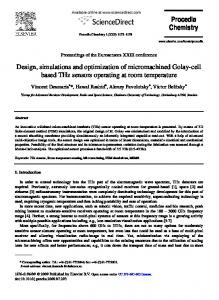
Transfer of different plasmids to B. polymyxa The main objective of this study was to introduce plasmid DNA into nitrogenfixing Bacillus, primarily B. polymyxa. First, B. polymyxa SCE2 harboring pFS 1 was transformed with different plasmid DNAs using the protocol supplied by Bio-Rad laboratories and applying a pulse of 6250 V/cm to cells in Hepes-sucrose-electroporation buffer (HEB 1 ×). Transformants were obtained with pC194, pE194, pBD64, pFT30 and pBC16 at transformation efficSencies showed in Table 2. Transfer of these 5 plasmids to SCE2 was confirmed by extraction of plasmid DNA from representative isolates (Fig. 1). Acquisition of different plasmids by B. polymyxa SCE2 resulted in displacement of the native plasmid after consecutive subculturing of SCE2 harboring different plasmids in selective media. However, pFS1 was always observed in DNA preparations from transformants newly isolated (Fig. 1A, lane 11).
Stability of plasmids transformed to B. polymyxa SCE2 Experiments were carried out to determine whether transformed cells promoted an
5 TABLE 2 Transformation of B. polymyxa SCE2 with different plasmids by using the general transformation procedure Plasmid
Transformants/#g of plasmid D N A a
pBC16 pT127 pFT30 pC194 pBD64 pE194 pRJ5 pRJ22
4.3 x 10³ 1.2 1.4 1.1 6.5 -
× x × ×
103 105 104 103
Cells were exposed to a single electric pulse (peak voltage, 2.5 kV; capacitance, 25 pF) which generated a peak field strength of 6.25 kV/cm. Electroporation buffer: HEB 1 x. aEfficiency of transformation was calculated using the average number of antibiotic resistance colonies recovered from at least two independent experiments.
A
B 1
2
3
4
5
6
7
8
9
10
11
1
2
3
4
5
6
7
8
9
10 11 12
Fig. 1. Agarose gel electrophoresis of plasmid DNAs isolated from the donor strains and from the different transformants obtained by electroporation of B. polymyxa SCE2 or SCE2-43. Lanes 1 in panels A and B are controls of SCE2 harboring pFSI and lanes 2 in both panels are controls of SCE2-43. (A) Lane 3, pBD64 isolated from B. subtilis BDI70; 4 and 5, pBD64 isolated from SCE2; 6, pC194 isolated from B. subtilis C; 7 and 8, pC194 isolated from SCE2; 9 and 10, pC194 isolated from SCE2-43; 11, pC194 newly isolated from SCE2 showing pFS1. (B) Lane 3, pE194 isolated from S. aureus RN2442; 4 and 5, pE194 isolated from SCE2; 6, pE194 isolated from SCE2-43; 7, pBC16 isolated from B. subtilis 168; 8 and 9, pBC16 isolated from SCE2; 10, pFT30 isolated from B. subtilis F6; 11 and 12, pFT30 isolated from SCE2.
6 TABLE 3 Stability of the different plasmids transformed to B. polymyxaSCE2 after 10 days in non-selective media Plasmid
pCl94
Recipient
SCE2 SCE2-43
pE194
SCE2 SCE2-43
Transformant
% of cells cured of plasmid aftera: 1 day
6 days
1 2 1 2
23 37.5 21.2 36
92 89 94 89.4
1 2 l 2
100 100 100 94.2 30 4
10 days
pBD64
SCE2
1 2
100 98.3
pBCI6
SCE2
l 2
I. 1 4.1
7 6.5
7.8 33
pFT30
SCE2
l 2
2.2 3.6
5 7.5
11.5 10
aTwo transformants harboring each plasmid were inoculated up to 10 times in GB broth when they were plated on GB agar without antibiotic. At least two hundred colonies of each transforrnant were tested for resistance to chloramphenicol (pC194 and pBD64), to tetracycline (pBC16 and pFT30) and to erythromycin (pE194).
elevated frequency o f curing o f transferred plasmids. Table 3 shows the stability o f these plasmids in B. polymyxa SCE2. pE194 was lost by the host cells after the first transfer to m e d i u m without erythromycin. This loss was independent o f the presence o f plasmid pFS1 as b o t h strains (SCE2 and SCE2-43) were cured o f p E 1 9 4 at the first growth on non-selective medium. Plasmids conferring resistance to chloramphenicol (pC194 and pBD64) showed an elevated frequency o f curing after 6 transfers in G B broth ( m a x i m u m o f 11% o f stability). Once again, this unstability could not be attributed to the presence o f p F S 1 as B. polymyxa SCE2-43 showed approximately the same frequency o f curing o f pC194 as observed in SCE2 (Table 3). Concerning the plasmids pBC 16 and pFT30, conferring tetracycline resistance, they showed to be relatively stable in SCE2 even after 10 transfers in non-selective medium.
Optimization of transformation procedure for B. polymyxa SCE2 The basic electroporation protocol was modified to allow identification o f conditions that would improve transformation efficiencies. Plasmid pC 194 isolated from B. subtilis C was chosen for these studies, unless otherwise stated.
Field strength. A n electric pulse o f 6250 V/cm at 25 ItFD, the m a x i m u m which the Gene Pulser can produce, typically resulted in a survival o f 5.9 to 10.4% (data from
7 TABLE 4 Effect of the electroporation buffer and its ionic strength on efficiency of transformation of B. polymyxa SCE2 with pC194 Electroporation buffer HEBa
PEB b
Tris-EB¢
Time constant ( m s )
Transformants/#g of DNAe
3.9 4.0 3.6
3.2 x 10⁵ 1.0 × 10⁵ 2.6 × l03
1×
2.7
3.8
2.5 ×
3.1
× 104 5.0 × 10³
2.9
3.6 × 10⁴
1× 1.5x d 2.5 x
a272 mM sucrose, 7 mM HEPES (ultra-pure, BioRad), pH 7.3, 1 mM MgC12. b272 mM sucrose, 7 mM potassium phosphate, pH 7.4, 2 mM MgCI2. c10% sucrose, 10 mM TrisoHCl, pH 7.5, 2 mM MgC12. d × -fold increase of all buffer components. eB. polymyxa was permeabilized in the presence of 10 ng of pC194 from B. polymyxa SCE2-43. Average of 4 experiments.
4 experiments) o f the original SCE2 culture. However, higher transformation efficiencies (transformants/#g plasmid D N A ) and frequencies (transformants/viable cells recovered) were generally o b t a i n e d with this voltage. T r a n s f o r m a t i o n frequencies were 1.4 × 10 - 4 at 6250 V/cm, 2.0 × 10 - 5 at 5000 V/cm, 0.6 × 10 - 5 at 3750 V/cm and 1.2 × 10 - 7 at 2500 V/cm when SCE2 cells were resuspended in HEB, 1 ×. Respectively, transformation efficiencies observed were 3.0 × 10⁵, 1.0 × 10⁵, 0.8 × 10⁵ and 2.2 × 10³/#g o f plasmid D N A . These data were calculated using the results o f two independent electroporation experiments. A l t h o u g h the highest transformation efficiency was obtained using the highest field strength, we c a n n o t attribute this to the field strength alone as the pulse duration and current flow vary with the peak voltage in the Gene Pulser used. The greatest n u m b e r o f transformants was consistently recovered following electroporation using 6250 V/ cm at 25 # F D also by Brigidi et al. [21] in B. subtilis, by L u c h a n s k y et al. [19] in L. acidophilus and by Powell et al. [22] in lactic streptococci. In B. cereus cells lower field strengths and capacitances m a y be required to obtain maximal transformation [231.
Electroporation buffer and buffer strength. The effects o f varying the electroporation buffer and its ionic strength on transformation efficiency is summarized in Table 4. W h e n SCE2 cells were resuspended in H E B 1 × , the highest transformation efficiency was obtained c o m p a r e d to PEB 1 × or Tris-EB. Both H E B and PEB yielded a greater n u m b e r o f transformants at a buffer strength o f 1 ×. The buffer concentrations for H E B and PEB 2.5 × resulted in approximately the same efficiencies o f transformation: 2.6 × 10³/#g and 5.0 × 10³//~g, respectively. All buffers p r o d u c e d time constants ranging from 2.7 to 4.0 ms. Brigidi et al. [21] also
8 TABLE 5 Effect of DNA concentration on the efficiencyof transformation of B. polymyxa SCE2 #g of pC194 DNA a
Transformants/#g of DNA,
0.05 0.1 0.25 0.5 1.0 2.5 5.0
1.0 1.8 1.0 1.5 12 34 32
x 10 3
aDNA was added in 12.5 #1 TE.
observed that the lowest ionic strength gave rise to more transformants but the preferred buffer was PEB. No significant difference was observed with HEB and PEB by Luchansky et al. [19].
Amount of plasmid DNA. Strain SCE2 was permeabilized in the presence of various amounts of pC194 (Table 5). The number of transformants recovered was not directly proportional to the amount of D N A included in the cuvette. Up to 0.5 #g of pC 194, the transformation efficiency showed to be almost the same, while the highest values were obtained with more than 1 #g of plasmid D N A added. Between the amounts of 0.5 and 1 /zg of pC194, an increase of approximately 10× in transformation efficiency of B. polymyxa SCE2 can be observed. However, different works describe the linear increase of transformation frequencies over a wide range of D N A concentrations (Miller et al. [24], 0.02 to 10 #g/ml; Taketo [20], 500 pg/ml to 5 #g/ml and McIntyre and Harlander [25], 10 ng to 1.0/~g/ml). Source of plasmid DNA and purity of DNA preparation. The source of plasmid D N A affected the efficiency of B. polymyxa transformation. When pC194 isolated from B. subtilis was used to transform B. polymyxa, we usually detected about 104 transformants/100 ng of plasmid D N A , while approximately the same number of transformants was obtained with 5 ng of plasmid D N A isolated from B. polymyxa SCE2-43. Restriction and/or modification of transferred plasmid could be responsible for the difference in transformation efficiency. The same was observed by Miller et al. [24] in Campylobacter. Furthermore, D N A preparation from strain SCE2-43 was not purified by CsC1-EtBr gradient but was only dialyzed against 10 m M Tris p H 7.8, 0.5 m M EDTA; indicating that highly purified plasmid D N A was not necessary to obtain high efficiencies of transformation.
Transfer of pC194 to different nitrogen-fixing Bacillus. Using conditions that reproducibly enhanced transformation efficiencies for B. polymyxa SCE2, transfer of pC194 into other nitrogen-fixing Bacillus strains was evaluated (Table 6). B. polymyxa strains were transformed at efficiencies varying from 1.2 × 1 0 4 to 6.6 × 10⁵/#g of plasmid DNA. Plasmid pC194 was transferred to B. azotofixans at much
TABLE 6 Transformation of B. polymyxa SCE2 and other nitrogen-fixing Bacillus with pC194 Strains B. polymyxa
SCE2 43 Loutit LMD 30.7.1 DSM 356
B. azolofixans
P3L5 RBN4 TEl0 F102
Transformants//~g of plasmid DNAa 2.5 × 10⁵b 3.3 × 10⁵ 6.3 × 10 4 1.2 × 10 4 6.6 × 10⁵ 3.5 x 101 2.5 × 10I 0 1.5 × 10²
B. macerans
RM2 LMD 24.3 53
0 0 0
aA total of 100 ng of pC194 was used in each experiment. bMean of 3 experiments.
lower efficiencies than those obtained for B. polymyxa, with 1.5 × 10²/#g D N A being the best result obtained. In this case, electrotransformation offers an unique advantage as other D N A introduction techniques have failed to succeed. No transformants were detected in B. macerans strains. Although the electroporation protocol was not optimized for each of these species, the results demonstrated that B. macerans cannot be transformed via electroporation using the same protocol used to other nitrogen-fixing Bacillus strains. Electroporation-induced transformation was shown to be a reliable method for the development of techniques for B. polyrnyxa genetic manipulation, with its use as a host for plasmid vectors. We also believe that with the optimized electroporation protocol the rapid advance of the genetics of the nitrogen-fixing Bacillus group may be possible.
Acknowledgements We are particularly indebted to Dr. Wim M. Degrave for the use of the electroporation apparatus and for all researchers that kindly provided strains. Thanks are due to all members from the Laboratory for Providing Biotechnology Products, Oswaldo Cruz Foundation, for their helpful discussion. This work was supported by the National Research Council of Brazil (CNPq) and FINEP.
References 1 Brown, B.J. and Carlton, B.C. (1980) Plasmid-mediated transformation in Bacillus megaterium. J.
10 Bacteriol. 142, 508 512. 2 Chang, S. and Cohen, S.N. (1979) High frequency transformation of Bacillus subtilis protoplasts by plasmid DNA. Mol. Gen. Genet. 168, 111 115. 3 Imanaka, T., Fujii, M., Aramori, I. and Aiba, S. (1982) Transformation of Bacillus stearothermophilus with plasmid DNA and characterization of shuttle vector plasmids between Bacillus stearothermophilus and Bacillus subtilis. J. Bacteriol. 149, 824-830. 4 Mc Donald, K.O. and Burke, Jr., W.F. (1984) Plasmid transformation of Bacillus sphaericus 1593. J. Gen. Microbiol. 130, 203-208. 5 Mallonee, D.H. and Speckman, R.A. (1989) Transformation of Bacillus polymyxa with plasmid DNA. Appl. Environ. Microbiol. 55, 2517-2521. 6 Seldin, L., Van Elsas, J.D. and Penido, E.G.C. (1984) Bacillus azotofixans sp. nov., a nitrogen-fixing species from Brazilian soils and grass roots. Int. J. Syst. Bacteriol. 34, 451-456. 7 Witz, D.F., Detroy, R.W. and Wilson, P.W. (1967) Nitrogen fixation by growing cells and cell-free extracts of the Bacillaceae. Arch. Mikrobiol. 55, 369-381. 8 Debabov, V.G. (1982) The industrial use of Bacilli. p. 331-363. In: The Molecular Biology of the Bacilli. D.A. Dubnau (ed.) Academic Press, New York. 9 Laube, V.M., Groleau, D. and Martin, S.M. (1984) 2,3-Butanediol production from xylose and other hemicellulosic components by Bacillus polymyxa. Biotechnol. Lett. 6, 257 262. 10 Mallonee, D.H. and Speckman, R.A. (1988) Development of a mutant strain of Bacillus polymyxa showing enhanced production of 2,3-butanediol. Appl. Environ. Microbiol. 54, 168 171. 11 Trevors, J.T. (1991) Electrotransformation of Bacteria. Methods Mol. Cell. Biol. 2, 247 253. 12 Chang, D.C., Chassy, B.M., Saunders, J.A. and Sowers, A.E. (1992) Guide to Electroporation and Electrofusion. Academic Press, San Diego, CA. 13 Seldin, L., Van Elsas, J.D. and Penido, E.G.C. (1983) Bacillus nitrogen fixers from Brazilian soils. Plant Soil 70, 243-255. 14 Van Elsas, J.D., Govaert, J.M. and Van Veen, J.A. (1987) Transfer of plasmid pFT30 between bacilli in soil as influenced by bacterial population dynamics and soil conditions. Soil Biol. Biochem. 19, 639~47. 15 Giambiagi-Marval, M., Mafra, M.A., Penido, E.G.C. and Bastos, M.C.F. (1990) Distinct groups of plasmids correlated with bacteriocin production in Staphylococcus aureus. J. Gen. Microbiol. 136, 1591-1599. 16 Niaudet, B. and Ehrlich, S.D. (1979) In vitro genetic labeling of Bacillus subtilis cryptic plasmid pHV400. Plasmid 2, 48-58. 17 Novick, R.P., Murphy, E., Gryczan, T., Baron, E. and Edelman, I. (1979) Penicilinase plasmids of Staphylococcus aureus: restriction~leletion maps. Plasmid 2, 109 129. 18 Maniatis, T., Fritsch, E,F. and Sambrook, J. (1982) Molecular Cloning: A Laboratory Manual. 2nd ed. Cold Spring Harbor Laboratory, Cold Spring Harbor, NY. 19 Luchansky, J.B., Muriana, P.M. and Klaenhammer, T.R. (1988) Application of electroporation for transfer of plasmid DNA to Lactobacillus, Lactococcus, Leuconostoc, Listeria, Pediococcus, Bacillus, Staphylococcus, Enterococcus, and Propionibacterium. Mol. Microbiol. 2, 637 646. 20 Taketo, A. (1988) DNA transfection of Escherichia coli by electroporation. Biochim. Biophys. Acta 949, 318 324. 21 Brigidi, P., De Rossi, E., Bertarini, M.L., Ricciardi, G. and Matteuzzi, D. (1990) Genetic transformation of intact cells of Bacillus subtilis by electroporation. FEMS Microbiol. Lett. 67, 135 138. 22 Powell, I.B., Achen, M.G., Hillier, A.J. and Davidson, B.E. (1988) A simple and rapid method for genetic transformation of lactic streptococci by electroporation. Appl. Environ. Microbiol. 54, 655 660. 23 Belliveau, B.H. and Trevors, J.T. (1989) Transformation of Bacillus cereus vegetative cells by electroporation. Appl. Environ. Microbiol. 55, 1649 1652, 24 Miller, J.F., Dower, W.J. and Tompkins, L.S. (1988) High-voltage electroporation of bacteria: genetic transformation of Campylobacterjejuni with plasmid DNA. Proc. Natl. Acad. Sci. USA 85, 856-860. 25 Mc Intyre, D.A. and Harlander, S.K. (1989) Genetic transformation of intact Lactococcus lactis subsp, lactis by high-voltage electroporation. Appl. Environ. Microbiol. 55, 604-610.
11 26 Line, M.A. and Loutit, M.W. (1971) Non-symbiotic nitrogen fixing organisms from some New Zealand Tussock-grassland soils. J. Gen. Microbiol. 66, 309-318. 27 Seldin, L. (1992) Primary characterization of the bacteriophage BA-4 from a nitrogen-fixing Bacillus azotofixans strain. Microbios 71, 167-177. 28 Van Elsas, J.D. and Pereira, M.T.P.R.R. (1986) Occurrence of antibiotic resistance among bacilli in Brazilian soils and the possible involvement of resistance plasmids. Plant Soil 94, 213-226.