© Birkhäuser Verlag, Basel, 2001 Inflamm. res. 50 (2001) 585– 591 1023-3830/01/120585-07 $ 1.50+0.20/0
Inflammation Research
Original Research Papers Estrogens ameliorate remote organ inflammation induced by burn injury in rats E. S. Özveri 1, A. Bozkurt 2, G. Haklar 3, S¸. Çetinel 4, S. Arbak 4, C. Yeg˘en 1 and B. Ç. Yeg˘en 2 1 2
3 4
Marmara University Hospital, Dept of General Surgery, Altunizade, TR-81190, Istanbul, Turkey Marmara University School of Medicine, Department of Physiology, Haydarpas¸a, TR-81326, Istanbul, Turkey, Fax: ++ 90 216 418 33 27, e-mail:
[email protected] /
[email protected] Marmara University School of Medicine, Dept of Biochemistry, Haydarpass¸a, TR-81326, Istanbul, Turkey Marmara University School of Medicine, Dept of Histology & Embryology, Haydarpas¸a, TR-81326, Istanbul, Turkey
Received 24 August 2000; returned for revision 23 November 2000; accepted by M.J. Parnham 18 July 2001
Abstract. Objective and design: The present study was designed to investigate the role of sex steroids in burninduced remote organ injury. Material or subjects: Male Wistar albino rats were given burn trauma (n=39), and underwent castration or sham operation at 2 h following the burn injury. Treatment: Rats were injected sc with either 17b estradiol benzoate (E2 , 10 mg/kg) or an androgen receptor blocker cyproterone acetate (CPA, 25 mg/kg) or vehicle, immediately after burn and at 12 h. Methods: At 24 h of burn insult, rats were decapitated. Blood samples for RIA of testosterone, estradiol and tumor necrosis factor (TNF)-a and the tissue samples for myeloperoxidase activitiy (MPO) were taken. ANOVA student’s t test was used for statistical analysis. Results: Castration, antiandrogen and E2 treatments increased plasma estradiol levels and depressed burn-induced elevation in serum TNF-a levels. In the liver and lung, burninduced increase in MPO was reduced by E2 and castration, while CPA was effective in reducing neutrophil infiltration only in the liver. Conclusion: We propose that treatment with estrogens or antiandrogens might be applicable in clinical situations to ameliorate systemic inflammation induced by burn. Key words: Castration – Androgen depletion – Estradiol – Myeloperoxidase activity
Correspondence to: B. Ç. Yeg˘en
Introduction Thermal injury may cause damage to multiple organs distant from the original burn wound and may lead to multiorgan failure [1]. Based on current research findings in animals and men, it was demonstrated that a local burn insult produces oxidant-induced organ changes as evidenced by increased lipid peroxidation in lung, liver and gut [2]. Generalized tissue inflammation is present in uninjured organs within hours of injury, even in the absence of shock. The local tissue trauma also activates a number of systemic mediator cascades, e.g. a complement activation, arachidonic acid release and cytokine production (interleukin 1, IL-1; tumor necrosis factor (TNF)-a, resulting in a generalized neutrophil sequestration and a ‘priming’ of local and systemic neutrophils and macrophages. Endotoxins and other bacterial by-products, which become evident as a result of burn wound colonization are potent activators of the primed neutrophils and macrophages [3]. This leads to the release of massive amounts of oxidants, arachidonic acid metabolites and proteases which cause further tissue damage [4]. An alternative hypothesis explaining leukocyte activation in patients with burn is that events in the microcirculation of injured skin, exacerbated by inadequate perfusion and resuscitation, result in leukocyte activation and production of further deleterious mediators [5]. Although the inflammation is initiated almost immediately after injury, the systemic response progresses with time. This concept of “postburn inflammation-induced disease” is essential for designing appropriate prevention and treatment modalities [4]. Several recent clinical studies in trauma patients have verified that infection can be considered to initiate sepsis and subsequent organ dysfunction and the susceptibility to and death from sepsis are higher in men than in women [6–8]. The suppressive effects of androgens on immunity have been reported on normal immune functions
586
as well as in autoimmune diseases [9, 10]. Experimental studies have demonstrated that female animals show normal or enhanced immune response after trauma-hemorrhage as opposed to a markedly decreased immune response in male mice [11]. Furthermore, castration of male mice before the induction of trauma or hemorrhage prevents the occurrence of immune depression, suggesting that testosterone is involved in producing immunodepression [12]. It has been demonstrated that testosterone is involved in other physiological events such as enhancing vasoconstriction, which may play a role in producing organ dysfunction following trauma [13]. Despite the aforementioned differences between males and females, it remains unknown whether sex steroids have any effect on systemic inflammation following trauma. Therefore, the aim of the present study was to determine the role of endogenous testosterone or exogenous estradiol in remote organ injury following burn trauma in rats.
E. S. Özveri et al.
Inflamm. res.
The liver, right lung and stomach were removed and the tissue samples were immediately frozen in liquid nitrogen and stored at –70 °C until they were assayed for myeloperoxidase activities. Another set of the samples was also obtained for histological analysis.
Radioimmunoassays Plasma testosterone and estradiol concentrations were determined using the double-antibody radioimmunoassay (RIA) kits (Rat testosterone RIA kit-Immunotech Co., Marseille, France and rat estradiol RIA kitImmunotech Co.). 50 (testesterone) and 100 (estradiol) ml plasma samples were assayed in duplicate. The cross-reactivity of the RIA for testosterone or the RIA for estradiol was 100%. The test values were determined by interpolation from the standard curves. The lowest detectable levels of testosterone and estradiol were 0.1 ng/mL and 20 pg/mL, respectively.
Measurement of serum TNF-a levels Materials and methods Animals Male Wistar Albino rats (250– 300 g) were kept in a light- and temperature-controlled room on a 12:12 h light-dark cycle, where the temperature (22 ± 0.5 °C) and relative humidity (65– 70%) were kept constant and fed a standard diet and water ad libitum. Experiments were approved by the Marmara University, School of Medicine, Animal Care and Use Committee. Surgical procedures and burn trauma were conducted under anesthesia performed by intraperitoneal (ip) injection of a mixture of ketamine (100 mg/kg) and chlorpromazine (12.5 mg/kg).
Surgery Rats were castrated or sham operated at 2 h after the induction of burn trauma. Bilateral orchidectomy was undertaken through a small longitudinal incision in the scrotum and testicular covering, the testis was then squeezed through this opening [14]. The spermatic vessels and the vas deferens passing to cranial pole were ligated. The scrotum was closed by absorbable sutures. In sham-operated animals, the skin of the scrotum was incised without any ligations.
A TNF-a enzyme-linked immunasorbent assay kit (Endogen, Mass, USA) was used with the manufacturer’s instructions without any modifications. Optical densities were read on a plate reader set at 450 nm. The concentration of TNF-a in the serum samples was calculated from the standard curve, multiplied by the dilution factor and was expressed as pg/mL.
Measurement of myeloperoxidase activity Tissue associated MPO activity – as an index of neutrophil accumulation- was determined in 0.2–0.5 g samples. The tissue samples were homogenized in 10 vol of ice-cold potassium phosphate buffer (20 mM K2HPO4 , pH 7.4). The homogenate was centrifuged at 10000 rpm for 20 min at 4 °C. The pellet was homogenized with an equivalent volume of 50 mM acetic acid (pH 6.0) containing 0.5% (wt/vol) hexadecyltrimethylammonium bromide (HETAB). MPO activity was assessed by measuring the H2O2-dependent oxidation of 3,3¢,5.5-tetramethylbenzidine. One unit of enzyme activity was defined as the amount of the MPO present that caused a change in absorbance of 1.0/min at 655 nm and 37°C [16].
Histologic changes Burn injury Under anesthesia, the dorsum of the rat was shaved and exposed to 90 °C water bath for 8 sec, to induce full-thickness burn involving 30 % of the total body surface area [15]. Fluid resuscitation was made (20 ml/kg saline; subcutaneously, sc) and the animals were placed on a heating pad until they recovered from anesthesia.
The samples of lung, liver, stomach and skin were fixed in 10 % buffered formalin and dehydrated in increasing concentrations of ethanol then cleared in toluene and embedded in paraffin. Sections 5 mm thick were stained with hematoxylin and eosin (HE) and the severity of tissue damage was scored according to previously defined criteria [17], (Table 1). Light microscopic evaluation of the skin samples revealed partial-thickness second degree burn injury, as characterized by superficial injury to varying portions of epidermis, but not extending to the entire dermis [18].
Experimental design Rats were treated sc twice – immediately after burn and at 12 h – with either 17b estradiol benzoate (10 mg/kg, in olive oil; Sigma) or an androgen receptor blocker cyproterone acetate (CPA, 25 mg/kg; Schering A.G.) or oil alone (vehicle, 1 mL/kg; Komili) before they were decapitated at 24 h. Trunk blood obtained by decapitation was taken into tubes, which were kept on crushed ice. The heparinized tubes were then centrifuged at 12 000 rpm for 10 min at 4 °C. Plasma was separated, placed in 1.5 mL microcentrifuge tubes, immediately frozen, and stored at –80 °C until the day of assays of the plasma hormones. On the other hand, serum samples were kept at –50°C until TNF-a levels were assayed.
Statistical analysis All data are expressed as means ± SEM with eight to ten rats per group. Instat statistical package (GraphPad Software, San Diego, CA, USA) was used. Following the assurance of normal distribution of data, oneway analysis of variance (ANOVA) with the Tukey-Kramer post-hoc test was used for multiple comparison. Values of p < 0.05 were regarded as significant.
Vol. 50, 2001
Estrogens in burn-induced inflammation
587
(723.24 ± 157.89 pg/ml; p < 0.001) treatment elevated plasma estradiol to significantly higher levels, but this increase was not as high as that reached by estradiol.
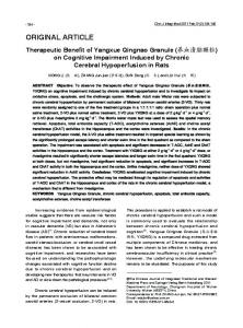
Results Plasma testosterone and estradiol levels Testosterone levels were significantly decreased in shamoperated (0.48 ± 0.05 ng/ml) and vehicle-treated (0.46 ± 0.12 ng/ml) animals subjected to burn injury, when compared with control rats (3.62 ± 1.30 ng/ml; p < 0.05). Neither the administration of CPA (0.61 ± 0.05 ng/ml) or estradiol (0.43 ± 0.05 ng/ml) nor castration (0.19 ± 0.07 ng/ml) had a further effect on reduced testosterone levels found after burn injury. In contrast to testosterone levels, plasma levels of estradiol were not affected by burn injury in the vehicle or sham operated groups (Fig. 1). Estradiol, as anticipated, increased the plasma estradiol levels (2500 ± 30 pg/ml; p < 0.001). Similarly, castration (131.15 ± 26.99 pg/ml; p < 0.05) or CPA
Fig. 1. Comparison of plasma estradiol levels in sham operated and castrated rats which were treated with oil (vehicle) or estradiol (E2 ; 10 mg/kg, sc) or cyproterone acetate (CPA; 25 mg/kg, sc) following burn injury. +p < 0.05 and +++ p < 0.001, compared to sham operated and vehicle treated rats, respectively.
Serum TNF-a levels In the sham operated and vehicle-treated burn groups (54.77 ± 0.90 pg/ml; p