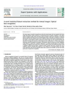
Particle Method for Sub-Voxel Extraction of. Cerebral Surface in Neonatal MR images. Daisuke Yokomichi*1, Syoji Kobashi*1,*2, Yuki Wakata*3, Kumiko ...
Particle Method for Sub-Voxel Extraction of Cerebral Surface in Neonatal MR images Daisuke Yokomichi*1, Syoji Kobashi*1,*2, Yuki Wakata*3, Kumiko Ando*3, Reiichi Ishikura*3, Kei Kuramoto*1,*2, Tomomoto Ishikawa*4, Shozo Hirota*3, Yutaka Hata*1 *1
Himeji Initiative in
*2
WPI Immunology Computational Medical and Health Frontier Research Center, Care Technology, Osaka University, Osaka, Japan Graduate School of Engineering, University of Hyogo, Hyogo, Japan
Abstract—Cerebral contour extraction from magnetic resonance (MR) images is a fundamental work to analyze brain MR images. The methods can be roughly classified into three approaches, voxel-based, mesh-based and particle-based. Each method has advantages and disadvantages. Especially, particle based method can extract the complicated sulci with sub-voxel accuracy. The remained work is to develop a method for estimating probability of particle transition among gray matter, white matter and cerebrospinal fluid. This paper proposes a new method for calculating the particle transition probability based on fuzzy inference technique. The proposed method was applied to computer synthesized MR images and neonatal brain MR images of volunteers. Keywords- neonate, MR image, cerebral surface, particle method, fuzzy inference
I.
INTRODUCTION
Brain extraction from magnetic resonance (MR) images is a fundamental procedure to analyze brain MR images. There are some automated extraction methods, and they can be classified into voxel base, mesh base, and particle base. Each of them has advantages and disadvantages. Most of brain extraction methods are classified into voxel based method. For example, Smith’s method segments the brain region using a deformable model [1]. This method determines a threshold value of dividing the brain region and the non-brain region from a histogram of MR images, and then the boundary is optimized with the deformable model. There have been also proposed some other related method [2][3][4][5][6]. These methods label the voxels by determining whether brain region or non-brain region. Thus, they cannot extract a smooth cerebral surface and the detailed shape of the intricate sulci. Few methods are based on a mesh model [7][8]. Cerebral surface extraction is a method to extract the continuous boundary surface between the cerebrospinal fluid (CSF) and the cerebrum. However, it is difficult to apply these methods to
*3
Department of Radiology, Hyogo College of Medicine, Hyogo, Japan
*4
Ishikawa Hospital, Hyogo, Japan
neonatal MR images because there are the large differences between adult and neonatal brains. To extract the cerebral surface from the neonatal MR images, Ref. [9] proposed a method based on a mesh model called thick rubber model (TRM) [9]. This method extracts the cerebral surface by deforming a rubber model with a constant thickness, called a TRM. However, this model is based on a mesh model, and the intricate cerebral sulcus is not extracted due to limitations in mesh size. Particle method, which can analyze fluid and architectural without a mesh model [10], has been applied to brain extraction. Xu's method assigns particles on the cerebral surface uniformly, and particles are pushed to vertically [11]. However, the complex and intricate cerebral sulci cannot be extracted because this method is designed for adult cerebral surface extraction. Ref. [12] has proposed a method to extract cerebral surface for neonatal MR images using three classes of particle, which were CSF particles, gray matter (GM) particles and WM particles. The particles are assigned according to the brain structure, and the assigned particles are transited based on MR signal and moved based on some forces. Although they showed the higher accuracy in comparison with voxel-based method, the remained work is to estimate the probability to change the particle classes. This paper proposes a new method to estimate probabilities of changing particle class based on fuzzy inference technique. Three features are defined from to evaluate a priori knowledge on particle classes. They are the ratios of particles in a voxel, particle density, and the thickness of cerebral cortex. Fuzzy degrees are estimated by using the calculated features. The proposed method is validated using computer-synthesized MR images and neonatal MR images. II.
PROPOSED METHOD
Fig. 1 is an overall procedure of extracting cerebral contour based on particle method [12]. The method reconstructs voxels of MR images by using three classes of particles, CSF, GM and
probability. In the following, the details of estimating class change probability are described.
Start
Skull Stripping
MR Image
Initialize particles
Iterated times > X
No
A. Overview of estimating probability When the estimated MR signal is not equal to the MR signal given by MR images, some particles should be changed into the other classes. To estimate the probability, this study focuses on the following priori knowledge of particles.
Transit particles
(1) MR signal calculated from ratio of particles should be equal to MR signal of the evaluating MR image.
Move particles
(2) Particles should connect with same class particles. And the surface of same class particles should be smooth.
Yes
(3) Thickness of cerebral cortex should be smaller than a threshold.
No
All particles converge
The above knowledge is evaluated by using fuzzy inference technique with three features, Rvoxel; ratio of particles, Rdensity; particle density , and Rthickness; the GM thickness defined bellow.
Yes
Correct isolated particles
Boundary extraction
Finish
Figure 1. Overall procedure of the proposed method.
WM particles. Each particle has an MR signal, and an intensity of voxel is determined by ratios of particles in the voxel according to partial volume effect (PVE) as follows.
I = γ CSF ICSF + γ GM IGM + γ WM IWM ,
(1)
γ CSF + γ GM + γ WM = 1 where I is MR signal of the voxel, γCSF, γGM, and γWM are the
current ratios of CSF particle, GM particle, and WM particle, respectively. ICSF, IGM and IWM are the ideal MR signal of CSF, GM and WM, respectively, and are preliminary determined by MR imaging parameter. That is, the cerebral contour will be extracted by assigning appropriate particles on appropriate positions. The classes of particle are changed according to a deg 1.0
B. The First Feature: Ratio of particles MR signal depends on ratio of tissues in the voxel according to Eq. (1). And, assume that a voxel consists tissues whose number of classes are less than or equal to two; i.e., the classes are either of (1) CSF, (2) CSF and GM, (3) GM, (4) GM and WM, or (5) WM. Thus, the ideal ratio of particles in a voxel, γ CSF , γ GM , and γ WM , are estimated from the value of MR signal. When the difference of the ratio of particles between the current ratio and the ideal ratio is high, the class of particles should be changed with a high probability. If the particle class is GM, a feature value Rvoxel is calculated by
(
Rvoxel = max γ GM − γ GM ,0
(2)
).
(3)
If the particle class is CSF or WM, Rvoxel is calculated by
(
Rvoxel = max γ GM − γ GM ,0 deg
L
M
H
1.0
0.0
L
M
H
0.0 0.2
0.8
0.6 0.4 Rvoxel
1.0
0.2
(a) Rvoxel.
0.6 0.4 Rdensity
0.8
1.0
(b) Rdensity.
deg 1.0
).
deg
L
M
H
1.0
0.0 2.0
3.0
4.0
VL L M
H
VH
0.0
Rthickness [mm] (c) Rthickness.
0.125 0.25 0.75 0.5 Transition probability (d) Probability of changing classes.
Figure 2. Fuzzy membership functions.
1.0
Rvoxel L L M L L L M M M H H L L L M M M H H H L M H H M H H
TABLE I. FUZZY IF-THEN RULES TO CALCULATE PROBABILITY OF CHANGING PARTICLE CLASS (a) GM particle (b) CSF and WM particles IF THEN IF Rdensity Rthickness Rprobability Rvoxel Rdensity Rthickness L L VL L L L L M VL L L H L M VL M L H L H L L L M M L L L M L M M L L M H L L L M L L L H L M L M M M L M M H L L L H L L L M L H L H M H M L M M H L M L H L H M M L H H M L M M M L M H M M M M H M M M H H L H M H L M M L M H M L M M M H M H H H H L H M H L H M H L M H H H M M H M H H H H H H VH M H M H L VH H H L H H VH H H M
Value of Fvoxel is described by using fuzzy linguistic values defined by fuzzy membership functions shown in Fig. 2(a). C. The second feature: particle density The homogeneous particle should locate continuously because the brain tissue locates continuously. Therefore, when a class of a particle is not equal to classes of neighboring particles, the class of particle should be changed with a high probability. To investigate distribution of other class particles, particle density of the same class σsame and the different class σother are calculated by; σ same ( p ) = ∑ δ same ( p, q ) × w ( r ) q
, ⎪⎧0 ( A ( p ) ≠ A ( q ) ) δ same ( p, q ) = ⎨ ⎪⎩1 ( A ( p ) = A ( q ) ) σ other ( p ) = ∑ δ other ( p, q ) × w ( r )
(4)
⎧⎪1 ( A ( p ) ≠ A ( q ) ) ,
(5)
q
δ other ( p, q ) = ⎨
⎪⎩0 ( A ( p ) = A ( q ) )
where w(r) is a weight function, and r is Euclidean distance from the interest particle p to particle q. The weight function is defined by; ⎧ re ⎪ w(r ) = ⎨ r ⎪ 0 ⎩
( 0 < r ≤ re ) ( r > re )
,
(6)
where re is a constant parameter. Using σsame and σother , feature value Rdensity is calculated by
Rdensity =
σ other . σ same + σ other
THEN Rprobability VL VL VL L L L L L L L L M M M M M M M M M H H H H VH VH VH
(7)
Value of Fdensity is described by using fuzzy linguistic values defined a fuzzy membership functions shown in Fig. 2(b). D. The third feature: GM thickness Cerebral cortex is the cerebral surface tissue, and is composed by GM. Because the cerebral cortex thickness is more than about 2 mm, probability of changing particle class is also considered. The current GM thickness from the processing particle is estimated from the spatial distribution of GM particles. The GM thickness of eight directions for every 45 degree on the basis of x-axis is estimated from GM particle distribution. The GM thickness for each direction is estimated by TGM = max ( r × raxis ) , (8) where raxis is the ratio of closeness with the basis of axis in each direction. The GM thickness is calculated to add the estimated thicknesses of twinned directions TGMα and TGMβ. The feature value Rthickness is calculated by
Rthickness = max (TGM α + TGM β ) .
(9) Value of Fthickness is described by using fuzzy linguistic values defined fuzzy membership functions shown in Fig. 2(c). E. Calculate probability of changing particle class Using the defined features, knowledge on probability of changing particle class is described by fuzzy IF-THEN rules.
(a) Ground truth (red line).
(b) Initial curve (red line).
(c) Extracted curve (red line).
Figure 3. Experimental result of synthesized MR image.
Value of probability is also described by fuzzy linguistics defined by fuzzy membership functions shown in Fig. 2(d). Consider a particle whose current class is GM. Let the three features extracted from the particle is xvoxel, xdensity, and xthickness. For example, if difference between the class ratio of particle and the ideal ratio is relatively large, class of neighboring almost particle is not equal to the class of the processing particle, and the GM thickness estimated from GM particle distribution is relatively high, the particle class should be changed into either CSF class or WM class which is existed near the GM particle. In this case, probability of changing particle class is given by a fuzzy IF-THEN rule ; IF Rvoxel is M, Rdensity is H, and Rthickness is M THEN Rprobabiltiy is M. M and H are fuzzy membership functions defined by Fig. 2. According to min-max-center of gravity method, a fuzzy degree of belonging with antecedent part of fuzzy IF-THEN rule is calculated by
μvoxel = max ( s ( Rvoxel ) ∧ M ) .
(
μdensity = max s ( Rdensity ) ∧ H
)
μthickness = max ( s ( Rthickness ) ∧ M )
III.
EXPERIMENTAL RESULTS
The proposed method was implemented in two dimensions, and was applied to artificially synthesized MR images to evaluate qualitative performance. The artificial MR image was synthesized from a closed curve represented by a Catmull-Rom spline curve. The closed curve was used as the truth curve, and ICSF, IGM and IWM were set to 50, 150, and 100, respectively. Fig. 3 shows the experimental results for the synthesized MR image. The experimental results were evaluated by using two measures, RMSEn and RMSEp shown in Fig. 4. RMSEn is defined as a root-mean-square distance from the ground truth curve to the nearest boundary particle. RMSEp is defined as a root-mean-square distance from a boundary particle to the nearest ground truth curve. RMSEn and RMSEp evaluate the degrees of underestimation and of overestimation, respectively. Both measures take the lower value for the better results. Next, the proposed method was applied to neonatal MR images. Revised age of subject is -2 week. Imaging parameters are a repetition time (TR) of 2000ms, an echo time (TE) of 165.25ms, and a matrix of 320 by 320 voxels. 200 sagittal MR
(10) TABLE II.
μ A = min ( μvoxel , μdensity , μthickness ) where s (α ) is a singleton function in a domain x which is defined as s(x) = 1 if x = a and s(x) = 0 otherwise, μA is the fuzzy degree belonging to the antecedent part. Then, the fuzzy membership function of consequent part shown in Fig. 2(c) is modified by alpha—cut with μA.
Synthesized Slice #85 Slice #90
RMSEN AND RMSEP
RMSEn 2.31mm 2.87mm 0.49mm
RMSEp 0.45mm 0.35mm 0.41mm
The other fuzzy IF-THEN rules are tabulated in Table I, and each fuzzy IF-THEN rule is evaluated by the similar manner described above. And, the max of the modified fuzzy membership functions for each domain is calculated. Finally, a gravity value of the obtained fuzzy membership function is obtained, and outputted as possibility of changing classes. Figure 4. Definitions of RMSEn (left) and RMSEp (right). Blue lines indicate boundary particles and red lines indicate the ground truth curve. Arrows indicate lines for measuring the distances.
(a) Ground truth (red line).
(b) Initial curve (red line).
(c) Extracted curve (red line).
Figure 5. Experimental results of neonatal MR image (slice #85).
(a) Ground truth (red line).
(b) Initial curve (red line).
(c) Extracted curve (red line).
Figure 6. Experimental results of neonatal MR image (slice #90).
images were acquired with a slice thickness of 1.5 mm and a spacing between slices of 0.75 mm. The ground truth curve of MR images were delineated by a physician. Fig. 5 and Fig. 6 show experimental results for sagittal MR images.
RMSEn and RMSEp for the obtained results are tabulated in Table 2. Because RMSEn and RMSEp are smaller than voxel size, the cerebral surface was extracted with good accuracy. However, RMSEn was larger than voxel size because a part of sulcus was not extracted well. Converged particle distributions are shown in Fig. 7. Blue particle is CSF particle, red particle is GM particle, and green particle is WM particle. GM layer was composed of GM particle. However, a part of WM particles did not changed to CSF particle in sulcus area. IV.
ACKNOWLEDGMENT This work was supported in part by Grants-in-Aid for Scientific Research, Japan. REFERENCES [1] [2]
[3]
[4]
CONCLUSION
This paper proposed an extraction method using particle method for neonatal MR images. The proposed method was applied to computer-synthesized MR image and neonatal MR images. In experimental results, the most of the cerebral surface was extracted. In the future, we will implement this method in 3-D, and consider a new parameter to extract cerebral surface better.
[5]
[6]
S. M. Smith, “Fast Robust Automated Brain Extraction,” Human Brain Mapping, Vol. 17, No. 3, pp. 143-155, 2002. K. Im, J. M. Lee, O. Lyttelton, S. H. Kim, A. C. Evans, and S. I. Kim, “Brain Size and Cortical Structure in the Adult Human Brain,” Cerebral Cortex, Vol. 18, No. 9, pp. 2181-2191, 2008. K. Boesen, K.Rehm, K. Schaper, S. Stoltzner, R. Woods, and D. Rottenberg, “Quantitative comparison of three brain extraction algorithms,” 9th Annual Meeting of the Organization for Human Brain Mapping, New York City, 2003. C. Fennema-Notestine, I. B. Ozyurt, C. P. Clark, S. Morris, A. BischoffGrethe, M. W. Bondi, T. L. Jernigan, B. Fischl, F. Segonne, D. W. Shattuck, R. M. Leahy, D. E. Rex, A. W. Toga, K. H. Zou, M. BIRN, and G. G. Brown, “Quantitative evaluation of automated skull-stripping methods applied to contemporary and legacy images: effects of diagnosis, bias correction, and slice location,” Human Brain Mapping, Vol. 27, No. 2, pp. 99-113, 2006. M. S. Atkins and B. T. Mackiewich, “Fully automatic segmentation of the brain in MRI,” IEEE Trans. On Medical Imaging, Vol.17, No.1, pp. 98-107, 1998. Y. Hata, S. Kobashi, S. Hirano, H. Kitagaki, and E. Mori, “Automated segmentation of human brain MR images aided by fuzzy information granulation and fuzzy inference,” IEEE Trans. Syst., Man, Cybern. C, Vol. 30, No. 3, pp. 381-395, 2000.
(a) Synthesized MR image
(b) neonatal MR image (slice #85)
:CSF particle :GM particle :WM particle
(c) neonatal MR image (slice #90) Figure 7. Converged particle distribution. [7]
D. MacDonald, N. Kabani, D. Avis, and A. C. Evans, “Automated 3-D Extraction of Inner and Outer Surfaces of Cerebral Cortex from MRI,” NeuroImage, Vol. 12, No. 3, pp. 340-356, 2000. [8] J. S. Kim, V. Singh, J. K. Lee, J. Lerch, Y. Ad-Dab’bagh, D. MacDonald, J. M. Lee, S. I. Kim, and A. C. Evans, “Automated 3-D Extraction and Evaluation of the Inner and Outer Cortical Surfaces Using a Laplacian Map and Partial Volume Effect Classification,” NeuroImage, Vol. 27, No. 1, pp. 210-221, 2005. [9] T. Oshiba, S. Kobashi, M. Ogawa, K. Ando, R. Ishikura, Y. Hata, S. Imawaki, and Y. Hata, “Subpixel Extraction of Neonatal Cerebral Surface from 3.0T MR Images Using a Thick Rubber Model,” Radiological Society of North America Scientific Assembly and Annual Meeting, 2008. [10] S. Koshizuka and Y. Oka, “Moving Particle Semi-implicit Method for Fragmentation of Incompressible Fluid,” Nuclear Science and Engineering, Vol. 121, No. 12, pp. 29-42, 1999.
[11] M. Xu, P. M. Thompson, and A. W. Toga, “Adaptive Reproducing Kernel Particle Method for Extraction of the Cortical Surface,” IEEE Transactions on Medical Imaging, Vol. 25, No. 6, pp. 755-767, 2006. [12] S. Kobashi, D. Yokomichi, Y. Wakata, K. Ando, R. Ishikura, K. Kuramoto, S. Hirota, and Y. Hata, “Cerebral Contour Extraction with Particle Method in Neonatal MR Images,” Journal of Advanced Computational Intelligence and Intelligent Informatics, pp. 362-369, 2011. [13] K. Yamaguchi, Y. Fujimoto, S. Kobashi, Y. Wakata, R. Ishikura, K. Kuramoto, S. Imawaki, S. Hirota, and Y. Hata, “Automated fuzzy logic based skull stripping in neonatal and infantile MR images,” IEEE World Congress on Computational Intelligence, pp. 800-806, 2010