www.nature.com/scientificreports
OPEN
Received: 19 July 2018 Accepted: 13 November 2018 Published: xx xx xxxx
Pathway sensor-based functional genomics screening identifies modulators of neuronal activity Alexander Herholt1,2, Ben Brankatschk2, Nirmal Kannaiyan2, Sergi Papiol 1, Sven P. Wichert2, Michael C. Wehr 1 & Moritz J. Rossner1 Neuronal signal transduction shapes brain function and malfunction may cause mental disorders. Despite the existence of functional genomics screens for proliferation and toxicity, neuronal signalling has been difficult to address so far. To overcome this limitation, we developed a pooled screening assay which combines barcoded activity reporters with pooled genetic perturbation in a dual-expression adeno-associated virus (AAV) library. With this approach, termed pathScreener, we comprehensively dissect signalling pathways in postmitotic neurons. This overcomes several limitations of lentiviralbased screens. By applying first a barcoded and multiplexed reporter assay, termed cisProfiler, we identified the synaptic-activity responsive element (SARE) as top performance sensor of neuronal activity. Next, we targeted more than 4,400 genes and screened for modulatory effects on SARE activity in primary cortical neurons. We identified with high replicability many known genes involved in glutamatergic synapse-to-nucleus signalling of which a subset was validated in orthogonal assays. Several others have not yet been associated with the regulation of neuronal activity such as the hedgehog signalling members Ptch2 and Ift57. This assay thus enhances the toolbox for analysing regulatory processes during neuronal signalling and may help identifying novel targets for brain disorders. Functional genomics screens are the gold-standard for dissecting gene networks at the genome-wide level and contribute to target identification and mode-of-action studies in the biomedical field1,2. They are frequently used to identify genes with roles in proliferation, cell viability, viral infection, gene-drug interaction, and most recently cellular signalling3–6. The development has been mainly driven by cancer-related phenotypes (e.g. proliferation) in relatively easy accessible huge cell populations7. Unfortunately, these screening protocols are not suitable for the study of signalling in postmitotic cell types like primary neurons, as many disease-related neuronal phenotypes rely more on cell-to-cell communication (e.g. synaptic transmission) and the formation of complex morphologies and networks8. Existing protocols require high cell numbers per individual perturbation to gain sensitivity and robustness, but cell numbers are very limited for most primary postmitotic cell types9. The dissection of signalling pathways currently requires the dissociation of the cell population for cell sorting or droplet-based single cell analysis10. While this was successful in the case of immune cells and neuronal stem cells4,11–14, it is not a favourable procedure for mature neuronal networks and might generate preparation associated artefacts15. We reasoned that a protocol avoiding any trituration followed by cell sorting would greatly facilitate the screening process and enable pooled genetic screenings of so far inaccessible cell-types, such as primary neurons. Neuronal activity-dependent signalling, e.g. modulating local protein synthesis and gene expression, is key to higher brain function and disturbed neuronal excitation and signalling has been associated with many brain diseases16,17. While descriptive methods such as proteomics and transcriptomics have delivered insight into the synaptic architecture and neuronal activity-dependent gene expression, respectively8,18,19, the empirical association of gene functions with a dedicated neuronal phenotype is a tedious endeavour and high-throughput techniques are not yet available. The activity state of neurons has long been visualized using reporter proteins controlled by activity-dependent promoters or enhancer elements20–23. We and others have demonstrated that signalling can be robustly measured by deep sequencing using pathway-specific reporters expressing short synthetic RNA barcode sequences24,25. Thus, we hypothesized that barcoded genetic sensors might provide the required sensitivity 1
Department of Psychiatry and Psychotherapy, University Hospital, LMU Munich, Munich, Germany. 2Systasy Bioscience GmbH, Munich, Germany. Correspondence and requests for materials should be addressed to M.J.R. (email:
[email protected])
SCientifiC RePorts |
(2018) 8:17597 | DOI:10.1038/s41598-018-36008-9
1
www.nature.com/scientificreports/ to perform comprehensive pooled screens for disturbed signalling in primary neuron cultures. In addition, the sensor approach would also circumvent the need for cell trituration and sorting. As a proof-of-principle, we first identified a top neuronal activity sensor to build a sensor-coupled shRNA effector library targeting over 4,400 genes. Next, we applied this screening technology to silenced versus activated cortical neuron cultures to identify modulators of synapse-to-nucleus signalling.
Results
Pathway profiling identifies top performance neuronal activity sensor. The sensitivity and robust-
ness of a genetic screen is likely as dependent on the performance and dynamic range of the reporter as in cellbased compound screens26. Therefore, a panel of 70 cis-regulatory pathway reporters was generated to identify a sensor with excellent performance in a given cellular paradigm. The library consists of clustered transcription factor binding sites, enhancers, or short promoters coupled to a luciferase reporter and individually to unique RNA barcodes which we collectively term cisProfiler (Fig. 1a and Supplementary Table S1). The cisProfiler pool was packaged into AAV particles and used to infect postmitotic cortical cultures which were treated either with a cocktail including tetrodotoxin (TTX) to silence neuronal activity, or with a cocktail containing bicuculline (BIC) raising neuronal activity (Fig. 1b, see Methods for details). Online monitoring of luciferase activity from the cisProfiler pool revealed a strong increase upon BIC stimulation, while TTX decreased the signal below baseline (Fig. 1c). To identify individual reporters mediating this response and in particular those displaying the largest dynamic range between an inactivated and activated state, we measured sensor activities by deep sequencing of the RNA barcodes (Fig. 1b) (see Methods for details). We identified a cluster of reporters which reacted to silencing and stimulation, containing regulatory elements of several prototypical neuronal-activity coupled genes such as Fos and Egr1 among others (Fig. 1d, Supplementary Fig. S1, and Supplementary Table S2). Several sensors displayed kinetics with a sustained response, amplified response, or transient response, essentially reflecting corresponding categories described for endogenous genes induced by neuronal activity27. The synaptic activity-responsive element (SARE) gave the largest dynamic range 4 hrs after BIC stimulation and outperformed classical neuronal activity reporters (Fig. 1d), in line with a previous report28. The SARE region is a conserved enhancer of the immediate-early gene Arc, consisting of binding sites for the transcription factors CREB, MEF2, and SRF/TCF, which can be found in promoters of multiple neuronal activity-dependent genes and is therefore likely integrating transcriptional responses elicited by different neuronal activity-dependent signaling pathways29,30. Given the high fold-change activation and well characterized structure of SARE, we decided to use this cis-regulatory element for developing the pathScreener assay. A cluster of four SARE repeats proximal to the 420 bp Arc core promoter (ArcMin) gave maximal dynamic range in luciferase reporter gene assays (Supplementary Fig. S2). This reporter is hereafter called E-SARE in accordance to the construct generated by Kawashima and colleagues and was subsequently used as final sensor element28.
Construction of the AAV pathScreener library by sensor-effector coupling. Instead of a lentiviral screening vector, we used the AAV system as it has certain advantages. AAV particles have a natural tropism for neuronal cells and do not elicit cytotoxicity31. In addition, AAV-based systems have been extensively investigated for human gene therapy purposes and the availability of many natural as well as synthetic capsid proteins allows broad applications in many different tissues and cell types32. Furthermore, the AAV genome persists extrachromosomally which avoids effects on reporter performance depending on the genomic locus as with integrated lentiviruses. The AAV screening vector presented here was designed as a dual-expression vector containing the E-SARE reporter (=sensor component) and the shRNA cassette (=effector component) in opposing directions yielding the final sensor-effector coupled design (Fig. 2a). The E-SARE reporter is driving the expression of the Firefly luciferase (luc2) and a 35 bp barcode sequence (BC) in response to neuronal activity as in the cisProfiler assay (Fig. 1). The shRNA is constitutively expressed by a human U6 promoter, which performed better compared to neuronal promoters (Supplementary Fig. S3). We verified that the proximity of the shRNA expression cassette does not affect the E-SARE sensor performance (Supplementary Fig. S4). Run-through transcripts from the hU6 promoter due to insufficient termination have been a concern, as they would bias the barcode pool for sequencing if transcribed into cDNA33. A comparison of random primers with oligo(dT) primers for cDNA synthesis indicated that the oligo(dT) primers are superior in this set-up and we show that transcriptional run-through by RNA polymerase is not interfering with the assay (Supplementary Fig. S4). We used a shRNA collection focused on 4,625 signalling pathway targets as template for library construction (see Methods for details). The shRNA cassette was amplified, extended by a short synthetic poly-adenylation signal (SpA) and a semi-random barcode sequence and cloned into the AAV vector containing the E-SARE reporter cassette (Supplementary Fig. S5). The proximity of barcode and shRNA within a window of less than 400 bp allowed assigning each shRNA with its barcode by deep sequencing (Supplementary Fig. S5) recovering ~97% of all target genes (see Methods for details). Pooled genetic interference screen in postmitotic cortical cultures. Within the library, the neu-
ronal activity-dependent E-SARE sensor controls the expression of a multitude of unique RNA barcodes. Each barcode is linked to a specific shRNA expressed from the same AAV vector (Fig. 2a). Thereby, deep sequencing of the barcode pool during screening is sufficient to identify the corresponding shRNA and to measure the sensor activity simultaneously. We performed two screens with 2–3 replicates per condition. For each replicate sample, 10 or 5 million primary cortical neurons plated on 15 or 10 cm dishes, termed screen A and B respectively, were infected with the AAV library pool at an infection rate of ~60% at day-in-vitro (DIV) 6 avoiding multiple infections per cell (Supplementary Fig. S6). The maturation of the neuronal network was continued until DIV12 to allow formation of active synapses and spontaneous network activity34,35. Half of the cultures were subsequently
SCientifiC RePorts |
(2018) 8:17597 | DOI:10.1038/s41598-018-36008-9
2
www.nature.com/scientificreports/
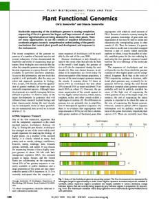
Figure 1. Pathway profiling in cortical neurons: Selection of a high-performance neuronal activity sensor. (a) Schematic map of the AAV cisProfiler vectors, used to simultaneously monitor multiple signalling events in neurons. See Supplementary Table S1 for details. (b) Workflow for the cisProfiler assay in primary cortical neurons. (c) Continuous live-cell recordings of the cisProfiler response in primary cortical neurons (DIV11–13) upon treatment with TTX cocktail (1 µM TTX, 100 µM APV) or BIC cocktail (50 µM BIC, 100 µM 4-AP, 100 µM glycine, 1 µM strychnine). Recording was performed in parallel to the assay shown in (d). (d) cisProfiler sensor responses at the indicated time points in primary cortical neurons as log2 fold-changes between treated (BIC, TTX) and untreated samples, measured by barcode sequencing. Sensors are ranked by the dynamic range between 4 hrs BIC cocktail stimulation and TTX cocktail treatment (timepoint used for pathScreener assay). Workflow as shown in (b). Treatment conditions as in (c). See Supplementary Table S2 for details. ITR, inverted terminal repeat; minMLP, minimal Major Late Promoter; BC, barcode; WPRE, Woodchock hepatitis virus posttranscriptional regulatory element; pA, poly-adenylation signal. BIC, bicuculline; TTX, tetrodotoxin; APV, (2R)-amino-5-phosphonovaleric acid; (2R)-amino-5-phosphonopentanoate; 4-AP, 4-Aminopyridine.
silenced using a TTX cocktail to reduce the E-SARE activity to baseline level to quantify the relative abundance of each functional library vector. In the remaining cultures, network activity was stimulated by a BIC cocktail in order to reveal genes which participate in neuronal activity-dependent signalling (see Methods for details). From each sample, total RNA was purified, and barcode pools were prepared for deep sequencing. The effect of each gene knockdown on E-SARE activity was determined by comparing all barcodes of the stimulated samples with each other after normalization to the silenced reference samples (Fig. 2b). It was expected that a knockdown of a positive regulator of activity-dependent neuronal signalling will decrease the relative E-SARE activity, whereas a knockdown of a negative regulator will increase the relative E-SARE activity. The induction of the E-SARE activity upon stimulation of synaptic activity by BIC was monitored in sister cultures by live cell online luciferase recordings (21 fold-change, BIC vs TTX) (Fig. 2c,d). The induction of barcode expression from the SCientifiC RePorts |
(2018) 8:17597 | DOI:10.1038/s41598-018-36008-9
3
www.nature.com/scientificreports/
Figure 2. Pooled sensor-effector coupled interference screen in cortical neurons. (a) Schematic map of the pathScreener library vector. (b) Workflow for the pathScreener experiments in primary cortical neurons. (c) Continuous live-cell recordings of the pathScreener library response in primary cortical neurons (DIV12) upon treatment with TTX cocktail (1 µM TTX, 100 µM APV) or BIC cocktail (50 µM BIC, 100 µM 4-AP, 100 µM glycine, 1 µM strychnine). Recording was performed in parallel to screen A/B. n = 3, ±s.e.m. ***p 103 cells per construct9. We used an enhanced Z-score ranking (ZS) and the Bioconductor DESeq2 package as two independent analysis methods for hit nomination (Table 1, Supplementary Table S3)4,36. Rankings from the two analysis methods were highly correlated (Spearman rank coefficient rho > 0.972, Screen A/B; Supplementary Fig. S7) and we observed a considerable number of shared hits among the top 100 positive regulators (54/100, p