Phase map retrieval in digital holography: avoiding the under-sampling effect by a lateral shear approach P. Ferraro1*, C. Del Core1, L. Miccio1, S. Grilli1, S. De Nicola1, A. Finizio1, and G. Coppola2 1
CNR–Istituto Nazionale di Ottica Applicata & Istituto di Cibernetica “E. Caianiello,” & LENS European Laboratory for Non Linear Spectroscopy, Via Campi Flegrei 34, 80078 Pozzuoli (Napoli), Italy 2 CNR–Istituto Microelettronica e Microsistemi, Via P. Castellino 111, 80100 Napoli, Italy
ABSTRACT In Digital Holography (DH) the numerical reconstruction of the whole wavefront refracted or reflected by a sample object allows one to extract the wrapped phase map mod, 2π. In fact, since the hologram is coded numerically as a digitized image, both the wavefront amplitude and phase can be reconstructed simultaneously to provide amplitude and phase contrast imaging. The resolution in the image plane is the reconstruction pixel size that depends on wavelength, reconstruction distance and the size of the CCD recording area. Efforts to improve the resolution of DH reconstructions have been accomplished, following various strategies: increasing of the hologram aperture by moving the camera in different positions or even by using synthetic aperture approaches, using a diffraction grating to record digital holograms with a wider solid angle in the object beam, or using multiple sources and/or multiple acquisitions. Although all of these methods allow one to increase the spatial resolution, one more complication exists concerning the loss of resolution that occurs in the usual DH reconstruction approaches. It can occur that the reconstructed wrapped phase map in the image plane is undersampled because of the limited pixel size which limits the spatial bandwidth of the reconstructed image. In such a case the phase distribution cannot be retrieved correctly by the usual unwrapping procedures. We show that the use of the digital Lateral-Shearing Interferometry (LSI) approach in DH provides the correct reconstruction of the phase map in the image plane, even in extreme cases where the phase profile changes very rapidly. We demonstrate the effectiveness of the method in a particular case where the profile of a highly curved silicon micro-electromechanical system membrane has to be reconstructed. Keywords: Lateral Shear Interferometer, Digital Holographic Microscope, Interference Microscopy.
1. INTRODUCTION DH compared to other interferometric methods and to conventional holography, offers new improvements in optical metrology. In particular, DH has been demonstrated to be a useful tool to study microelectromechanical systems (MEMS) [1], microcorner cubes [2], biological matter [3,4], vibration analysis [5], and particles [6,7]. The possibilities offered by DH are due to its intrinsic features. In fact, since the hologram is coded numerically as a digitized image, both amplitude and phase of the reflected o refracted complex wavefront can be reconstructed simultaneously. The spatial resolution in DH is limited by both the number and the size of the detector pixels. Efforts to improve the resolution of DH reconstructions have been accomplished, following various strategies: increasing the hologram aperture by moving the CCD in different positions [8], by using synthetic aperture approaches [9], using a diffraction *
[email protected]
phone +39.081.8675040
fax +39.081.8675118
Optical Micro- and Nanometrology in Microsystems Technology II edited by Christophe Gorecki, Anand K. Asundi, Wolfgang Osten Proc. of SPIE Vol. 6995, 699506, (2008) · 0277-786X/08/$18 · doi: 10.1117/12.782780 Proc. of SPIE Vol. 6995 699506-1 2008 SPIE Digital Library -- Subscriber Archive Copy
grating to record holograms with a wider solid angle [10], or using multiple sources and/or multiple acquisitions [11– 14]. In this paper we want to discuss a method to avoid the loss of resolution that occurs, by means of the undersampling effect (US), in the usual DH reconstruction process. In fact, it can occur that the wrapped phase map of the reconstructed wavefront is under-sampled, due to the reconstruction pixel (RP) size, which limits the spatial bandwidth of the reconstructed image. The US effect is very common in interferometry, and two procedures can be found in literature to retrieve the correct phase [15,16] but they doesn’t work in DH. These methods allow to recover the correct phase under specific assumptions on the expected phase function and, moreover, they are based on the applying of appropriate numerical procedures to the aliased mod 2π-wrapped phase, while the approach presented here operates just to avoid the aliasing effect. A DH method for the reconstruction of the correct phase has been presented in Ref. [17]. The resolution in the reconstructed image plane is recovered by increasing synthetically the aperture of the digital hologram, this is accomplished by padding the hologram with zeros to reduce the RP size and to avoid the US of the wrapped phase mod 2π. However, the above method requires a large number of pixels to be processed as a function of the expected resolution. The same problem could occur in reconstructing the hologram by the highest-resolution reconstruction approach, named the convolution transformation method, in which the RP size is the same as the CCD pixel size [3]. Recently it has been presented a method that allows to retrieve the phase by a single-image acquisition it combines DH with digital LSI. This technique avoids any cumbersome and tedious adjusting procedure, such as the double image acquisition and/or the adjusting numerical procedure [2], to obtain a phase map free of defocus aberration.
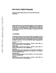
2.EXPERIMENTAL SETUP Fig.1(a) shows the DH setup. The wavelength λ of the laser was 532 nm. The samples was observed through a 20×, 0.4 numerical aperture microscope objective. A digital hologram was recorded with 1024×1024 pixels by CCD camera, the pixel size of the detector was 6.7 µm. The profile of the object is retrieved numerically on the basis of DH theory. We tested a MEMS having a large curvature due to the presence of residual stress [17]. Fig.1(b) shows a scanning electron microscope image of a MEMS structure. Reference beam BS
MO
Object beam
S
CCD
(a)
(b)
Figure 1
3.THEORY Holography is based on the theory of scalar diffraction in the Fresnel approximation. The hologram recorded by the CCD is processed to reconstruct the complex wavefield of the object beam in the image plane. From the knowledge of the complex field is possible to recover the amplitude and the phase of the light refracted by the sample. The reconstructed diffracted field Q(ξ ,η ) in the reconstruction plane (ξ ,η ) at distance d from the hologram plane is obtained by the Rayleigh-Sommerfield diffraction integral and can be written as:
Q (ξ ' ,η ' ) =
∞
1 2π π exp(i ) ∫ R ( x, y ) I ( x, y ) exp[i [(ξ '− x) 2 + (η '− y ) 2 ]]dxdy iλ d λd − ∞ λd
(1)
R ( x, y ) is the reference digital wave and I ( x, y ) is the interference pattern recorded by the CCD camera. The intensity I (ξ ,η ; d ) and the phase distribution φ (ξ ,η ; d ) of the reconstructed image can be determined as follow: where
Proc. of SPIE Vol. 6995 699506-2
I (ξ ' ,η ' ; d ) =| Q(ξ ' ,η ' ) |2 Im[Q(ξ ' ,η ' )] ϕ (ξ ' ,η ' ; d ) = arctan Re[Q(ξ ' ,η ' )]
( 2)
In case the contributions of the higher order optical aberrations are considered negligible, the phase retardation introduced by the sample is embedded in a parabolic term due to the microscope objective, namely the defocus term. Lateral Shearing Interferometry consists in the digital shear of the computed phase map and then in the subtraction of the sheared maps to obtain a phase free of the parabolic term. The difference between the two sheared maps is:
∆ϕ ξ ' = φ (ξ ' ,η ' ) − φ (ξ '− sξ ' ,η ' ) and, for small shearing,
(3)
∆ϕ ξ ' approaches to the phase derivative minus a tilted plane: ∂ϕ (ξ ' ,η ' ) ∆ϕ ξ ' (ξ ' ,η ' ) ikξ ' − ≈ sξ ' sξ ' ∂ξ '
We remove digitally the tilted plane so that:
∂ϕ ∆ϕ ξ ' ≈ ∂ξ ' sξ '
(4)
(5)
and the phase profile of the sample is obtained by applying a standard trapezoidal integration rule to Eq.(3), without the contribution of the defocus aberration. In Fig.2 intermediate step in the reconstruction process is showed. Retrieved phase values are affected by integration errors of the order of the difference values ∆φξ ' < π , averaged between consecutive pixels of the phase profile. This technique besides being suitable in the case of specimen which does not present flat area for the reference hologram recording it is applicable to avoid the US effect. Indeed it can happen that, retrieving the phase of the reconstructed wavefront in the appropriate image plane, the wrapped phase map is undersampled, due to the Reconstruction Pixel size (RP), which limits the spatial bandwidth of the reconstructed image. The consequence of such a problem is that in some critical circumstances the phase cannot be reconstructed correctly. The RP size is the resolution in the reconstruction plane. For the Fresnel transformation reconstruction process RP size is given by ∆x=dλ/N∆ξ, where d is the distance between the hologram and the image plane; λ is the laser wavelength; and N and ∆ξ are, respectively, the number of pixels and the pixel size of CCD array [3]. The RP size can affect the correctness of the reconstructed phase map because the value of the Nyquist frequency fN=1/2∆x can be lower than that of the fringe spatial frequency, thus causing the so-called “aliasing” phenomenon. In other words, where the fringe frequency is greater than the Nyquist frequency, the phase is mapped back to a spatial frequency lower than the Nyquist one, i.e., (US) occurs, and consequently the unwrapping algorithms fail where the phase difference between two adjacent pixels exceeds π rad. This problem is well known in interferometry, and two procedures have been proposed in the literature to retrieve the correct phase [15,16] but not in DH. Moreover, these methods allow one to recover the correct phase but only under specific assumptions on the expected phase function and, more important, by applying the appropriate numerical procedures to the aliased mod 2 π wrapped phase, while the approach presented here operates just to avoid the aliasing effect.
Proc. of SPIE Vol. 6995 699506-3
Shear along two directions
H
H SHEAR DIRECTIOS
Shift. 1 pixel
SHEAR DIRECTIOS
i i
Hsx
4T(H) - 4T(Hsy)
4T(H) - 4T(Hsx)
Fig.2: Digital lateral shearing step in the reconstruction process applied to biological sample.
4.EXPERIMENTAL RESULTS In this paper we will show that DH, together with LSI, allows to obtain the correct phase profile of a silicon MEMS structure by a single-image-processing procedure, avoiding both the US effect and the unwrapping procedure. We present results obtained on a MEMS having a large curvature due to the presence of residual stress [17]. We apply the proposed method and demonstrate how it can be effective for the Fresnel transformation method reconstruction process even if it can be applied to the convolution one. The shape of the MEMS was affected by deflection mainly due to the residual stress present in micromachined double-layer structures, having a parabolic profile given by
y≅ where
ϑ1
1 2 x + (ϑ0 + ϑ1 ) x 2R
(6)
ϑ0 + ϑ1 is the MEMS initial slope and R is its radius of curvature. ϑ0 is the contribute due to mean stress, while due to the stress gradient. This profile is simply related to ϕ ( x , y ) , which is the phase retardation of the
is complex wave field reflected by the MEMS, given by:
y( x) =
λ φ ( x, y ) 4π
(7)
It is important to note that the eventual US of the phase map has to be avoided to obtain the correct MEMS profile. From Eqs.(6,7) and using Nyquist criterion we calculate the maximum range x max , along which we are sure that correct sampling occurs. The Nyquist criterion establishes that the minimum phase increment between two consecutive pixels that can be retrieved is less than π ( δϕ < π ). The maximum distance is
⎡ N∆ξ ⎤ − (ϑ0 + ϑ1 )⎥ x max = R ⎢ ⎣ 4d ⎦
Beyond this range the phase values are not real because the Nyquist criterion is not verified, and the US effect will certainly occur. In [17] the maximum sampled phase ϕ ( x max , y ) in accordance with Eq. (6) was 104 rad. Fig.3(a) shows the wrapped phase map of the MEMS, and Fig.3(c) shows the corresponding plot of the unwrapped phase along the x direction. Clearly the Nyquist limit was exceeded and the US effect occurred in the wrapped phase map as evidenced by the appearance of the circular fringe on the tip of the corner. To overcome the US effect the hologram was
Proc. of SPIE Vol. 6995 699506-4
padded with zeros by enhancing the CCD pixel number in a fictitious way from 1024 × 1024 pixels up to 2048 × 2048. This numerical process increases the Nyquist frequency and thus the spatial resolution, by reducing the RP size. Fig. 3(b),3(d) show the results. The US is clearly removed since the maximum sampled phase in that case was 401 rad and was higher than the value of the maximum of the phase due to the MEMS, namely about 160 rad. Fig. 3(e),3(f) show the computed MEMS surface when US is occurring and when it is removed, respectively. 400
400
•
Pixel
Pixel
V
200
0
(a) 0
100 Pixel
0
200
80 40
200 Pixel
0
200 Pixel
400
100
(c) 0
100 Pixel
(d) 400
200 rad
80 40 0 100 Pixel
0
200
120 rad
(b) 0
200 rad
rad
120
0
200
(e)
(f)
0 200
0
100
100 Pixel
Pixel
200 0
200
400
Pixel
Fig. 3. MEMS wrapped phase maps from: (a) 1024 × 1024 pixels hologram and (b) 1024 × 1024 pixels padded to 2048 × 2048 pixels; (c) and (d) are the unwrapped phase along the x direction for the original and the padded hologram, respectively. (e) is the surface profile obtained from the US wrapped phase map (a); (f) is the surface profile as obtained from wrapped phase map (b) free of the US effect.
We show here that the correct MEMS phase profile, ϕ ( x , y ) can be obtained by an alternative approach based on DH in combination with LSI. We performed the numerical reconstruction of the complex wavefront in the image plane. Then we obtained a replica of the wavefield by numerically shifting it in that plane as we said in the previous section. The intermediate step of the digital shearing procedure are presented in Fig.4. The phase difference ∆ϕ ξ ' of the shearogram given by Eq.(3) is shown in Fig.4(a) The linear term in the Eq.(4) can be easily removed and the result is showed in Fig.4(b). The phase profile of the MEMS is obtained by applying a standard trapezoidal integration rule to Eq.(5). In Fig.4(d) the surface profile of MEMS is presented, while Fig.4(c) shows the plot profile of the corresponding phase along the x direction of the MEMS. We can note that the shape obtained by this procedure is parabolic as expected, and the maximum phase value is identical with that obtained by the padding method described in Ref.[17].
Proc. of SPIE Vol. 6995 699506-5
200
Pixel
Pixel
200
100
0
(a) 0
100 Pixel
(b) 100 Pixel
0
200
200
rad
rad
200 100 0
100
0
200
10 mm
100
(e)
(c) 0
100 Pixel
200
Pixel 100
0
200 100 Pixel
Fig. 4. (a) shearogram of the phase map of the MEMS; (b) Derivate of the phase map of the MEMS without the linear term; (c) profile of the phase of the MEMS along a central row of the phase map; (d) phase map of the MEMS as obtained by the DH–LSI approach.
5.REFERENCES 1. P. Ferraro, G. Coppola, S. De Nicola, A. Finizio, and G. Pierattini, Opt. Lett. 28, 1257 2003. 2. J. Kühn, E. Cuche, Y. Emery, T. Colomb, F. Charrière, F. Montfort, M. Botkine, N. Aspert, and C. Depeursinge, Proc. SPIE 6188 618804 2006. 3. S. Grilli, P. Ferraro, S. De Nicola, A. Finizio, G. Pierattini, and R. Meucci, Opt. Express 9, 294 2001. 4. C. Mann, L. Yu, C.-M. Lo, and M. Kim, Opt. Express 13, 8693 2005. 5. P. Marquet, B. Rappaz, P. J. Magistretti, E. Cuche, Y. Emery, T. Colomb, and C. Depeursinge, Opt. Lett. 30,468 2005. 6. F. Dubois, N. Callens, C. Yourassowsky, M. Hoyos, P. Kurowski, and O. Monnom, Appl. Opt. 45, 864 2006. 7. J. Garcia-Sucerquia, W. Xu, M. H. Jericho, and H. J. Kreuzer, Opt. Lett. 31, 1211 2006. 8. F. Le Clerc, M. Gross, and L. Collot, Opt. Lett. 26, 1550 2001. 9. J. H. Massig, Opt. Lett. 27, 2179 2002. 10. C. Liu, Z. Liu, F. Bo, Y. Wang, and J. Zhu, Appl. Phys. Lett. 81, 3143 2002. 11. V. Mico, Z. Zalevsky, P. García-Martínez, and J. García, J. Opt. Soc. Am. A 23, 3162 2006. 12. V. Mico, Z. Zalevsky, and J. García, Opt. Express 14, 5168 2006. 13. V. Mico, Z. Zalevsky, P. García-Martínez, and J. García, Appl. Opt. 45, 822 2006. 14. S. A. Alexandrov, T. R. Hillman, T. Gutzler, and D. D. Sampson, Phys. Rev. Lett. 97, 168102 2006.
15. J. E. Greivenkamp, Appl. Opt. 26, 5245 1987. 16. J. Muñoz, M. Strojnik, and G. Páez, Appl. Opt. 42, 6846 2003. 17. P. Ferraro, S. De Nicola, A. Finizio, G. Coppola, and G. Pierattini, Appl. Phys. Lett. 85, 2709 2004.
Proc. of SPIE Vol. 6995 699506-6