The photo-oxidative stress on human melanoma cells was generated in the presence of two types of nano-TiO2: Degussa. P25 nanoparticles (80% anatase and ...
Nanotoxicology, 2011; Early Online, 1–12 © 2011 Informa UK, Ltd. ISSN: 1743-5390 print / 1743-5404 online DOI: 10.3109/17435390.2011.625129
Photocatalytic and phototoxic properties of TiO2-based nanofilaments: ESR and AFM assays Katarzyna Pierzchała1, Małgorzata Lekka2, Arnaud Magrez1, Andrzej J. Kulik3, László Forró1 & Andrzej Sienkiewicz1 Laboratory of Complex Matter Physics, Ecole Polytechnique Fédérale de Lausanne, Switzerland, 2Laboratory of Biophysical Microstructures, The Henryk Niewodniczan´ski Institute of Nuclear Physics, Polish Academy of Sciences, Poland and 3Laboratory of Physics of Living Matter, Ecole Polytechnique Fédérale de Lausanne, Switzerland
Nanotoxicology Downloaded from informahealthcare.com by 128.178.179.135 on 10/13/11 For personal use only.
1
Abstract There is uncertainty in understanding of the relationship between physico-chemical parameters of nanosized titanium dioxide (nano-TiO2) and its toxicity when brought into contact with living cells. This study provides a multidisciplinary experimental insight into the toxicity and phototoxicity of the custom-made TiO2-based nanowires (TiO2-NWs). We employed electron spin resonance (ESR) to detect reactive oxygen species (ROS) generated in aqueous suspensions of TiO2-NWs and combined these results with atomic force microscopy (AFM) to trace the onset of toxic effects towards human melanoma cells. The cells were treated with low concentrations (~2.5 mg/ml) of TiO2-NWs and Degussa P25. High-resolution AFM surface topography and cell elasticity measurements revealed toxic effects both in cells incubated with TiO2-NWs in the dark and exposed to the photo-oxidative stress under UVA radiation. In contrast to ROS generation efficacy in the absence of cells in vitro, no direct correlation was found between the physical parameters of nano-TiO2 and cell toxicity.
care formulations and sun-blocking creams (Zvyagin et al. 2008). Since the discovery of TiO2-mediated photolysis of water in the 1970s by Fujishima and Honda, nano-TiO2 has also been intensively investigated as a photocatalytic material (Fujishima & Honda 1972; Fujishima et al. 2000). In particular, nano-TiO2 has found applications as a very efficient photocatalyst for environmental purification, such as decomposition of organic compounds in polluted air and waste waters (Mills & Wang 1999a, 1999b; Sopyan et al. 1996; Tachikawa et al. 2007). NanoTiO2 has also been widely studied for sensing applications and its chemical specificity towards many gases (Zuruzi et al. 2006). It has been demonstrated that the photocatalytic activity of nano-TiO2 can be tailored by judicious use of surface activation and doping with various impurities (Lin et al. 1998; Ruiz et al. 2002; Thevenot et al. 2008; Rengifo et al. 2010). The two most commercially important polymorphs of TiO2 are rutile and anatase. Both rutile and anatase crystallise in the tetragonal system (4/m 2/m 2/m), with six-coordinated titanium atom (TiO6). In both structures, TiO6 octahedra share edges and corners. However, in anatase, there are zig-zag chains of TiO6 octahedra, whereas the more tightly packed rutile has straight chains of TiO6 octahedra. These crystallographic differences are reflected in the optical band gap (Eg) and refracting indexes (n) for both materials, being 3.2 eV and 2.49 and 3.0 eV and 2.9, for anatase and rutile, respectively. It is customarily believed that anatase possesses superior photocatalytic properties as compared with rutile (Chen et al. 2007). There is a growing evidence that photocatalytic and phototoxic properties of nano-TiO2 strongly depend on their crystal structure, degree of crystallinity, crystallite size and shape, surface morphology and chemistry, nature of bulk and surface crystal defects, specific surface area, as well as on aggregation state in aqueous suspensions (Watson et al. 2003; Braydich-Stolle et al. 2009). Recently, there has been a widespread interest to systematically manipulate the shape and size of anatase TiO2 nanoparticles to control and improve their chemical,
Keywords: electron spin resonance, atomic force microscopy, oxidative stress, reactive oxygen species, cell protein staining, nanotoxicity, nano-engineered materials, titanium dioxide (TiO2)
Introduction Titanium dioxide (TiO2) is one of the top 50 chemicals produced worldwide. Micronised TiO2, a commonly employed white pigment and opacifier, is a key ingredient to provide whiteness and opacity to products such as paints, paper, inks, food, several pharmaceuticals and plastics. Nanosized TiO2 (nano-TiO2) is one of the most widely used nanoscale materials to date (Robichaud et al. 2009). Due to its efficient absorption of the ultraviolet radiation (UV), nano-TiO2 is incorporated into numerous cosmetic products, such as several topical skin
Correspondence: Andrzej Sienkiewicz, Institute of Physics of Complex Matter, EPFL, Station Physique 3, 1015 Lausanne, Switzerland. Tel: +(41 21) 693 4337. Fax: +(41 21) 693 4470. E-mail: andrzej.sienkiewicz@epfl.ch (Received 23 March 2011; accepted 1 September 2011)
Nanotoxicology Downloaded from informahealthcare.com by 128.178.179.135 on 10/13/11 For personal use only.
K. Pierzchała et al. physical and photophysical properties. Titania nanotubes and nanowires are especially interesting, as they combine the high aspect ratio and high specific surface area with versatile chemistry. Thus, it opens new avenues for numerous applications as sensitive and selective chemical or biological sensors, which could potentially be massively multiplexed in devices of small size (Kolmakov & Moskovits 2004; Liao et al. 2008; Mor et al. 2004), and building blocks to construct novel photovoltaic devices (Kamat 2008; Kuang et al. 2008). The importance of TiO2 nanotubes in the field of biomimetic materials and implants technology is becoming increasingly recognised (Park et al. 2007). A marked effect of TiO2 nanotubes on the clotting kinetics of the whole blood has also been found. In particular, it has been observed that the high aspect ratio (~1000) of TiO2 nanotubes facilitated a more rapid formation of the fibrin network, thus leading to the clot formation at reduced clotting times. By contrast, particular nano-TiO2 have revealed the opposite effect on blood clotting process (Roy et al. 2007). Potential improvement of the photocatalytic properties towards UV light-induced decomposition of organic compounds has also been reported for supported TiO2 nanotubes (Macak et al. 2007). It has also been shown that fibrous TiO2 had much higher toxic effect than particulate nano-TiO2 on alveolar macrophages (Mitsuyasu et al. 2002). Interestingly, in contrast to particulate nanomaterials, the toxic impact of nanowires and nanotubes has been less studied. In particular, the correlations between the size, aspect ratio, aggregation state and toxicity of nanofibrous materials remain the subject of a strong debate. While early studies, focused mostly on particulate materials, pointed to such correlations (Oberdörster et al. 1992, 2004), there is growing evidence of a lack of direct and simple correlations between the above physical particle parameters (Warheit et al. 2006, 2007). This latter statement seems to be intriguing, especially in the context of nanofibrous materials having large aspect ratios and resulting different physico-chemical properties. Numerous studies have demonstrated that under illumination with UV light, nano-TiO2 causes severe oxidative damage to living cells (Cai et al. 1991; Saito et al. 1992; Sakai et al. 1994). It is believed that this significantly enhanced cell phototoxicity is mediated by exogenous reactive oxygen species (ROS), which are photogenerated by nano-TiO2 under UV illumination (Sayes et al. 2006). Cells contain a variety of biomolecules that are potential targets for oxidation by various types of ROS. ROS include . free radicals like hydroxyl radical (OH ) and superoxide • anion ( O2 ), and molecules like hydrogen peroxide (H2O2), hypochlorous acid (HClO) and singlet oxygen ( 1Δ g ), which is an electronically excited state of molecular oxygen (O2). Nucleic acids, polyunsaturated fatty acids and proteins via their amino acids residues, such as methionine, histidine, cysteine, tyrosine and tryptophan, all react appreciably with ROS (Davies, 2005). Owing to their short lifetimes, ROS are limited to short radii of action. Therefore, in the context of nano-TiO2-mediated phototoxicity, the extent of ROS-mediated oxidative damage strictly depends on the distance between the photocatalytic particles and cellular/ subcellular targets (Gogniat et al. 2006; Redmond & Kochevar, 2006). It has been also shown that on internalisation of
nano-TiO2, living cells such as brain macrophages, monocytes, neutrophils and microglia can release substantial amounts of ROS (Xiao et al. 2009; Colton & Gilbert 1987). The excessive formation of ROS can modify the cell metabolism, thus leading to disruption of the cellular oxidative/antioxidative balance and deleterious lipid and protein oxidation (Wang et al. 2008). Especially, when nano-TiO2 accumulates in mitochondrial compartments, the excessive production of ROS can overwhelm the antioxidant defence of the cell and induce mitochondrial apoptotic mechanisms (Long et al. 2006; De Lorenzo 1970; Xia et al. 2006). The excellent photocatalytic properties of TiO2-mediated toxicity have been shown to eradicate cancer cells, thus rendering this material suitable for therapeutic applications in photodynamic therapy (PDT), a minimal-invasive cancer treatment modality (Huang et al. 1997; Zhang & Sun, 2004; Verma et al. 2007). Recently, strong cytotoxic effects of TiO2-based nanowires (TiO2-NWs) and nanotubes on human lung tumour cells have been reported (Magrez et al. 2009). In this latter comparative study, the overall dark cytotoxicity of the filamentous nanoTiO2 was found substantially higher than for multi-walled carbon nanotubes (MWCNTs). Toxicity of nanofilamentous TiO2 is of particular importance since, independent of emerging applications in electronics and biomedicine, this material is considered as a possible replacement of filamentous forms of asbestos, which are legally prohibited in many countries. In contrast to the large number of studies related to the dark toxicity, the problem of light-induced toxicity of different nano-TiO2 polymorphs, including nanofilamentous TiO2, has been addressed less frequently. Therefore, in this work, we compared the differences in the phototoxic effects of filamentous and particulate nano-TiO2 on living human cells. The interaction of nanoparticles with cells is cell typedependent (Sohaebuddin et al. 2010). Since malignant cells are easy to culture, as well as tumours are interesting potential targets for in vivo applications of nanoparticles, for this study, we selected the malignant human melanoma 1205Lu cell line, which represents the highly invasive behaviour (Smalley et al. 2006). In particular, we monitored the response of 1205Lu cells exposed to low concentrations (2.56 mg/ml) of custommade TiO2-NWs and commercially available particulate nano-TiO2 photocatalyst, Degussa P25 (80% anatase and 20% rutile). Since multiple lines of evidence suggest that exposure of cells to stress translates into cell elasticity changes (Blake et al. 1999; Vileno et al. 2007), we implemented atomic force microscopy (AFM). AFM has emerged in recent years as a powerful tool for studying structure, dynamics and function of living cells under nearly physiological conditions with ultrahigh resolution and in real time. Moreover, AFM working in a force spectroscopy mode provides direct insight into the changes occurring to the cell structure and dynamics via quantitative studies of cellular elastic properties (represented by the relative change of the cell’s Young’s modulus). In this work, the AFM force spectroscopy revealed marked changes in the cell elasticity for 1205Lu cells exposed to nano-TiO2 both in the dark and under illumination with low intensities of UVA light (1.0 mW/cm2). The light-induced decrease of cell elasticity correlates well with marked changes in cell
Nanotoxicology Downloaded from informahealthcare.com by 128.178.179.135 on 10/13/11 For personal use only.
Nanotoxicity of TiO2-based nanofilaments morphology revealed by optical and AFM imaging as well as with changes in actin fibre organisation visualised using fluorescence microscopy. In this work, we also used electron spin resonance (ESR), a method of choice for direct ROS detection, to characterise in vitro the photocatalytic properties of TiO2-NWs. Although ESR pointed to relatively low efficacy of ROS formation in the presence of TiO2-NWs, their cell toxicity was found similar to that of the nanoparticulate reference material, Degussa P25. Overall, the results of this work pinpoint the damages that occur quickly (e.g., within 240 s) to cells exposed to the photooxidative stress in the presence of nano-TiO2. In particular, AFM measurements of cell elasticity and cell surface topography suggested that the initial ROS-mediated deleterious processes could be correlated with changes in the cytoskeleton organisation. These results corroborated with the optical microscopy showing marked changes in cell morphology and also with fluorescent microscopy pointing to changes in the organisation of actin fibre network for cells exposed to the photooxidative stress.
Materials and methods Cell lines The human melanoma cell line 1205Lu (ATCC CRL-2812) selected for this study was kindly provided by Prof. Piotr Laidler, Chair of Medical Biochemistry, Jagiellonian University Medical College, Kraków, Poland. This cell line has been derived from WM793 cells (lung metastases) after subcutaneous injection into immune-deficient mice. The 1205Lu cells are highly invasive and exhibit spontaneous metastasis to lung and liver (Juhasz et al. 1993). Cells were grown at 37� C in 95% humidified air containing 5% CO2 incubator (Nuaire), in RPMI 1640 (pH 7.4, Sigma) culture medium, which was supplemented with 10% of FBS (foetal bovine serum, Sigma) and 1% of antibiotics (50.5 units/ml penicillin, 50.5 mg/ml streptomycin and 101 mg/ml neomycin, Sigma, NuAire Laboratory Equipment Supply., Plymouth MN, USA and Sigma-Aldrich, Schnelldorf, Switzerland).
Nano-TiO2 The photo-oxidative stress on human melanoma cells was generated in the presence of two types of nano-TiO2: Degussa P25 nanoparticles (80% anatase and 20% rutile, average crystallite size of 26 nm) and synthesised TiO2-NWs (Na2Ti3O7). Powders of nano-TiO2 were suspended in 10 mM phosphate buffered saline (PBS, pH = 7.4, containing 150 and 27 mM of NaCl and KCl, respectively; (SigmaAldrich, Schnelldorf, Switzerland) at the concentration of 2.564 mg/ml.
Synthesis of TiO2 nanowires TiO2-NWs have been synthesised by hydrothermal treatment of anatase with a highly concentrated NaOH solution according to method proposed by Armstrong et al. (2004, 2005). Briefly, the TiO2-NWs were prepared by adding 6 g of anatase (99.9%, Alfa Aesar GmbH & Co KG, Schiltigheim), to 33 ml of a 15 M solution of NaOH followed by 72 h of heating at 170� C.
Transmission electron microscopy Small amounts of nano-TiO2 were dispersed in ethanol using an ultrasound bath for 40 min. Next, the suspension was dropped onto a holey carbon films on 300-nm meshsize copper grids, Plano S147-3 (Plano GmbH, Wetzlar, Germany). After drying it in air, the specimens were observed by transmission electron microscopy (TEM) model TEM Philips/FEI CM20 (FEI Company Eindhoven, The Netherlands) at an accelerating voltage of 200 kV.
X-ray powder diffraction The X-ray powder diffraction (XRD) patterns of nano-TiO2 powdered samples were recorded using XRD diffractometer Rigaku D/max-2400 (Rigaku Corp., Tokyo, Japan) and Cu Ka radiation.
Dynamic light scattering Malvern Instruments Zetasizer dynamic light scattering (DLS) system was used to measure hydrodynamic size of the aggregates in the culture media of RPMI 1640 (pH 7.4) supplemented with 10% of FBS and 1% of antibiotics.
Electron spin resonance The equal amounts of 32 mg of two types of nano-TiO2, nanoparticles (NPs) and NWs, were suspended in 90 ml of PBS. Next, the TiO2 suspensions were homogenised in an ultrasound bath for 40 min. To detect the presence of ROS in vitro we used two commercially available chemical agents: spin trap, 5,5¢dimethylpyrroline-1-oxide (DMPO, Sigma-Aldrich, Schnelldorf, Switzerland) and stable nitroxide radical, 4-hydroxy2,2,6,6-tetramethylpiperidine 1-oxyl (TEMPOL, Sigma-Aldrich, Schnelldorf, Switzerland). DMPO was used for ESR detection of hydroxyl and superoxide radicals while TEMPOL, as a target for ROS. Before its application, DMPO was carefully purified by filtration on charcoal. Then, the stock solutions of 0.5 M DMPO and 50 mM TEMPOL were prepared in ultrapure water and stored at -20 and 4� C, correspondingly. Prior to ESR measurements, 900 ml aliquots of nano-TiO2 suspensions were mixed either with 100 ml of the stock solution of spin trap DMPO or with 100 ml of TEMPOL. The resulting final concentrations of spin-trap DMPO and TEMPOL were 50 mM and 50 mM, respectively. Subsequently, the 1 ml aliquots of prepared suspensions containing either DMPO or TEMPOL and nano-TiO2 were transferred into small Pyrex beaker (5 ml volume, 20 mm outer diameter (OD) and 30 mm height) and exposed to UVA illumination (wavelength ~ 365 nm) using a UV spot light source Lightingcure�, model LC-8, from Hamamatsu Photonics, France. The Lightingcure source incorporates a 200 W MecuryXenon lamp (model L7212-01) and a quartz fibre light guide, which is optimised for high transmittance in the UV spectral region. The light guide termination was positioned at 0.5 cm distance over the open face of the beaker, thus yielding illumination power density of 10 mW/cm2 (for the lamp power setting at 20%). The actual quantity of UVA light illuminating the aqueous suspension of nano-TiO2 was measured with a
Nanotoxicology Downloaded from informahealthcare.com by 128.178.179.135 on 10/13/11 For personal use only.
K. Pierzchała et al. UV-metre, model C6080 from Hamamatsu Photonic (France). To avoid sedimentation during illumination, the suspension of nano-TiO2 was constantly stirred with the use of a small magnetic stirrer. Immediately after subsequent exposures to UVA, aliquots of ca. 7 ml of illuminated suspensions were transferred into 0.7 mm inner diameter (ID) and 0.87 mm OD quartz capillary tubes (VitroCom, USA, sample height of 25 mm) and sealed on both ends with Cha-Seal� tube sealing compound (Medex International, Inc., USA). ESR experiments were carried out at room temperature using an ESP300E spectrometer (Bruker BioSpin GmbH, Rheinstetten, Germany), equipped with a standard rectangular mode TE102 cavity. Routinely, for each experimental point of ROS detection with DMPO or TEMPOL, five-scan fieldswept ESR spectra were recorded. The typical instrumental setting were: microwave frequency ~9.38 GHz, microwave power 10 mW for DMPO and 2 mW for TEMPOL, sweep width 120 G, modulation frequency 100 kHz, modulation amplitude 0.4 G for DMPO and 0.5 G for TEMPOL, receiver gain 4 � 104, time constant 40.9 ms, conversion time 81 ms and time per single scan 81 s.
Photo-oxidative stress protocol for melanoma 1205Lu cells For the viability test and AFM measurements, melanoma cells were cultured on a glass coverslips (18 mm � 18 mm, Deckgläser) placed in the Petri dish for 2 days in the incubator until they formed semi-confluent layer. Then, the oxidative stress was applied in the following way. Cells were incubated with nano-TiO2 materials, that is, nanowires – Na2Ti7O3, and nanoparticles – Degussa P25, suspended in the growing medium for 30 min, and then exposed to the UVA irradiation (1.0 mW/cm2) for 60, 240 and 460 s. Afterwards, cells were placed again in the incubator for 10 min followed by washing them with PBS buffer in order to remove nano-TiO2. At this point, cells were taken for the AFM measurements. In case of viability test, cells after the exposure to ROS were removed from glass coverslips using cell scraper (Saarstedt AG & Co., Numbrecht, Germany), and immediately placed into the cassette used in a cell counter (NucleoCounter�, ChemoMetec).
Viability test To assess the effect of various polymorphs of nano-TiO2, that is, nanowires and nanoparticles, on 1205Lu cells, the viability assay implementing propidium iodine (PI) was performed. NucleoCounter� is a compact fluorescence microscope with a green light emitting diode source exciting the characteristic red fluorescence of the PI intercalating with DNA. The CCD camera registers the red light and the signals are automatically correlated to the cell number. Cell viability V (the number of living cells) was calculated using the following formula:
V =
Ct − Cnv Ct
where Ct is the total cell count in the sample and Cnv is the number of non-viable cells.
Atomic force microscopy The AFM, Model Xe-120, from Park Systems Corp., Suwon, South Korea, equipped with “liquid cell” setup was used for topography imaging and force spectroscopy measurements performed directly on living cells under nearly physiological conditions. The measurements were carried out in PBS buffer at room temperature using commercially available silicon nitride cantilevers with the nominal spring constant of 0.01 N/m and tip radius of 50 nm (MLCT-AUHW, Veeco, Santa Barbara, CA, USA). Force curves, that is, dependence between the cantilever deflection and the relative scanner position, were recorded at the approach and retrace velocities of 5 mm/s. The determination of the Young’s modulus is based on the force–indentation curve that reflects the cell stiffness (Weisenhorn et al. 1993). The average value of the Young’s modulus was determined by fitting the Gaussian distribution to the histogram of the values obtained for each curve following the method described elsewhere (Lekka et al. 1999).
Phalloidin staining To visualise the actin cytoskeleton, cells were stained using the following protocol. Cells were cultured on glass coverslips. After formation of a semi-confluent layer, they were exposed to the photo-oxidative stress (460 s of UVA irradiation for both types of titania nanocompounds). After the exposure, cells were fixed using 4% formaldehyde solution in PBS buffer for 15 min, followed by rinsing them in PBS buffer. Next, they were permeabilised at 4 degree with 0.2% solution of Triton X-100 for 5 min and washed again with PBS buffer. Actin filaments were labelled with phalloidin–FITC solution (phalloidin fluorescently labelled with fluorescein isothiocyanate, Sigma, Sigma-Aldrich, Schnelldorf, Switzerland 300 units/ ml of PBS) for 30 min at room temperature. Such prepared samples were imaged using fluorescent microscope (Meiji Techno Co., Ltd., Chikumazawa, Japan TC5600 Inverted Biological Microscope, MA865 Blue Excitation Filter).
Optical microscopy Optical microscopy imaging of cell morphological features, after the exposure to the photo-oxidative stress, was performed for fixed cells (4% formaldehyde for 15 min) using Meiji Techno TC5600 Inverted Biological Microscope.
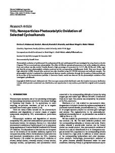
Results TiO2 nanowires characterisation The morphological properties of TiO2-NWs were studied using TEM and XRD. They were accompanied by the comparison with the commercially available, nanocrystalline form of TiO2, Degussa P25, a mixture of anatase and rutile. Figure 1 presents typical TEM images of both nano-TiO2 forms: (a) Degussa P25, which is in a form of nanoparticles with the diameter of 26 ± 1 nm, (b) the synthesised nanowires with average diameter of 35 ± 5 nm and length of 0.5– 1.0 mm. The summary of properties of the studied nano-TiO2 forms is presented in Table I. To verify the form of the synthesised TiO2-NWs, the XRD measurements were performed (Figure 2). The obtained nanowires showed distinct XRD pattern as compared with
Nanotoxicity of TiO2-based nanofilaments A
B
Nanotoxicology Downloaded from informahealthcare.com by 128.178.179.135 on 10/13/11 For personal use only.
Figure 1. TEM images of nano-TiO2 particles: (A) Degussa P25, (B) Na2Ti3O7 nanowires.
those collected for commercially available nano-TiO2 composed of known crystal structure. The way of the synthesis yielded, most probably, nanowires with NaxHyTiO2+d composition as suggested in the original report by Armstrong et al. (2004, 2005). Thus, the synthesised nanowires have the Ti-O skeleton of the layered structure, which consists of TiO6 octahedrons sharing edges and corners. Subsequent washing of the NaxHyTiO2+d materials with hydrochloric acid promotes the complete exchange of Na+ by H+, thus yielding Hx+yTiO2+d nanowires without drastic modification of their morphological characteristics. Further heat treatment of the Hx+yTiO2+d materials leads to the formation of TiO2-NWs with different polymorphism depending on the technology conditions (Armstrong et al. 2004, 2005). Our XRD spectra show no phase transformation from the starting material. Moreover, we observed the peak at about 10 degree, which was identified by Chen et al. (2002) as the (200) peak of the H2Ti3O7. Therefore, we conclude that the synthesised nanowires are titanate rather than anatase TiO2.
function of illumination time of the ESR signal (DMPO-OH spin adduct) represent the efficiency of ROS generation by TiO2 nanoparticles (Degussa P25, here treated as a reference material; blue triangles), and the custommade TiO2-NWs (red dots). The DMPO control (black rhombus) is also shown in the Figure 3A. The insets show the typical ESR spectrum of the paramagnetic product, DMPOOH and photocatalytic degradation of the water-soluble nitroxide radical TEMPOL, respectively. As can be seen in Figure 3B, the TiO2-NWs generated ROS. However, under similar experimental conditions, their ROS generation capacity was markedly smaller when compared with Degussa P25. The ESR measurements revealed photocatalytic properties of custom-made TiO2-NWs showing their ability to . generate at least two types of ROS: hydroxyl (OH ) and/or • superoxide ( O2 ) radicals. However, the efficiency was smaller than that obtained for the commercially available compound (Degussa P25).
Photocatalytic properties of nano-TiO2 The ESR technique offers an ideal method for the study of formation of ROS which are photosynthesised in the presence of light-excited nanoparticles (Hirakawa et al. 2001). Therefore, for the detection of ROS photogenerated in the presence of TiO2-NWs, we applied ESR spin-trapping technique with a spin-trap DMPO, which is known to scavenge . both OH and O2•- radicals. TEMPOL, a stable nitroxide radical and well-known superoxide dismutase (SOD) mimicking agent, was used as a target for ROS. The results of photocatalytic properties of custom-made TiO2-NWs obtained with ESR spin-trapping with DMPO and TEMPOL are shown in Figure 3. The intensity plots as a Table I. Properties of nano-TiO2 forms. Primary particle DLS size Name size (TEM) (mm) Degussa P25 26 nm 3.435 35 nm � 0.8 Na2Ti3O7 NW 0.5–1.0 mm
TiO2 nanowires
R-HD2 - rutile
JA-1 Tayca - anatase
P25 Degussa QY (A.U.)* 0.02714 0.0041
% of QY of P25 100 15.12
*QY is relative quantum yield, that is, photocatalytic efficiency of nano-TiO2 forms. A.U.: arbitrary unit; DLS: dynamic light scattering; NW: nanowire; TEM: transmission electron microscopy; TiO2: titanium dioxide.
10
20
30
40
50
60
70
80
2 theta [deg] Figure 2. XRD patterns of commercially available nano-TiO2 (R-HD2 Huntsman – rutile, JA-1 Tayca – anatase and Degussa P25) that were compared with the spectrum obtained for TiO2-based custom-made nanowires.
K. Pierzchała et al. 20 G 0.008
P25 Time
0.007
ESR intensity [A.U.]
0.006
UV light
A
0.003
0.002 Nanowires 0.001 Control
Nanotoxicology Downloaded from informahealthcare.com by 128.178.179.135 on 10/13/11 For personal use only.
0.000 0
50
100
150
200
250
Illumination time [sec]
Control B
20 G
P25
1.2
UV light
Nanowires
ESR signal intensity I\I0
1.0
0.8
0.6
0.4
0.2
0.0 0 20
300
600
900
1200
1500
1800
2100
Illumination time [sec] Figure 3. (A) The ESR-derived efficiency of the photoproduction of ROS for three types of nano-TiO2: (i) Degussa P25; (ii) TiO2 nanowires (Na2Ti3O7); (iii) control: the ESR signal intensity of DMPO-OH as a function of illumination time in the absence of nanomaterials. All nano-TiO2 materials were suspended in PBS buffer at the concentration of 32 mg/100 ml and 50 mM of DMPO and illuminated with UVA light. Inset: typical ESR trace of the paramagnetic spin adduct, DMPO-OH. (B) Photocatalytic degradation of TEMPOL (50 mM) in the presence of custom-made sodiumcontaining TiO2 nanowires and Degussa P25. Control: the ESR signal intensity of TEMPOL as a function of illumination time in the absence of nanomaterials. Inset: typical ESR trace of decay of TEMPOL.
Cell survival It has been known that different types of TiO2 are toxic for living matter if the large concentration is applied (Sayes et al. 2006). Thus, in our studies as a first step, the cell viability test was performed in order to estimate the cytotoxic action of the studied materials: Na2Ti3O7 nanowires and Degussa P25 nanoparticles (Figure 4). The results clearly showed different cell survival rate depending on the type of nanoTiO2 applied to living cells after 230 s of exposure to UVA light. The fraction of surviving cells in the controls (nontreated cells in culture medium without exposure to UVA
light and to nano-TiO2) was 90%, which corresponds to the natural (normal) level of dead cells in population. Once TiO2-NWs were added and UVA exposure was performed, the certain fraction of cells was killed. From all studied nanomaterials, the most toxic appeared in Degussa P25, for which 23% of cells were killed (C). In the presence of the custom-made Na2Ti3O7 nanowires, the fraction of viable cells decreased by ca. 4% (B). These results indicate proportionality between the efficiency of the ROS generation by nano-TiO2, measured by ERS in vitro, and the observed cytotoxic effect (Degussa P25 > Na2Ti3O7
Nanotoxicity of TiO2-based nanofilaments A
100
Lu 1205 control (A)
80
P 25
Nanowires
40
Elasticity of living cells exposed to oxidative stress Lu 1205 exposed nanowires (B) C
20
Nanotoxicology Downloaded from informahealthcare.com by 128.178.179.135 on 10/13/11 For personal use only.
melanoma cells are nicely spread on a glass coverslip while the illumination of UVA (460 s) and the presence of nano-TiO2 change their morphology. Cells tend to shrink their volume what was accompanied by the presence of blebs at the edges. Similar behaviour was observed in the presence of Degussa P25 (data not shown). Thus, one can clearly conclude that the photo-oxidative stress induces large and fast changes in the organisation of actin filaments.
60 Control
Viability [%]
B
0 Melanoma Lu 1205 cells
Lu 1205 exposed to P25 (C)
Figure 4. Viability test used for the evaluation of toxic effects of nanocrystalline titanium dioxide on 1205Lu cells exposed for 230 s to UVA light (left panel). The visualisation of dead cells, performed using propidium iodine staining of cell nucleus, is presented in right panel: (A) untreated cells, (B) those incubated with Na2Ti3O7 nanowires and (C) Degussa P25.
nanowires) and the biological effect (Degussa P25 > Na2Ti3O7 nanowires).
Morphology of melanoma cells after photo-oxidative exposure In culture conditions, without any applied external stress, melanoma 1205Lu cells form semi-confluent layer composed of nicely spread, flat cells. The phalloidin–FITC staining of actin reveals well-organised fibres (Figure 5, upper panel “1205Lu control”). They were attributed to stress fibres, which consist of a bunch of single actin filaments organised mostly in parallel lines (a diameter of a single actin filaments is around 10 nm) (Milligan et al. 1990). After 30 min of incubation with nano-TiO2 in the dark, the organisation of actin filaments remained similar to untreated cells (Figure 5, upper row of the middle panel “Nanowires no UVA” and upper row of the lower panel “P25 no UVA”). Once UVA was applied to the cells to generate the photo-oxidative stress under the exposure of 230 s, regardless of the type of the nano-TiO2, the quasi-parallel organisation of stress fibres was lost (Figure 5, lower row of the middle panel “Nanowires + UVA” and lower row of the lower panel “P25 + UVA”). The diameter of the observed fibres was in the range of 50– 660 nm as determined by the AFM topography (Figure 6), taking into account the convolution effect of sample topography and tip shape (Engel et al. 1997). The larger exposure time of 460 s (applied prior to the elasticity measurements) induced heavy changes in cell morphology, which precluded imaging. The analysis of cell shape using optical microscopy confirmed the morphology change revealed by phalloidin–FITC staining (Figure 7). The different distribution of actin filaments was accompanied by distinct shape of cell undergoing oxidative stress induced by the presence of TiO2-NWs. Non-treated
In our previous work, we have reported that cell damage induced by ROS can be quantitatively estimated via the Young’s modulus value (Vileno et al. 2006). It has been shown that AFM could be used as a potential marker of cellular toxicity, which was sensitive to damages occurring at a single-cell level. Figure 8B presents the relative change of the cell stiffness as a function of UVA exposure time, calculated for all studied nano-TiO2. For non-treated cells, that is, control (Figure 8A, a), the Young’s modulus value equalled to 1.02 ± 0.20 kPa. To quantify the relative change in cell stiffness all data were normalised to this value. Melanoma cells, incubated for 30 min with the corresponding nano-TiO2 (either with Degussa P25 nanoparticles or Na2Ti3O7 nanowires) in the dark, appeared more rigid as compared with the control cells. The effect was more pronounced for nanowires then for Degussa P25 nanoparticles. The Young’s modulus value raised by ~75% and 50% for Na2Ti3O7 nanowires and Degussa P25 nanoparticles, correspondingly. This can be explained by the distinct rate of nanoTiO2 uptake and their possible presence in the cell interior after 30 min of the incubation. The growth of the elasticity modulus can also be associated with modification of the rate and extent of microtubule assembly/disassembly process in the presence of nano-TiO2 (Gheshlaghi et. al. 2008). When UVA irradiation was applied to induce the generation of ROS, the Young’s modulus for cells treated with custom-made nanowires dropped significantly only after 460 s of irradiation. Shorter exposure resulted in no sufficient ROS production that could induce the cell softening, that is, the Young’s modulus remained at the level of the cells incubated with nanowires in the dark. The effect of Degussa P25 on melanoma cells was quite different. Up to 240 s of UVA illumination, the cell stiffness increased almost two times as compared with the control. Then, after further exposure (460 s), it dropped significantly. A 4.5-fold decrease was observed as compared with the value obtained for the cells incubated with nano-TiO2 without UVA illumination (Figure 8B, a). In parallel, the effect of UVA alone has been also observed (Figure 8A). However, the UVA influence on melanoma cells was smaller (~13% of the Young’s modulus decrease) than the effect of UVA light and the presence of Degussa P25 (~80%).
Discussion One of the materials selected for this study were anatasebased TiO2-NWs. The motivation for evaluating the photocatalytic and toxic and phototoxic properties of this type of nano-TiO2 is driven by an increasing interest in this novel class of nanoparticles, combining a high degree of
K. Pierzchała et al.
Lu 1205 control 50 µm
50 µm
50 µm
50 µm
50 µm
50 µm
50 µm
50 µm
50 µm
50 µm
50 µm
50 µm
50 µm 50 µm
50 µm
50 µm
Nanowires no UVA
P25 no UVA
P25 + UVA
Figure 5. The fluorescent images of the metastatic melanoma 1205Lu cells stained with phalloidin–FITC after exposure to the photo-oxidative stress for 230 s in the presence of TiO2 nanowires and P25. From the top to the bottom are shown: non-treated cells (control), cells after 0.5 h incubation with nano-TiO2 in the dark and cells exposed to the photo-oxidative stress for 230 s, for Na2Ti3O7 nanowires and P25, respectively.
Lu 1205 cells 15×15 [µm]
5×5 [µm]
4 µm 2
0
0 10
0
10
µm
20
30
20
30
0
2.5
5
7.5 µm
10
12.5
15
2.5
5
7.5 µm
10
12.5
15
15
0
0
1
2
0
1
2
µm
3
4
5
3
4
5
µm
0
0
2.5
1
10
5
2
µm
µm 7.5
20
3
10
4
12.5
30
B
5
0
2.5
1
10
5
µm
µm 7.5
20
3
10
30
12.5
A
5
15
35×35 [µm]
0
Nanotoxicology Downloaded from informahealthcare.com by 128.178.179.135 on 10/13/11 For personal use only.
Nanowires + UVA
µm
0
µm
Figure 6. The AFM images of the metastatic melanoma 1205Lu cells. (A) Non-treated cells (control); (B) cells after 0.5 h incubation with TiO2 nanowires, which was then followed by 230 s exposure to UVA light (1 mW/cm2). The panels from the left to the right show decreasingly smaller scanned cell surface areas, that is, 35 � 35 mm, 15 � 15 mm and 5 � 5 mm.
Nanotoxicity of TiO2-based nanofilaments
A
B
Figure 7. The optical images of the metastatic melanoma 1205Lu cells line: (A) non-treated cells (control) and (B) cells exposed to the photo-oxidative stress for 460 s after 0.5 h incubation with TiO2 nanowires (UVA light exposure, 1 W/cm2). Scale bar 25 mm.
Lu 1205 cells 3.5
B
Control + UVA
3.5
3.0
3.0
2.5
2.5
2.0
2.0
E/E0
A
E/E0
Nanotoxicology Downloaded from informahealthcare.com by 128.178.179.135 on 10/13/11 For personal use only.
crystallinity with a high aspect ratio. Anatase-based TiO2NWs are considered as ideal candidates for future efficient photocatalytic and electronic materials, with potential applications in advanced oxidation processes (AOPs), biosensing and photovoltaics. Thus, the study was focused on photocatalytical and toxic properties of custom-made anatasebased TiO2-NWs, with a diameter of 35 nm and length of 0.5–1.0 mm. The obtained results were compared with the well-known photocatalyst, Degussa P25 (80% anatase and 20% rutile, with an average grain size of 26 nm). The ESR spin-trapping detection of ROS with DMPO performed for anatase-based TiO2-NWs revealed a marked photocatalytic . generation of two types of radicals, OH and O2•- . However, the efficiency of ROS production was substantially weaker (reaching only 20%) than that of Degussa P25. The concentrations of photoproduced ROS in the presence of nanowires selected for this study were sufficiently high to cause adverse effects on living melanoma cells. In addition to spectroscopic studies of ROS formation in the presence nanoTiO2, cell viability tests were performed. Staining of cells illuminated with UVA (230 s, 1.0 mW/cm2) with PI, which detects necrotic cells. It was found that Degussa P25 had higher phototoxic potential than the anatase-based TiO2-NWs. This was also observed in cell morphology changes, such as the occurrence of membrane blebs and alterations in cell cytoskeleton. The presence of membrane blebs observed using both optical and AFM microscopy might be explained by the altered expression of the actin-binding family of proteins, as suggested by Cunningham (1995) and Cunningham et al. (1992). It is accepted that the loss of this abundant cytoplasmic protein indicates initiation of apoptosis (Kwiatkowski 1999). Its lower expression has also been associated with reduced cellular stiffness (Gardel et al. 2006). The incidence of membrane blebs was accompanied by disappearance of the actin cortex stress fibres. These alterations were noticeable on the cell surface already after 230 s,
Lu 1205 exposed to nanowires and UVA
Lu 1205 control
1.5
1.5
1.0
1.0
0.5
0.5
0.0
a
b
c
d
0.0
P25 Nanowires
a
b
c
d
Figure 8. Relative change of the Young’s modulus of melanoma 1205Lu cells determined from AFM measurements (all results were normalised to the value obtained for control cells, 1.02 ± 0.2 kPa). (A) Control experiments in the absence of nano-TiO2: non-treated cells without nano-TiO2 (a); cells after the irradiation with UVA only 60 s (b), 240 s (c) and 460 s (d). (B) Exposure to nano-TiO2: cells after 30 min of the incubation in the dark without UVA (a); cells after 0.5 h incubation with Na2Ti3O7 nanowires and P25 in the dark and then exposed to UVA for 60 s (b), 240 s (c) and 460 s (d).
Nanotoxicology Downloaded from informahealthcare.com by 128.178.179.135 on 10/13/11 For personal use only.
K. Pierzchała et al. which was visualised using AFM and fluorescent microscopy. Images of selectively stained actin showed features that corroborated with those observed in AFM topography. The AFM elasticity measurements detect early changes in cytoskeleton organisation in living 1205Lu melanoma cells exposed to external stimuli (Lekka et al. 1999, 2001). It is accepted that for small indentations (up to 500–1000 nm), the actin filaments are mostly responsible for the mechanical properties of cells (Wakatsuki et al. 2001). The AFM elasticity measurements performed in this work revealed that the Young’s modulus values increased for cells incubated for 0.5 h with nano-TiO2 in the dark. The observed increase in cellular stiffness was of 50–115%. For cells incubated for 0.5 h with nano-TiO2 in the dark, and subsequently exposed to UVA for a short time (60 and 240 s), the observed Young’s modulus values were similar to those measured for cells exposed to nano-TiO2 in the dark. Thus, in this work, the evolution of cell stiffness was found to be similar to that observed for glioblastoma cells incubated with low concentrations (147 mM) of fullerols in the dark (Vileno et al. 2006). Similar increase of the cell stiffness was also observed by Lu et al. (2008) for blebbistatin-treated cells. The observed increase of the Young’s modulus values for cells incubated with nano-TiO2 in the dark and followed by short time exposures to UVA light are intriguing. The increase in cellular stiffness could be associated with mechanisms related to early apoptosis. In particular, during this transient period, the nucleus and microtubules govern an increase in the cortical Young’s modulus (Pelling et al. 2009). In contrast to the above results, the cell stiffness dramatically decreased for longer exposures to UVA (460 s). The mechanical dynamics of apoptotic/necrotic cells are highly controlled by the cyto-architecture. The actin network appears to govern the cellular stiffness and the cortical Young’s modulus measured by AFM. As the cell continues through the apoptotic progression, the Young’s modulus values decrease, as the internal cyto-architecture is broken down. The ability of different particles to penetrate cells can explain some differences in their toxicity (Limbach et al. 2005). The most likely mechanism for cellular uptake of particles is endocytosis, foremost pinocytosis, where endocytic vesicles are formed (Singh et al. 2007). The results of the present study may partially be explained by the fact that both nanowires and nanoparticles migrate inside cells using different pathways, and hence are more or less available depending on whether they are in vesicles or free in the cytoplasm. Since cell stiffness reveals properties of the cytoskeleton, the large observed changes may be explained by damages induced by ROS in close proximity to the cell surface. The potential targets for such attack are membrane lipids, transmembrane proteins, or focal adhesion points, which provide anchoring sites for the cytoskeleton (Conlon et al. 2002). The damages induced in these structures would lead to alterations of cell cytoskeleton organisation, and thus, would lead to changes in cell elasticity (Janmey et al. 1995 and Janmey 1998). Our previous results demonstrated the potential of nano-TiO2 for generation of ROS in aqueous suspensions (Vileno et al. 2007). The custom-made TiO2-NWs studied in
this work also revealed a marked ROS generation in the aqueous media. Exposure to various nano-TiO2 and UVA radiation causes pronounced effects to the cell: the actin filamentous network disappears, cells lose their mechanical strength and shape and finally, their structural integrity. The results of this work also pinpoint the damages that occur quickly (e.g., within 240 s) to cells exposed to the photo-oxidative stress in the presence of nano-TiO2. In particular, AFM measurements of cell elasticity and cell surface topography suggested that the initial ROS-mediated deleterious processes could be correlated with changes in the cytoskeleton organisation. These results corroborated with the optical microscopy showing marked changes in cell morphology and also with fluorescent microscopy pointing to changes in the organisation of actin fibre network for cells exposed to the photo-oxidative stress. Overall, the cell morphology changes for cells exposed to photo-oxidative stress were noticeable on ROS generation by nano-TiO2 used in this study, with the most pronounced changes in cell morphology induced by Degussa P25. This work points also to the usefulness of nanoscale scanning tools, like AFM, to detect and quantify mechanisms of toxicity of nanomaterials.
Acknowledgements The authors are grateful to Prof. Piotr Laidler for providing cell cultures and to Dr. Mireille Crittin for technical advices in performing DLS measurements. The study was partially supported by SNSF project nos. 205320-112164 and IZ73Z0_128068/1 (SCOPES).
Declaration of interest The authors report no conflicts of interest. The authors alone are responsible for the content and writing of the paper.
References Armstrong AR, Armstrong J, Canales J, Bruce PG. 2004. TiO2-B nanowires. Angew Chem Int Ed 43:2286–2288. Armstrong G, Armstrong AR, Canales J, Bruce PG. 2005. Nanotubes with the TiO2-B structures. Chem Commun 19:2454–2456. Blake DM, Maness PC, Huang Z, Wolfrum EJ, Huang J. 1999. Application of the photocatalytic chemistry of titanium dioxide to disinfection and the killing of cancer cells. Sep Purif Methods 28:1–50. Braydich-Stolle LK, Schaeublin NM, Murdock RC, Jiang J, Biswas P, Schlager JJ, Hussain SM. 2009. Crystal structure mediates mode of cell death in TiO2 nanotoxicity. J Nanopart Res 11:1361–1374. Cai R, Hashimoto K, Itoh K, Kubota Y, Fujishima A. 1991. Photokilling of malignant cells with ultrafine titanium dioxide powder. Bull Chem Soc Jpn 64:1268–1273. Chen Q, Du GG, Zhang S, Peng LM. 2002. The structure of trititanate nanotubes. Acta Crystallogr B 58:587. Chen XD, Wang Z, Liao ZF, Mai YL, Zhang MQ. 2007. Roles of anatase and rutile TiO2 nanoparticles in photooxidation of polyurethane. Polymer Test 26:202–208. Colton CA, Gilbert DL. 1987. Production of superoxide anions by a CNS macrophage, the microglia. FEBS Lett 223:284–288. Conlon KA, Rosenquist T, Berrios M. 2002. Site-directed photochemical disruption of the actin cytoskeleton by actin-binding Rose Bengal-conjugates. J Photochem Photobiol B 68:140–146. Cunningham CC. 1995. Actin polymerization and intracellular solvent flow in cell surface blebbing. J Cell Biol 129:1589–1599. Cunninghan CC, Gorlin JB, Kwiatkowski DJ, Hartwig JH, Janmey PA, Byers HR, Stossel TP. 1992. Actin-binding protein requirement for cortical stability and efficient locomotion. Science 255:325–327.
Nanotoxicology Downloaded from informahealthcare.com by 128.178.179.135 on 10/13/11 For personal use only.
Nanotoxicity of TiO2-based nanofilaments Davies MJ. 2005. The oxidative environment and protein damage. Biochim Biophys Acta 1703:93–109. De Lorenzo AJ. 1970. The olfactory neuron and the blood–brain barrier. In: Wolstenholme G, Knight J. editors. Taste and Smell in Vertebrates London: J&A Churchill. pp. 151–176. Engel A, Schoenenberger CA, Müller DJ. 1997. High resolution imaging of native biological sample surfaces using scanning probe microscopy. Curr Opin Struct Biol 7:279–284. Fujishima A, Honda K. 1972. Electrochemical Photolysis of water at a semiconductor electrode. Nature 238:37–38. Fujishima A, Rao TN, Tryk DA. 2000. Titanium dioxide photocatalysis. J Photochem Photobiol C Photochem Rev 1:1–21. Gardel ML, Nakamura F, Hartwig JH, Crocker JC, Stossel TP, Weitz DA. 2006. Prestressed F-actin networks cross-linked by hinged filamins replicate mechanical properties of cells. PNAS 103:1762–1767. Gheshlaghi ZN, Gholam HR, Shahin A, Mahmoud G, Roya M. 2008. Toxicity and interaction of titanium dioxide nanoparticles with microtubule protein. Acta Biochim Biophys Sin 40(9):777–782. Gogniat G, Thyssen M, Denis M, Pulgarin C, Dukan S. 2006. The bactericidal effect of TiO2 photocatalysis involves adsorption onto catalyst and the loss of membrane integrity. FEMS Microbiol Lett 258:18–24. Hirakawa T, Kominami H, Ohtani B, Nosaka Y. 2001. Mechanism of photocatalytic production of active oxygens on highly crystalline TiO2 particles by means of chemiluminescence probing and ESR spectroscopy. J Phys Chem B 105:6993–6999. Huang NP, Xu MH, Yuan CW, Yu RR. 1997. The study of the photokilling effect and mechanism of ultrafine TiO2 particles on U937 cells. J Photochem Photobiol A Chem 108:229–233. Janmey P, Chaponnier Ch. 1995. Medical aspects of the actin cytoskeleton. Curr Opin Cell Biol 7:111–117. Janmey P. 1998. The cytoskeleton and cell signaling: component localisation and mechanical coupling. Physiol Rev 78:763–781. Juhasz I, Albelda SM, Elder DE, Murphy GF, Adachi K, Herlyn D, et al. 1993. Regulation of extracellular matrix proteins and integrin cell substratum adhesion receptors on epithelium during cutaneous human wound healing in vivo. Am J Pathol 143:528. Kamat PV. 2008. Quantum Dot Solar Cells. Semiconductor Nanocrystals as Light Harvesters. J Phys Chem C 112, 18737–18753. Kolmakov A, Moskovits M. 2004. Chemical Sensing and catalysis by One-dimensional metal-oxide nanostructures. Annu Rev Mater Res 34:151–180. Kuang D, Brillet J, Chen P, Takata M, Uchida S, Miura H, et al. 2008. Application of highly ordered TiO2 nanotube arrays in flexible dye-sensitized solar cells. ACS Nano 2:1113–116. Kwiatkowski DJ. 1999. Function of gelsolin: motility, signaling, apoptosis, cancer. Cur Opin Cell Biol 11:103–108. Lekka M, Laidler P, Gil D, Lekki J, Stachura Z, Hrynkiewicz AZ. 1999. Elasticity of normal and cancerous human bladder cells studied by scanning force microscopy. Eur Biophys J 28:312–316. Lekka M, Laidler P, Ignacak J, Labedz M, Lekki J, Struszczyk H, et al. 2001. The effect of chitosan on stiffness and glycolytic activity of human bladder cells. Biochim Biophys Acta Mol Cell Res 1540 (2):127–136. Liao DL, Badour CA, Liao BQ. 2008. Preparation of nanosized TiO2/ZnO composite catalyst and its photocatalytic activity for degradation of methyl orange. J Photochem Photobiol A Chem 194:11–19. Limbach LK, Li Y, Grass RN, Brunner TJ, Hintermann MA, Muller M, et al. 2005. Nanoparticle uptake in human lung fibroblasts: effects of particle size, agglomeration, and diffusion at low concentrations. Environ Sci Technol 39:9370–9376. Lin H, Kumon S, Kozuka H, Yoko T. 1998. Electrical properties of sol– gel-derived transparent titania films doped with ruthenium and tantalum. Thin Solid Films 315:266–272. Long TC, Saleh N, Tilton RD, Lowry G, Veronesi B. 2006. Titanium dioxide (P25) produces reactive oxygen species in immortalized brain microglia (BV2): implications for nanoparticle neurotoxicity. Environ Sci Technol 40:4346–4352. Lu L, Oswald SJ, Ngu H, Yin FC-P. 2008. Mechanical properties of actin stress fibers in living cells. Biophys J 95(12):6060–6071. Macak JM, Zlamal M, Krysa J, Schmuki P. 2007. Self-organized TiO2 nanotube layers as highly efficient photocatalysts. Small 3:300–304. Magrez A, Horvath L, Smajda R, Salicio V, Pasquier N, Forro L, Schwaller B. 2009. Cellular toxicity of TiO2-based nanofilaments. ACS Nano 3(8):2274–2280.
Milligan RA, Whittaker M, Safer D. 1990. Molecular structure of F-actin and location of surface binding sites. Nature 348:217–221. Mills A, Wang J. 1999a. The kinetics of semiconductor photocatalysis; light intensity effects. Z Phys Chem 213:49–58. Mills A, Wang J. 1999b. Photobleaching of methylene Blue sensitised by TiO2: an ambiguous system? Photochem Photobiol A Chem 127:123–134. Mitsuyasu W, Mitsushi O, Yuichiro K, Yoko T, Masato N, Toshihiko S, Yoshiharu A. 2002. Differences in the effects of fibrous and particulate titanium dioxide on alveolar macrophages of fischer 344 rat. J Toxicol Environ Health Part A 65:1047–1060. Mor GK, Carvalho MA, Varghese OK, Pishko MV, Grimes CA. 2004. A room-temperature TiO2-nanotube hydrogen sensor able to selfclean photoactively from environmental contamination. J Mater Res 19:628–634. Oberdörster G, Ferin J, Gelein R, Soderholm SC, Finkelstein J. 1992. Role of the alveolar macrophage in lung injury: studies with ultrafine particles. Environ Health Perspect 97:193–199. Oberdörster G, Sharp Z, Atudorei V, Elder A, Gelein R, Kreyling W, Cox C. 2004. Translocation of inhaled ultrafine particles to the brain. Inhal Toxicol 16:437–445. Park J, Bauer S, von der Mark K, Schmuki P. 2007. Nanosize and vitality: TiO2 nanotube diameter directs cell fate. Nano Lett 7:16860–1691. Pelling AE, Veraitch FS, Pui-Kei Chu C, Mason Ch, Horton MA. 2009. Mechanical dynamics of single cells during early apoptosis. Cell Motil Cytoskeleton 66:409–422. Redmond RW, Kochevar IE. 2006. Spatially resolved cellular responses to singlet oxygen. Photochem Photobiol B 82:1178–1186. Rengifo-Herrera JA, Pierzchała K, Sienkiewicz A, Forró L, Kiwi J, Moser JE, Pulgarin C. 2010. Synthesis, characterization, and photocatalytic activities of nanoparticulate N, S-Codoped TiO2 having different Surface-to-Volume Ratios. J Phys Chem C 114:2717–2723. Robichaud CO, Uyar AE, Darby MR, Zucker LG, Wiesner MR. 2009. Estimates of upper bounds and trends in Nano-TiO2 production as a basis for exposure assessment. Environ Sci Technol 43(12):4227–4223. Roy SC, Paulose M, Grimes CA. 2007. The effect of TiO2 nanotubes in the enhancement of blood clotting for the control of hemorrhage. Biomaterials 28:4667–4672. Ruiz A, Arbiol J, Cirera A, Cornet A, Morante JR. 2002. Surface activation by Pt-nanoclusters on titania for gas sensing applications. Mater Sci Eng C 19:105–109. Saito T, Iwase T, Horie J, Morioka T. 1992. Mode of photocatalytic bactericidal action of powdered semiconductor TiO2 on mutans streptococci. J Photochem Photobiol B 14:369–379. Sakai H, Ito E, Cai RX, Yoshioka T, Kubota Y, Hashimoto K, Fujishima A. 1994. Intracellular Ca2+ concentration change of T24 cell under irradiation in the presence of TiO2 ultrafine particles. Biochim Biophys Acta 1201:259–265. Sayes CM, Wahi R, Kurian PA, Liu Y, West JL, Ausman KD, et al. 2006. Correlating nanoscale titania structure with toxicity: a cytotoxicity and inflammatory response study with human dermal fibroblasts and human lung epithelial cells. Toxicol Sci 92:174–185. Singh S, Shi T, Duffin R, Albrecht C, van Berlo D, Hohr D, et al. 2007. Endocytosis, oxidative stress and IL-8 expression in human lung epithelial cells upon treatment with fine and ultrafine TiO2: role of the specific surface area and of surface methylation of the particles. Toxicol Appl Pharmacol 222:141–151. Smalley KSM, Haass NK, Brafford PA, Lioni M, Flaherty KT, Herlyn M. 2006. Multiple signaling pathways must be targeted to overcome drug resistance in cell lines derived from melanoma metastases. Mol Cancer Ther 5(5):1136–1144. Sohaebuddin SK, Thevenot PT, Baker D, Eaton JW, Tang L. 2010. Nanomaterial cytotoxicity is composition, size, and cell type dependent. Part Fibre Toxicol 7:22. Sopyan I, Watanabe M, Murasawa S, Hashimoto K, Fujishima A. 1996. An efficient TiO2 thin-film photocatalyst: photocatalytic properties in gas-phase acetaldehyde degradation. J Photochem Photobiol A Chem 98:79–86. Tachikawa T, Fujitsuka M, Majima TJ. 2007. Mechanistic insight into the TiO2 photocatalystic reactions: design of new photocatalysts. J Phys Chem C 111:5259–5275. Thevenot P, Cho J, Wavhal D, Timmons RB, Tang L. 2008. Surface chemistry influences cancer killing effect of TiO2 nanoparticles. Nanomedicine Nanotechnol Biol Med 4:226–236. Verma S, Watt GM, Mai Z, Hasan T. 2007. Strategies for enhanced photodynamic therapy effects. Photochem Photobiol 83:996–1005.
K. Pierzchała et al. Watson SS, Beydoun D, Scott JA, Amal R. 2003. The effect of preparation method on the photoactivity of crystalline titanium dioxide particles. Chem Eng J 95:213–220. Weisenhorn AL, Khorsandi M, Kasas S, Gotzos V, Butt HJ. 1993. Deformation and height anomaly of soft surfaces studied with an AFM. Nanotechnology 4:106–113. Xia T, Kovochich M, Brant J, Hotze M, Sempf J, Oberley T, et al. 2006. Comparison of the abilities of ambient and manufactured nanoparticles to induce cellular toxicity according to an oxidative stress paradigm. Nano Lett 6(8):1794–1807. Xiao-Bo L, Shun-Qing X, Zhi-Ren Z, Schluesener HJ. 2009. Apoptosis induced by titanium dioxide nanoparticles in cultured murine microglia N9 cells. Chin Sci Bull 54(20):3830–3836. Zhang AP, Sun YP. 2004. Photocatalytic killing effect of TiO2 nanoparticles on LS-174-T human colon cancer cells. World J Gastroenterol 10:3191–3193. Zuruzi AS, Kolmakov A, MacDonald NC, Moskovits M. 2006. Highly sensitive gas sensor based on integrated titania nanosponge arrays. Appl Phys Lett 88:102904 p1–3. Zvyagin Andrei V, Zhao X, Gierden A, Sanchez W, Ross JA, Roberts MS. 2008. Imaging of zinc oxide nanoparticle penetration in human skin in vitro and in vivo. J Biomed Opt 13(6):064031–064039.
Nanotoxicology Downloaded from informahealthcare.com by 128.178.179.135 on 10/13/11 For personal use only.
Vileno B, Lekka M, Sienkiewicz A, Jeney S, Stoessel G, Lekki J, et al. 2007. AFM study of early changes in single cell stiffness induced by UV in the presence of nano-TiO2. Environ Sci Technol 41:5149–5153. Vileno B, Marcoux PR, Lekka M, Sienkiewicz A, Fehér T, Forró L. 2006. Spectroscopic and photophysical properties of a highly derivatized C60 fullerol. Adv Funct Mater 16:120–128. Wakatsuki T, Schwab B, Thompson NC, Elson EL. 2001. Effects of cytochalasin D and latrunculin B on mechanical properties of cells. J Cell Sci 114:1025–1036. Wang J, Chen C, Liu Y, Jiao F, Li W, Lao F, et al. 2008. Potential neurological lesion after nasal instillation of TiO2 nanoparticles in the anatase and rutile crystal phases. Toxicol Lett 18:72–80. Warheit DB, Webb TR, Colvin VL, Reed KL, Sayes CM. 2007. Pulmonary bioassay studies with nanoscale and fine-quartz particles in rats: toxicity is not dependent upon particle size but surface characteristic. Toxicol Sci 95:270–280. Warheit DB, Webb TR, Sayes CM, Colvin VL, Reed KL. 2006. Pulmonary instillation studies with nanoscale TiO2 rods and dots in rats: toxicity is not dependent upon particle size and surface area. Toxicol Sci 91:227–236.
All in-text references underlined in blue are linked to publications on ResearchGate, letting you access and read them immediately.