Research Article J. Ginseng Res. Vol. 34, No. 2, 113-121 (2010) DOI:10.5142/jgr.2010.34.2.113
Physicochemical Characterization and NMR Assignments of Ginsenosides Rb1, Rb2, Rc, and Rd Isolated from Panax ginseng Jin-Gyeong Cho1, Min-Kyung Lee1, Jae-Woong Lee2, Hee-Jung Park1, Dae-Young Lee1, Youn-Hyung Lee2, Deok-Chun Yang1, and Nam-In Baek1* 1
Graduate School of Biotechnology and Department of Oriental Medicinal Materials & Processing, Kyung Hee University, Yongin 446-701, Korea 2 Department of Horticultural Biotechnology, Kyung Hee University, Yongin 446-701, Korea The fresh ginseng roots were extracted with aqueous methanol, and the obtained extracts were partitioned using ethyl acetate, n-butanol, and water, successively. The repeated silica gel and octadecyl silica gel column chromatogaraphy for n-butanol fraction afforded four diol ginseng saponins, ginsenosides Rb1, Rb2, Rc, and Rd. The physicochemical, spectroscopic, and chromatographic characteristics of these ginsenosides were measured and compared with those reported in the literature. Some of the peak assignments in previously published 1H- and 13C-nuclear magnetic resonance (NMR) spectra were inaccurate. This study employed two-dimensional NMR experiments, including 1H–1H correlation spectroscopy, heteronuclear single quantum correlation, and heteronuclear multiple bond connectivity, to determine exact peak assignments. Keywords: Diol ginsenoside, Fast atom bombardment/mass spectrometry, High-performance liquid chromatography, Nuclear magnetic resonance
INTRODUCTION Saponin is one of the most important compounds in Panax ginseng C.A. Meyer which exhibit various pharmacological activities. To date, approximately 70 kinds of saponin have been isolated from Panax ginseng. Most of them are protopanaxdiol and protopanaxtriol, which are aglycones of dammarane-type triterpenoids. Only a few ginsenosides, such as ginsenoside Ro, are aglycones of oleanolic acid [1,2]. Identification of ginsenosides is usually performed by nuclear magnetic resonance (NMR) analyses, but several discrepancies and/or inaccuracies exist in the published NMR data. Among ginseng saponins, we previously described the various physicochemical properties of ginsenoside Rg1, measured with standardized methods, and further identified signals in its
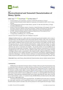
two-dimensional (2-D) NMR spectrum [3]. In the present study, we acquired physicochemical and spectroscopic data from four major diol-saponins, i.e., ginsenosides Rb1, Rb2, Rc, and Rd. The aglycone of ginseng diol-saponins is protopanaxdiol, which is a dammarane moiety hydroxylated via β-linkages to carbon atoms C3, C12, and C20 with a double bond between C24 and C25. In diol-saponins, a sugar group is attached to the hydroxyl groups at C3 and C20. In ginsenosides Rb1, Rb2, Rc, and Rd, sophorose [β-D-glucopyranosyl-(1→2)D-glucopyranose] is β-linked to the hydroxyl group at C3. In ginsenoside Rb1, β-D-glucopyranosyl-(1→6)D-glucopyranose is β-linked to the hydroxyl group at C20. α-L-Arabinopyranosyl-(1→6)-D-glucopyranose Received 8 Feb. 2010, Revised 30 Mar. 2010, Accepted 31 Mar. 2010
This is an Open Access article distributed under the terms of the Creative Commons Attribution Non-Commercial License (http://creativecommons.org/licenses/by-nc/3.0/) which permits unrestricted non-commercial use, distribution, and reproduction in any medium, provided the original work is properly cited.
ⓒ The Korean Society of Ginseng
*
Corresponding author E-mail:
[email protected] Tel: +82-31-201-2661, Fax: +82-031-201-2157
113
http://ginsengres.org pISSN: 1226-8453 eISSN: 2093-4947
J. Ginseng Res. Vol. 34, No. 2, 113-121 (2010)
is β-linked to the hydroxyl group at C20 in ginsenoside Rb2, and α-L-arabinofuranosyl-(1→6)-glucopyranose is β-linked in ginsenoside Rc, and β-D-glucopyranose is β-linked in ginsenoside Rd (Fig. 1). All four of these saponins are bisdesmoides, in which a sugar is attached at C3 and C20. NMR peak assignments for the hydroxyl group and the methyl-linked carbon atom, the olefine carbon atom, and protons linked to individual carbon atoms differ significantly in the literature [4-17]. Although ginsenosides were identified in the 1970s, the lack of 2-D NMR techniques limited the amount of structural information available. As a result, past reports may contain inaccurate peak assignments. In the present study, individual signals were identified using modern 2-D NMR techniques. These included heteronuclear single quantum correlation (HSQC), which provides information related to hydrogen atoms bound to a given carbon atom, and heteronuclear multiple bond connectivity (HMBC), which yields information on neighboring hydrogen and carbon atoms. Melting points, specific rotation, infrared (IR) absorbance, and fast atom bombardment/mass spectrometry (FAB/MS) data were also collected using standardized methods and data were discussed relative to literature values [17–23]. Retention factors (Rf) of each saponin in both normal and reversed-phase thin layer chromatography (TLC) experiments and the standardized retention times of each ginsenoside through a carbohydrate-based high-performance liquid chromatography (HPLC) column are also presented herein.
October 2007. Reagents, instrumentation, and measurement methods Kieselgel 60 was purchased from Merck Co. (Darmstadt, Germany). Kieselgel 60 F254 and RP-18 F254S were used as solid phases in TLC. The former utilized a mobile phase composed of CHCl3-MeOH-H2O (65:35:10); the latter mobile phase consisted of a MeOH-H2O blend (3:1). Detection of substances on the TLC plate was performed by observation under a UV lamp (ENF-240C; Spectronics Corp., New York, NY, USA) or by spraying the developed plate with 10% aqueous H2SO4 followed by heating and observing color development. HPLC was performed at 50°C and 30 psi using an LC-20A (Shimadzu, Kyoto, Japan) equipped with an evaporative light scattering detector (ELSD; Shimadzu). The HPLC analytical column was a Carbohydrate ES (4.6×250 mm, 5 µm; Grace, Deerfield, IL, USA). The column was eluted in a step-wise gradient at a flow rate of 0.8 mL/min using solvent A (acetonitrileH2O-isopropanol=80:5:15) and solvent B (acetonitrileH2O-isopropanol=60:25:15). The elution schedule was as follows: 25% solvent A for 28 minutes, 75% A for 75 minutes, 90% A for 50 minutes, and 100% A for 60 minutes. NMR spectra were recorded on a Varian Inova AS 400 spectrometer (400 MHz; Varian, Palo Alto, CA, USA). Then 0.0625 mol of each ginsenoside (69.3 mg Rb1, 67.4 mg Rb2, 67.4 mg Rc, and 59.1 mg Rd) was dissolved in 0.75 mL (0.083 M) pyridine-d5 and placed in a 5-mm-diameter NMR tube (Norell, Landisville, NJ, USA) with a tetramethylsilane internal-standard adjusted to 0 ppm. IR spectra were measured with an IR spectrometer (599B; Perkin Elmer, Waltham, MA, USA). Two milligrams of each sample was dissolved in 100 uL of MeOH and one drop of the solution was poured onto
MATERIALS AND METHODS Ginseng samples Six-year-old fresh ginseng roots were purchased from the Geumsan ginseng market in Chungnam, Korea in
Fig. 1. Chemical structures of ginsenosides Rb1, Rb2, Rc, and Rd isolated from the roots of Panax ginseng.
DOI:10.5142/jgr.2010.34.2.113
114
Cho et al. NMR Data of Diol Ginsenosides
a CaF2 salt plate (Spectral Systems, Hopewell Junction, NY, USA) and allowed to evaporate. Measurements were performed at room temperature. FAB/MS was carried out with a JMS-700 mass spectrometer (JEOL, Tokyo, Japan) using glycerol as the matrix. Optical rotation was measured with a P-1020 polarimeter (JASCO, Tokyo, Japan) on 10 mg of each ginsenoside, dissolved in MeOH in a 1 mL sample cell at a depth of 1 dm.
eluent (101 L) to obtain 20 fractions (PGB16-1– PGB16-20). PGB16-12 (3.85 g, Ve/Vt=0.62–0.72) was further fractionated over an octadecyl silica gel (ODS) column (φ 4.5×14 cm, MeOH-H2O=3:2, 25 L) to afford 18 additional fractions (PGB16-12-1–PGB16-1218) including ginsenoside Rb2 [PGB16-12-5, 70 mg, Ve/Vt=0.22–0.33, TLC Rf=0.32 (RP-18 F254S, MeOHH2O=3:1), Rf=0.46 (Kieselgel 60 F254, CHCl3-MeOHH2O=65:35:10)] and ginsenoside Rb1 [PGB16-12-10, 70 mg, Ve/Vt=0.5–0.61, TLC Rf=0.40 (RP-18 F254S, MeOH-H2O=3:1), Rf=0.42 (Kieselgel 60 F254, CHCl3MeOH-H 2 O=65:35:10)]. PGB16-7 (370 mg, Ve/ Vt=0.16–0.21) was also separated over an ODS column (φ 4×6 cm, 3:1 MeOH-H2O, 2.4 L) to yield 20 additional fractions (PGB16-7-1–PGB16-7-20) including ginsenoside Rd [PGB16-7-16, 104 mg, Ve/Vt=0.31–0.46, TLC Rf=0.25 (RP-18 F254S, MeOH-H2O=3:1), Rf=0.50 (Kieselgel 60 F254, CHCl3-MeOH-H2O=65:35:10)]. Fraction PGB16-9 (1.7 g, Ve/Vt=0.25–0.29) was also fractionated on an ODS colum (φ 4×6 cm) with 10 L of 3:1 MeOH-H2O to yield 36 fractions (PGB16-9-1– PGB16-9-36) including ginsenoside Rc [PGB16-9-26, 19 mg, Ve/Vt=0.49–0.53, TLC Rf=0.40 (RP-18 F254S, MeOH-H2O=3:1), Rf=0.48 (Kieselgel 60 F254, CHCl3MeOH-H2O=65:35:10)]. Physicochemical and spectroscopic data from each ginsenoside are shown in Tables 1-3.
Isolation of diol ginsenosides Twenty kilograms (fresh weight) of 6-year-old fresh ginseng roots were cut into pieces and extracted with 90% MeOH (50 L) for 24 hours at room temperature. The extracts were passed through filter paper and the residues were extracted twice more with 80% MeOH (6 L). The filtrate was evaporated under reduced pressure at 45°C to yield 2.2 kg of dried extract. The dried extract was partitioned between ethyl acetate (EtOAc, 3 L×3) and H2O (3 L). The remaining H2O layer was extracted again with normal butanol (n-BuOH, 2.8 L×3). Each layer was concentrated under reduced pressure to obtain EtOAc (25 g), n-BuOH (169 g), and H2O fractions. The BuOH extract (n-BuOH fraction of Panax ginseng, PGB) (160 g) was applied to a silica gel column (φ 10×24 cm) and eluted in three steps with a mixture of CHCl3-MeOH-H2O (step 1, 65 L of 10:3:1; step 2, 55 L of 8:3:1; step 3, 30 L of 6:4:1) to yield 24 fractions (PGB1–PGB24). Fractions PGB16 and PGB17 were combined (12 g, Ve/Vt=0.65–0.73, where Ve refers to the volume of eluent for the corresponding fraction and Vt represents the total elution volume). The combined fractions were separated on a silica gel column (φ 7×17 cm) with a CHCl3-MeOH-H2O (65:35:10)
RESULTS AND DISCUSSION The purity of the isolated compounds was over 99% as determined by HPLC and 1H-NMR. Most of the saponins were obtained as white powders. This agrees
Table 1. Physicochemical characteristics for ginsenosides Rb1, Rb2, Rc, and Rd Ginsenoside Rb1
Ginsenoside Rb2
Ginsenoside Rc
Ginsenoside Rd
Crystals
White powder(H2O-MeOH)
White powder (H2O-MeOH)
White powder (H2O-MeOH)
White powder (H2O-MeOH)
Mp (°C)
170-171
181-183
168-169
182-183
[a]D
+6.49° (20°C, c=0.50, MeOH)
+14.08° (20°C, c=0.50, MeOH)
+1.91° (20°C, c=0.50, MeOH)
+18.1° (20°C, c=0.50, MeOH)
IR (cm-1)
3365, 2919, 1590, 1079, 1032
3389, 2942, 1640, 1077, 1029
3367, 2943, 1649, 1078, 1031
3366, 2943, 1647,1077, 1034
FAB/MS (m/z)
1131, 789, 425, 407, 365, 325
1101, 789, 425, 407
1101, 789, 425, 407
969, 789, 425
TLC (Rf)
0.421), 0.402)
0.461), 0.322)
0.481), 0.402)
0.501), 0.252)
HPLC (r.t. min)
54.33)
47.73)
48.13)
36.53)
1)
Kieselgel 60 F254, CHCl3-MeOH-H2O (65:35:10). Kieselgel RP-18 F254S, MeOH-H2O (3:1). 3) Carbohydrate ES (4.6×250 mm, 5 µm), solvent A (acetonitrile-H2O-isopropanol=80:5:15), solvent B (acetonitrile-H2O-isopropanol=60:25:15), gradient elution: The concentration of A was 25 to 50% at 0 to 28 minutes, 50 to 75% at 28 to 47 minutes, 75 to 90% at 47 to 50 minutes, 90 to 100% at 50 to 52 minutes, and 100 to 0% at 52 to 60 minutes. The flow rate was 0.8 mL/min. Mp, melting point; IR, infrared; FAB/MS, fast atom bombardment/mass spectrometry; TLC, thin layer chromatography; HPLC, high-performance liquid chromatography. 2)
115
http://ginsengres.org
J. Ginseng Res. Vol. 34, No. 2, 113-121 (2010)
Table 2. 1H NMR data for ginsenosides Rb1, Rb2, Rc, and Rd (400 MHz, pyridine-d5, dH) H-No.
Ginsenoside Rb1
Ginsenoside Rb2
Ginsenoside Rc
1
1.511)
0.74, 1.56
0.70
Ginsenoside Rd 0.82, 1.47
2
1.81
1.81, 2.17
1.81, 2.18
1.79, 2.17
3
3.25 dd, 11.6, 4.42)
3.25, dd, 11.2, 3.62)
3.25, dd, 11.6, 4.42)
3.26, dd, 11.6, 3.62)
5
0.67
0.67
0.63
0.67
6
1.47
1.47, 1.36
1.46
1.46. 1.59 1.20, 1.46
7
1.20
1.21, 1.48
1.16, 1.43
9
1.33
1.35
1.33
1.95
11
1.30
1.97, 1.56
1.48, 1.95
1.00, 1.95
12
4.28
4.09
4.1
4.06
13
1.97
1.97
1.98
1.35
15
0.90, 1.97
1.00, 1.56
0.95, 1.48
1.00, 1.54
16
1.82, 2.18
1.36, 1.82
1.33, 1.81
1.35, 1.76
17
2.55
2.53
2.51
2.53
18
0.94
0.95
0.92
0.95
19
0.79
0.81
0.77
0.82
21
1.62
1.61
1.61
1.60
22
1.84, 2.36
1.83, 2.34
1.81, 2.33
1.83, 2.32
23
2.18, 2.55
2.34, 2.53
2.33, 2.51
2.46, 2.23
24
5.30
5.30
5.29
5.24
26
1.59
1.65
1.59
1.60
27
1.64
1.62
1.64
1.60
28
1.25
1.26
1.25
1.27
29
1.08
1.07
1.07
1.09
30
0.94
0.94
0.92
0.95
3-O-glc-1’
4.89 d, 7.62)
4.86 d, 7.62)
4.88 d, 7.22)
4.88 d, 7.22)
2’
4.18
4.13
4.84
4.10
3’
4.12
4.22
4.22
4.25 4.08
4’
4.01
3.97
3.95
5’
3.88
3.85
4.28
3.85
6’
4.42, 4.48
4.18, 4.37
4.53
4.25, 4.40
2’-O-glc-1”
5.33 d, 7.62)
55.29 d, 7.62)
5.35 d, 7.62)
5.31 d, 7.22)
2”
4.01
4.03
4.10
4.02
3”
4.12
4.11
4.15
4.14
4”
4.16
4.23
4.30
4.22
5”
4.12
4.17
3.91
4.16
6”
4.41, 4.49
4.31, 4.50
4.22, 4.46
4.40, 4.42
20-O-glc-1’”
5.08 d, 8.02)
5.07 d, 7.62)
5.11 d, 8.02)
5.14 d, 8.02)
2’”
3.88
3.87
3.94
3.92
3’”
4.28
4.24
3.91
4.26 4.08
4’”
4.09
4.09
4.10
5’”
4.06
3.98
4.10
3.85
6’”
4.42, 4.70
4.19, 4.62
4.08, 4.64
4.42, 4.47
6”’-O-sugar-1””
5.04 d, 8.02)
4.94 d, 6.02)
5.63 d, 7.22)
2””
3.99
4.37
4.21
3””
4.18
4.18
4.77
4””
4.29
4.29
4.73
5””
4.19
3.76, 4.28
4.18, 4.22, 4.45
6””
4.42, 4.49
1)
The signals, the coupling pattern of which was not described, were overlapped each other. 2) Chemical shift, coupling pattern, J in Hz.
DOI:10.5142/jgr.2010.34.2.113
116
Cho et al. NMR Data of Diol Ginsenosides
Table 3. 13C-NMR data for ginsenosides Rb1, Rb2, Rc, and Rd (100 MHz, pyridine-d5, dC) Carbon no.
Ginsenoside Rb1
Ginsenoside Rb2
Ginsenoside Rc
Ginsenoside Rd
1
39.241
39.635
39.256
39.589
2
26.707
27.062
26.722
27.060
3
89.021
89.332
89.006
89.301
4
40.075
40.059
39.771
40.082
5
56.400
56.802
56.438
56.764
6
18.510
18.866
18.540
18.866
7
35.176
35.555
35.199
35.540
8
39.756
40.438
40.090
40.431
9
50.251
50.592
50.243
50.700
10
36.951
37.314
36.973
37.307
11
30.824
31.134
30.862
31.309
12
70.209
70.565
70.299
70.557
13
49.508
49.826
49.477
49.840
14
51.654
51.790
51.472
51.805
15
30.824
31.134
30.862
31.187
16
26.707
27.062
26.722
27.055
17
51.441
52.078
51.699
52.048
18
16.091
16.439
16.371
16.394
19
16.356
16.667
16.091
16.697
20
83.281
83.842
83.372
83.652
21
22.468
22.748
22.460
22.816
22
36.268
36.571
36.230
36.518
23
23.287
23.605
23.264
23.643
24
125.889
126.161
125.964
126.191
25
131.007
131.340
130.969
131.166
26
25.895
26.160
25.880
26.153
27
18.062
18.282
17.979
18.183
28
28.162
28.503
28.193
28.511
29
16.697
17.000
16.712
17.023
30
17.494
17.819
17.478
17.789
3-O-glc-1’
105.082
105.286
105.081
105.362
2’
83.486
83.721
83.440
83.758
3’
77.973
78.397
77.988
78.564
4’
71.505
72.119
72.127
72.013
5’
78.140
78.246
78.307
78.382
6’
62.815
63.088
62.914
63.224
2’-O-glc-1”
105.923
106.203
105.969
106.294
2’’
77.124
77.298
77.131
77.412
3’’
78.337
79.368
79.255
79.542
4’’
71.665
72.043
71.710
72.013
5’’
78.337
78.200
78.132
78.261
6”
62.724
63.232
62.709
63.224
20-O-glc-1’”
98.068
98.363
98.083
98.568
2’”
74.880
75.198
75.069
75.471
3’”
78.337
78.595
78.132
78.648
4’”
71.665
71.998
71.710
72.013
5’”
77.124
76.919
76.578
78.564
6’”
69.746
69.480
68.540
63.080
6”’-O-sugar-1””
105.324
104.717
110.101
2””
75.251
72.392
83.372
3””
78.337
74.333
78.868
4””
71.665
68.752
86.003
5””
79.262
65.749
62.709
6””
62.815
117
http://ginsengres.org
J. Ginseng Res. Vol. 34, No. 2, 113-121 (2010)
with most literature sources in which ginsenosides were obtained as white or colorless powders [4,12,19]. Preliminary experiments showed that more precise and accurate melting points were obtained with the melting point apparatus supplied by Standard Research System as opposed to the Fisher-John unit that had been used previously. As a result, melting points determined in the current study often differed significantly from values found in the literature. The melting point of ginsenoside Rb1 has been reported in the literature anywhere from 168°C to 198°C [4,16,19]. The current study indicated a melting point at the lower end of this range at 170°C to 171°C. Literature values for ginsenoside Rb are 197°C to 199°C [4]; the current study yielded 181°C to 183°C. Liu et al. [12] and Wang et al. [23] state melting points of ginsenoside Rc of 193°C and 201°C, respectively, while the current study found 168°C to 169°C. Dong et al. [4] and Wang et al. [14] reported melting points of 204°C and 209°C, respectively, for ginsenoside Rd, while the current study gave 182°C to 183°C. Significant differences with literature sources were also found with optical rotation. Ginsenoside Rb1 has an optical rotation of +11.5° or +12.42° according to Dong et al. [4] or Sanada et al. [19], respectively, while the current study measured +6.49°. Likewise, the optical rotation of ginsenoside Rb2 has been stated as +15.1° or +28.8° according to Dong et al. [4] or Yahara et al. [20], respectively, while a value of +14.08° was obtained herein. The specific rotation of ginsenoside Rc was measured as +1.91°, similar to the literature value of +2.1 [23]. In the case of ginsenoside Rd, the literature value is +16.8° [4], while the result obtained herein was +18.1°. The hydroxyl groups on these ginsenosides render them marginally soluble in nonpolar solvents like CCl4 or CHCl3. Therefore, each compound was mixed with KBr and compressed under reduced pressure to form a pellet for IR absorbance measurements. However, due to water absorption by the pellet and the accompanying spectral interference, a new method was employed in which the saponin was dissolved in MeOH, cast onto KBr or CaF2 plates, and allowed to evaporate. Preliminary results showed less interference in the latter method. Ginsenosides Rb1, Rb2, Rc, and Rd all exhibited absorption peaks corresponding to the O–H stretch of each hydroxyl group (3365, 3389, 3367, 3366, respectively), C– H stretching (2919, 2942, 2943, 2943), C=C stretching (1590, 1640, 1649, 1647), C–H bending (1079, 1077, 1078, 1077), and C–O bending (1032, 1029, 1031, 1034).
DOI:10.5142/jgr.2010.34.2.113
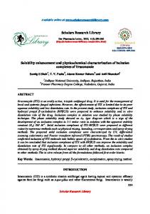
Glycosides like saponin contain multiple hydroxyl groups and exhibit very low volatilities. Thus, mass spectra are usually obtained with FAB/MS. Relative to EI/MS, FAB/MS is a soft ionization method that tends to yield high abundances of molecular ions and relatively smaller proportions of fragment ions. This study employed a positive ionization method. Ginsenoside Rb1 exhibited a molecular ion at m/z 1131 [M+Na]+ and fragment peaks at m/z 789, 425, 407, 365, and 325. The molecular ion of ginsenoside Rb2 was observed at m/ z 1101 [M+Na]+ with fragment peaks at m/z 789, 425, and 407. Ginsenoside Rc revealed m/z 1101 [M+Na]+ as a molecular ion peak and m/z 789, 425, and 407 were observed as fragment ion peaks. Ginsenoside Rd exhibited a molecular ion peak at m/z 969 [M+Na]+ with fragment peaks at m/z 789 and 425. NMR spectra were obtained at 40°C from 0.08 M solutions of each compound dissolved in pyridine-d5. Each spectrum represents the accumulation of 8 scans for 1HNMR and over 1024 scans for 13C-NMR. The chemical name of ginsenoside Rb1 is 3-O-[β-Dglucopyranosyl (1→2)-β-D-glucopyranosyl]-20-O[β-D-glucopyranosyl (1→6)-β-D-glucopyranosyl]3β,12β,20β-trihydroxydammar-24-ene. Since ginsenoside Rb1 contains four attached sugar moieties, it dissolved easily in methanol, pyridine, and dimethyl sulfoxide (DMSO). The few double bonds and many oxygen-linked carbon atoms made pyridine-d5 the better suited solvent for NMR measurements due to less overlap of ginsenosides and solvent-derived signals, although both methanol and DMSO have been used. 13CNMR measured in the former solvent exhibited peaks that were generally shifted to lower magnetic fields relative to those encountered when acquiring spectra in pyridine-d5 [8,16]. The extent of this shift was approximately 1.2 to 1.5 ppm and could be as high as 2.0 ppm. In particular, oxygen-linked carbon atoms C3 and C20 were shifted to lower magnetic fields with chemical shifts of 2.6 and 2.1 ppm, respectively. Among the eight methyl groups, C21 and C28 showed the largest shifts of 5.1 and 3.7 ppm, respectively, as confirmed by HSQC measurements. C21 and C28 were identified as the peaks at 22.468 ppm and at 28.162 ppm, respectively. In contrast, NMR spectra measured in DMSO-d6 were generally shifted to higher magnetic fields by 0.5 to 1.0 ppm [16]. Although C20 was observed at lower magnetic fields than those indicated in the literature when measured in pyridine-d5 [5,9,17], C2’ and C20 gave rise to peaks at δC 83.486 and δC 83.281 ppm, respectively, with C2’ at the lower magnetic field. In this
118
Cho et al. NMR Data of Diol Ginsenosides
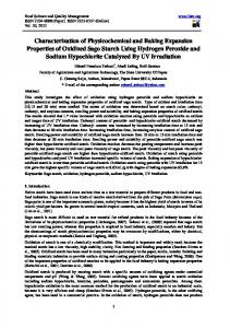
case, since these two carbon atoms were representative of a methine group and a quarternary carbon, respectively, identification was easily made via distortionless enhancement by polarization transfer measurements. Also, the methyl carbon atoms C18 and C19 corresponded to peaks at δC 16.091 and δC 16.356 ppm, respectively. However, the order of the chemical shift differed from that of literature sources [5,7,9,17]. The significant difference in chemical shift between H18 (0.94 ppm) and H19 (0.79 ppm) indicated that the signals at 16.091 and 16.356 ppm, which were correlated with each proton signal in the HSQC spectrum, corresponded to C18 and C19, respectively. In the 1H-NMR spectrum in reference [4], signals corresponding to H26 and H27; H28 and H29 yielded identical chemical shifts. In the current study, the allylic methyl protons H26 and H27 yielded peaks at 1.59 and 1.64 ppm, and H28 and H29 yielded peaks at 1.25 and 1.08 ppm, respectively. This relationship was confirmed by HMBC and HSQC spectra. Among the anomer hydrogen atoms H1’” and H1”” of the sugar moieties, the latter was reported to appear at lower magnetic fields [4]. However, in the current study H1’” and H1”” yielded peaks at 5.08 and 5.04 ppm, respectively, which differed significantly from the chemical shifts cited in the literature. The results of the current study were verified by a J3 correlation between H1’” and C20 (δC 83.281), and between H1”” and C6’” (δC 69.746) in the HMBC spectrum (Fig. 2). The chemical name of ginsenoside Rb2 is 3-O-[β-Dglucopyranosyl (1→2)-β-D-glucopyranosyl]-20-O[β-D-glucopyranosyl(1→6)-α-L-arabinopyranosyl]3β,12β,20β-trihydroxydammar-24-ene. The observed chemical shifts of C18 and C19 in the 13C-NMR spectrum differed from those in reference [17]. The significant difference in the chemical shifts of H18 (0.95 ppm) and H19 (0.81 ppm) indicated that the 13C peaks at 16.439 ppm and 16.667 ppm, which were correlated with both protons in the HSQC spectrum, corresponded to C18 and C19. Although Oura et al. [17] gives 124.80 ppm as the chemical shift of the olefine methine carbon, C24, the current study yielded δC 126.16, a difference of 1.36 ppm. This finding was verified with correlations of C24 between H23 (δH 2.34, 2.53) via J2 as well as among H26 (δH 1.65) and H27 (δH 1.62) via J3 in the HMBC spectrum (Fig. 3). In the 1H-NMR spectrum, the chemical shifts of the methyl protons H21, H26, and H27 were 1.61, 1.65, and 1.62 ppm, respectively, which represents a different order than that cited in reference [6]. However, since the chemical shifts of C21, C26, and C27 were absorbed at very different chemical
Fig. 2. Heteronuclear multiple bond connectivity spectrum of ginsenoside Rb1.
Fig. 3. Heteronuclear multiple bond connectivity spectrum of ginsenoside Rb2.
shifts such as 22.748, 26.160, and 18.282 ppm, respectively, three methyl groups were easily identified using the HSQC experiment. Dong et al. [4] reports the same chemical shifts for the methyl protons H28 and H29. But there was a marked difference between the chemical shifts of C28 (28.503 ppm) and C29 (17.000 ppm), HSQC spectrum indicated the signals observed at 1.26 and 1.07 ppm as H28 and H29, respectively. The chemical name of ginsenoside Rc is 3-O-[β-Dglucopyranosyl (1→2)-β-D-glucopyranosyl]-20-O[β-D-glucopyranosyl(1→6)-α-L-arabinofuranosyl]3β,12β,20β-trihydroxy dammar-24-ene. The methyl protons H21 and H28 were assigned to the signals at 1.61 and 1.25 ppm, respectively, Wang et al. [23], however, reports the opposite assignment. The assignment for H21 reported herein was made using correlations observed among C20 (J2, δC 83.372), C17 (J3, δC 51.699), and C22 (J3, δC 36.230) in the HMBC spectrum. Likewise, the assignment of H28 was based on
119
http://ginsengres.org
J. Ginseng Res. Vol. 34, No. 2, 113-121 (2010)
cluding of HMBC and HSQC, to accurately assign the chemical shifts of each signal. On a normal phase silica gel TLC plate (CHCl3-MeOHH2O=65:35:10), Rf of 0.42, 0.46, 0.48, and 0.50 were observed for ginsenosides Rb1, Rb2, Rc, and Rd, respectively. Reverse-phase ODS TLC (MeOH-H2O=3:1) yielded Rf values of 0.40, 0.32, 0.40, and 0.25, respectively. Although the ginsenosides did not exhibit fluorescence when excited at 254 or 365 nm, the location of each compound was revealed by a light purple color when sprayed with 10% H2SO4 and heating. HPLC retention times were 54.3, 47.7, 48.1, and 36.5 minutes for ginsenosides Rb1, Rb2, Rc, and Rd, respectively, by analysis methods described in Materials and Methods.
Fig. 4. Heteronuclear multiple bond connectivity spectrum of ginsenoside Rc.
ACKNOWLEDGEMENTS
observed correlations among C4 (J2, δC 39.771), C29 (J3, δC 16.712), C3 (J3, δC 89.006), and C5 (J3, δC 56.438) (Fig. 4). The chemical name of ginsenoside Rd is 3-O-[β-Dglucopyranosyl (1→2)-β-D-glucopyranosyl]-20-O-βD-glucopyranosyl-3β, 12β,20β-trihydroxydammar24-ene. NMR peaks corresponding to C18 and C19 were observed at δC 16.394 and δC 16.679, respectively, which differed from the assignments reported in the literature [10,11,14,17]. The significant difference in chemical shift between H18 (0.95 ppm) and H19 (0.82 ppm) indicated that the 13C peaks at 16.394 ppm and 16.697 ppm, which were correlated with both protons in the HSQC spectrum, corresponded to C18 and C19, respectively. The chemical shifts of the methyl carbon atoms C27, C29, and C30 differed from those in the literature [13-15]. The distinct difference in the chemical shifts of H27 (1.60 ppm), H29 (1.09 ppm), and H30 (0.95 ppm) indicated that the 13C peaks at 18.183 ppm, 17.023 ppm, and 17.789 ppm, which were correlated with these protons in the HSQC spectrum, were C27, C29, and C30, respectively. For the tetracyclic triterpene compounds characterized herein, many of the methine and methylene proton signals overlapped at higher magnetic fields. In addition, many of the oxygenated-methine proton signals of the four-sugar glycoside overlapped in the 1H-NMR spectrum. Thus, identification of each signal would be very difficult based solely on one-dimensional NMR techniques. To date, peak assignments in NMR data for these types of materials has been based on previously reported data. However, much of the earlier data may be erroneous due to instrument-resolution limitations. The current study employed 2-D NMR techniques, in-
DOI:10.5142/jgr.2010.34.2.113
This research was supported in part by the grant of the Korean Ginseng Center for Most Valuable Products of Kyung Hee University funded by Korea Institute of Planning & Evaluation for Technology in Food, Agriculture, Forestry & Fisheries (IPET) of Ministry of Agriculture and Forestry, Korea.
REFERENCES 1. Jung HK, Lim SK, Park MJ, Bae CS, Yoon KC, Han HJ, Park SH. The protective effect of ginseng saponin against high glucose-induced secretion of insulin-like growth factor (IGF)-I in primary cultured rabbit proximal tubule cells. J Ginseng Res 2009;33:26-32. 2. Hong HD, Choi SY, Kim YC, Lee YC, Cho CW. Rapid determination of ginsenosides Rb1, Rf, and Rg1 in Korean ginseng using HPLC. J Ginseng Res 2009;33:8-12. 3. Lee DY, Cho JG, Lee MK, Lee JW, Park HJ, Lee YH, Yang DC, Baek NI. Identification of NMR data for ginsenoside Rg1. J Ginseng Res 2008;32:291-299. 4. Dong A, Ye M, Guo H, Zheng J, Guo D. Microbial transformation of ginsenoside Rb1 by Rhizopus stolonifer and Curvularia lunata. Biotechnol Lett 2003;25:339-344. 5. Du Q, Jerz G, Waibel R, Winterhalter P. Isolation of dammarane saponins from Panax notoginseng by highspeed counter-current chromatography. J Chromatogr A 2003;1008:173-180. 6. Karikura M, Miyase T, Tanizawa H, Taniyama T, Takino Y. Studies on absorption, distribution, excretion and metabolism of ginseng saponin. VI. The decomposition products of ginsenoside Rb2 in the stomach of rats. Chem Pharm Bull 1991;39:400-404. 7. Kim YH, Lee YG, Choi KJ, Uchida K, Suzuki Y. Trans-
120
Cho et al. NMR Data of Diol Ginsenosides
glycosylation to ginseng saponins by cyclomaltodextrin glucanotransferases. Biosci Biotechnol Biochem 2001;65:875-883. 8. Zhao X, Wang J, Li J, Fu L, Gao J, Du X, Bi H, Zhou Y, Tai G. Highly selective biotransformation of ginsenoside Rb1 to Rd by the phytopathogenic fungus Cladosporium fulvum (syn. Fulvia fulva). J Ind Microbiol Biotechnol 2009;36:721-726. 9. Yang CR, Kasai R, Zhou J, Tanaka O. Dammarane saponins of leaves and seeds of Panax notoginseng. Phytochemistry 1983;22:1473-1478. 10. Cao XL, Tian Y, Zhang TY, Liu QH, Jia LJ, Ito Y. Separation of dammarane-saponins from notoginseng, root of Panax notoginseng. (Burk.) F. H. Chen, by HSCCC coupled with evaporative light scattering detector. J Liq Chromatogr Relat Technol 2003;26:1579-1591. 11. Cheng LQ, Kim MK, Lee JW, Lee YJ, Yang DC. Conversion of major ginsenoside Rb1 to ginsenoside F2 by Caulobacter leidyia. Biotechnol Lett 2006;28:1121-1127. 12. Liu C, Han J, Duan Y, Huang X, Wang H. Purification and quantification of ginsenoside Rb3 and Rc from crude extracts of caudexes and leaves of Panax notoginseng. Sep Purif Technol 2007;54:198-203. 13. Ma WG, Mizutani M, Malterud KE, Lu SL, Ducrey B, Tahara S. Saponins from the roots of Panax notoginseng. Phytochemistry 1999;52:1133-1139. 14. Wang W, Zhao Y, Rayburn ER, Hill DL, Wang H, Zhang R. In vitro anti-cancer activity and structure-activity relationships of natural products isolated from fruits of Panax ginseng. Cancer Chemother Pharmacol 2007;59:589-601. 15. Zhao X, Wang J, Li J, Fu L, Gao J, Du X, Bi H, Zhou Y, Tai G. Highly selective biotransformation of ginsenoside Rb1 to Rd by the phytopathogenic fungus Cladosporium
fulvum (syn. Fulvia fulva). J Ind Microbiol Biotechnol 2009;36:721-726. 16. Teng R, Li H, Chen J, Wang D, He Y, Yang C. Spectral assignments and reference data. Magn Reson Chem 2002;40:483-488. 17. Oura H, Kumakai A, Shibata S, Takaki K. Chemical constituents of Panax ginseng. In: Panax ginseng: it’s study and progress. Tokyo: Kyoritz Press Ltd., 1981. p. 46-47. 18. Karikura M, Miyase T, Tanizawa H, Taniyama T, Takino Y. Studies on absorption, distribution, excretion and metabolism of ginseng saponins. VII. Comparison of the decomposition modes of ginsenoside-Rb1 and -Rb2 in the digestive tract of rats. Chem Pharm Bull 1991;39:23572361. 19. Sanada S, Kondo N, Shoji J, Tanaka O, Shibata S. Studies on the saponins of ginseng. I. Structures of ginsenoside-Ro, -Rb1, -Rb2, -Rc and -Rd. Chem Pharm Bull 1974;22:421-428. 20. Yahara S, Kaji K, Tanaka O. Further study on dammarane-type saponins of roots, leaves, flower-buds, and fruits of Panax ginseng C.A. Meyer. Chem Pharm Bull 1979;27:88-92. 21. Dong A, Ye M, Guo H, Zheng J, Guo D. Microbial transformation of ginsenoside Rb1 by Rhizopus stolonifer and Curvularia lunata. Biotechnol Lett 2003;25:339-344. 22. Teng R, Ang C, McManus D, Armstrong D, Mau S, Bacic A. Regioselective acylation of ginsenosides by Novozyme 435 to generate molecular diversity. Helv Chim Acta 2004;87:1860-1872. 23. Wang H, Tong YX, Ye WC, Zhao SX. Studies on chemical constituents in roots of Polygala tenuifolia. Zhongguo Zhong Yao Za Zhi 2003;28:828-830.
121
http://ginsengres.org