4821 The Journal of Experimental Biology 209, 4821-4827 Published by The Company of Biologists 2006 doi:10.1242/jeb.02567
Commentary Plasticity and stability in neuronal output via changes in intrinsic excitability: it’s what’s inside that counts David J. Schulz Biological Sciences, University of Missouri–Columbia, Columbia, MO 65211, USA e-mail:
[email protected]
Accepted 2 October 2006 Summary The nervous system faces an extremely difficult task. It must be flexible, both during development and in adult life, so that it can respond to a variety of environmental demands and produce adaptive behavior. At the same time the nervous system must be stable, so that the neural circuits that produce behavior function throughout the lifetime of the animal and that changes produced by learning endure. We are only beginning to understand how neural networks strike a balance between altering individual neurons in the name of plasticity, while maintaining long-term stability in neural system function. The balance of this plasticity and stability in neural
networks undoubtedly plays a critical role in the normal functioning of the nervous system. While mechanisms of synaptic plasticity have garnered extensive study over the past three decades, it is only recently that more attention has been turned to plasticity of intrinsic excitability as a key player in neural network function. This review will focus on this emerging area of research that undoubtedly will contribute a great deal to our understanding of the functionality of the nervous system.
Key words: neuronal excitability, ion channels, plasticity.
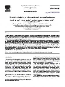
Introduction Intrinsic excitability refers to the propensity of a neuron to fire action potentials when exposed to an input signal, be it presynaptic activity or a direct input from a particular sensory modality. In other words, the intrinsic excitability of neurons is responsible for the translation of synaptic input to the particular output function of a given neuron. Neuronal excitability is directly attributable to the suite of ion channels inserted into the membrane of the cell, as well as the biochemical properties and kinetics of those channels. Because of their fundamental role in generating neuronal output, it is not surprising that alterations in the properties, spatial distribution, or abundance of these ion channels can result in considerable plasticity for neurons and the networks to which they belong. Plasticity in intrinsic excitability may play multiple important roles in the functioning nervous system. For example, increasing the likelihood that an action potential will be triggered by summed synaptic inputs is a de facto mechanism for strengthening connections between cells independent of synaptic strength (Fig.·1A). Thus mechanisms that alter neuronal excitability can lead to plasticity in responses to synaptic stimulation, ultimately affecting processes such as learning and memory and other activitydependent forms of neural plasticity.
Alternatively, alterations in intrinsic excitability also can have a stabilizing influence on neural function. Some neurons may respond to changes in synaptic input or endogenous activity by altering their excitability to maintain a given firing pattern or firing rate (Fig.·1B). For example, a pacemaker neuron that experiences a decrease in synaptic drive may independently increase its own excitability by up-regulating depolarizing conductances in order to maintain a fixed level of firing output. This ‘homeostatic plasticity’ has more recently been implicated as a mechanism of stabilizing neuronal function. Finally, it is possible that changes occur in the intrinsic properties of neurons in networks with disrupted innervation as a result of injury or disease. Such alterations may result in activity-dependent changes in the intrinsic properties of the cells in these networks, which then go on to form functionally different, isolated networks. It is not clear what the long-term effects of changes in excitability are in these networks. However, as our focus for treatments for such problems as spinal cord injury are geared towards re-establishing contact between higher systems and peripheral motor networks, it behooves us to understand how the distal networks have been altered as a result of deprivation of descending inputs. It is these three aspects of plasticity in neuronal excitability
THE JOURNAL OF EXPERIMENTAL BIOLOGY
4822
D. J. Schulz
in mature nervous systems that will be discussed in this review: long-term potentiation and depression of intrinsic excitability, homeostatic plasticity of neuronal excitability, and changes in excitability following perturbation of afferent input. It should be noted that a great deal of plasticity in intrinsic properties is found in the developing nervous system (see Moody and Bosma, 2005). This developmental aspect of the story, while of great importance, is beyond the scope of this review. Learning and long-term potentiation and depression of intrinsic excitability Synaptic plasticity and long-term alteration of synaptic strength have been at the heart of research into the cellular mechanisms of learning and memory for three decades. Yet while synaptic activity is responsible for the conveyance of information between neurons, the ‘decision’ of what to do with that information is the result of integrated activity in the dendrites and soma and ultimately the generation (or not) of action potentials in the spike initiation zone. Therefore, the intrinsic excitability of a neuron is critical for determining the ultimate output of any synaptic input. Thus the question becomes whether action potential generation is a static
A
Potentiation of intrinsic excitability
Potentiating event Period of potentiation
Excitability of post-synaptic neuron Post-synaptic spike frequency Time
B
Compensation of intrinsic excitability
Change in pre-synaptic activity Period of compensation
Excitability of post-synaptic neuron Post-synaptic spike frequency
Time
component of synaptic throughput with no bearing on mechanisms of learning and memory, or whether experiencedependent changes in neuronal activity also may be the result of modification of intrinsic excitability. Some of the best evidence that learning may in part be the result of changes in neuronal excitability come from conditioning paradigms. One of the first examples of plasticity in intrinsic excitability as a function of behavioral training (and presumably via synaptic drive due to this training) was described in the nudibranch mollusk Hermissenda crassicornis. Persistent increases in intrinsic excitability of type B photoreceptors can be elicited via pairing of light and rotation stimuli (Alkon, 1984). This change is due, at least in part, to a decrease of the transient K+ current (IA). Decreases in hyperpolarizing K+ currents such as IA act to increase neuronal excitability. These increases in excitability in Hermissenda photoreceptors persist for days or weeks, thus may have long-term effects on learning and memory. Similar changes in intrinsic excitability are known from other invertebrate conditioning paradigms, such as conditioned pneumostome closure in the snail Helix (Gainutdinov et al., 1998) and conditioned gill/siphon withdrawal in Aplysia (Antonov et al., 2001). While these correlative examples of changes in intrinsic excitability suggest that this mechanism plays a role in learning, it is not clear whether changes in excitability represent a functional portion Fig.·1. Examples of plasticity in intrinsic neuronal excitability. (A) Diagram illustrating the concept of potentiation of intrinsic excitability. The circles represent two different neurons connected by an excitatory synapse (triangle). In the case of potentiation of excitability, one form of plasticity in intrinsic properties of neurons, activity in the pre-synaptic neuron potentiates the connection with the post-synaptic neuron. This potentiation is the result of an increase in the overall excitability of the post-synaptic neuron independent of changes in synaptic strength. One possible mechanism for this change in excitability is a decrease in action potential threshold. The result of this increased excitability is an increase in the input–output function of the postsynaptic cell: more spikes are elicited post-synaptically for the same amount of pre-synaptic input. This results in a de facto increase in synaptic strength mediated by changes in the intrinsic excitability of the postsynaptic neuron. Examples of this kind of plasticity have recently been published (Cudmore and Turrigiano, 2004; Li et al., 2004; Xu et al., 2005). (B) Diagram illustrating the concept of compensatory changes in excitability leading to conserved neuronal output. In this case, the post-synaptic neuron maintains a constant level of spiking activity as a result of compensatory decreases in excitability triggered by increased pre-synaptic activity. The outcome is a constantly maintained level of spiking activity in the postsynaptic cell, which could be the result of an increased action potential threshold, for example. For examples of this kind of plasticity, see recent publications (Aptowicz et al., 2004; Brickley et al., 2001; van Welie et al., 2004).
THE JOURNAL OF EXPERIMENTAL BIOLOGY
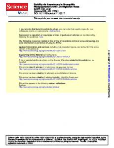
Plasticity in neuronal excitability of the memory trace or simply play a facilitative role in formation of memory. For example, during eye-blink conditioning in the rabbit (Oryctolagus cuniculus), a puff of air that elicits a reflex eye blink is coupled to a neutral stimulus (such as a sound tone) until the neutral stimulus itself is able to elicit the blink reflex. Pyramidal cells in areas CA1 and CA3 of the hippocampus show changes in intrinsic excitability 24·h after this conditioning, including increased spiking in response to current injection and decreases in the amplitude of the afterhyperpolarization evoked by bursts of action potentials (Moyer, Jr et al., 1996; Thompson et al., 1996). Yet after 7 days, while the memory trace of the conditioning is still intact (and indeed can last for months), the changes in excitability recorded after 24·h are no longer detectable (Moyer, Jr et al., 1996). These results suggest, at least in this system, that the increase in excitability does not form part of the memory trace itself but rather may facilitate the formation of a synaptic memory trace by increasing the likelihood of the induction of synaptic long-term potentiation in the hippocampus or elsewhere (Zhang and Linden, 2003). In the past 5 years, many reports of increases in neuronal excitability in response to high frequency synaptic stimulation have emerged. This phenomenon, collectively known as longterm potentiation of intrinsic excitability (LTP-IE), has been reported in cells of layer V visual cortex (Cudmore and Turrigiano, 2004), granule cells (Armano et al., 2000) and deep nuclear neurons of the cerebellum (Aizenman and Linden, 2000), and hippocampal CA1 neurons (Daoudal and Debanne, 2003; Xu et al., 2005), to name a few. Recently long-term depression of intrinsic excitability also has been reported in cultured hippocampal neurons and cells of the somatosensory cortex (Li et al., 2004). The mechanisms involved in altering excitability are as varied as the cell types implicated. These mechanisms include decreases in the magnitude and kinetics of A-type K+ currents (Frick et al., 2004), slowly inactivating K+ currents (Li et al., 2004), calcium-activated K+ currents (Sanchez-Andres and Alkon, 1991) and voltage-gated Na+ currents (Xu et al., 2005), and are thought to act through calcium-mediated activation of phosphorylation pathways (Cudmore and Turrigiano, 2004; Li et al., 2004; Xu et al., 2005). While the discovery of numerous sites and mechanisms of LTP-IE has further implicated this phenomenon as playing a key role in neural plasticity, what are badly needed are investigations of the functional consequences of this plasticity. In addition to the aforementioned conditioning paradigms, one of the best examples of a functional role for regulation of intrinsic neuronal excitability comes from work in the optic tectum of Xenopus tadpoles (see Fig.·2). In freely swimming tadpoles, a persistent visual stimulation regime increases the excitability of neurons in the optic tectum (Aizenman et al., 2003) (Fig.·2A). This increase in excitability is due at least in part to an increase in the peak amplitude of voltage-gated Na+ current (Fig.·2B), and is correlated with (and perhaps initiated by) a subsequent decrease in the synaptic drive onto these cells (Aizenman et al., 2002). The end result is an overall shift in the
4823
input–output relationship in these tectal neurons: tectal neurons with enhanced excitability exhibit enhanced responses to temporally coherent (i.e. bursts of) stimuli (Aizenman et al., 2003). Further, tectal neurons from intact animals subject to these stimulation protocols are more sensitive to subsequent visual stimuli, and yet these cells do not show changes in spontaneous activity (Fig.·2C). These data suggest that this enhancement of neuronal excitability plays a functional role by increasing signal-to-noise ratios for visual stimuli and thereby improve stimulus detection in vivo. Homeostatic plasticity and intrinsic excitability The regulation of intrinsic excitability may exist in parallel to synaptic plasticity and act in concert to control the throughput of synaptic signals. Yet when one considers that neurons are constantly subject to various mechanisms of plasticity and modulation, it becomes clear that in the absence of some form of stabilizing activity cells rapidly would be pushed outside of the parameters for normal neuronal function. The question then becomes, how do cells maintain the flexibility to respond to changes in the environment, yet constrain this flexibility such that functionally relevant output is maintained? Homeostatic processes are widespread throughout all living systems, and the nervous system is no exception. Individual neurons as well as neuronal networks must maintain levels of excitability and connectivity to ensure that consistent functional output is achieved. Evidence for such homeostatic mechanisms in neurons and neural circuits is steadily growing (Davis and Bezprozvanny, 2001; Turrigiano, 1999; Turrigiano and Nelson, 2004). For example, in both mice and Drosophila, the neuromuscular junction can compensate for decreased postsynaptic excitability by increasing presynaptic transmitter release to maintain a normal level of muscle depolarization (Petersen et al., 1997; Sandrock, Jr et al., 1997). Yet it is only more recently that homeostatic mechanisms involved in controlling intrinsic excitability have been investigated in detail. There are two lines of evidence that support the idea that ionic conductances are maintained in a homeostatic fashion to stabilize the excitability of a given neuron: measurements of endogenous membrane conductances, and manipulations that alter the excitability of a neuron. It is often assumed that all neurons of the same cell type that produce similar output have identical intrinsic properties, both within an animal and across individuals. Yet recent theoretical work has argued that similar neuronal activity can be obtained from different combinations of membrane conductances (Golowasch et al., 2002; Liu et al., 1998; Prinz et al., 2003; Prinz et al., 2004). These theoretical studies are supported by recently published work measuring the variability of membrane conductances and the corresponding levels of ion channel expression in single identified neurons of the crab stomatogastric ganglion (Schulz et al., 2006). In lateral pyloric (LP) neurons, three- to fourfold inter-animal variability is seen for three potassium currents measured in two-electrode voltage
THE JOURNAL OF EXPERIMENTAL BIOLOGY
4824
D. J. Schulz Control
A * P>0.02
Average number of spikes
4
3 Visually stimulated 2 20 mV
1
100 ms 0 0
B
40 80 120 160 Injected current (pA)
50 I (pA) Vm (mV) –60 –40 –20 20 40 –50 –100 –150
I (pA) 800 I(K+) trans. + I(K ) sust. 600 I-plateau 400 200
–200 –250 –300
C
Control
10 pA 200 ms
–60 –40 –20 –200
20 40 Vm (mV)
Visually stimulated
20 pA 200 ms
clamp (Fig.·3B), even though the firing pattern of LP neurons is highly conserved from animal to animal (Fig.·3A). These same neurons showed a similar amount of variability in mRNA levels for the ion channels corresponding to these conductances (Fig.·3C), demonstrating that this variability exists at multiple functional levels, and is not simply an artifact of ‘noise’ in voltage clamp recordings (Fig.·3D). Presumably the variance seen in these neurons reflects, in part, the result of experiencedependent plasticity. Therefore, this variability may be a manifestation of compensatory mechanisms that allow nervous systems to maintain stable function over time. There also is a growing body of evidence from experiments that manipulate the excitability of a neuron and look for compensatory shifts in membrane conductances that allow the neuron to maintain stable output properties. In neurons of the crustacean stomatogastric ganglion, overexpression of the channel responsible for a transient K+ current (IA) leads to a
Fig.·2. Visually driven regulation of intrinsic neuronal excitability in Xenopus tectal neurons improves stimulus detection in vivo. (A) Tectal neurons from tadpoles exposed to a visual stimulation protocol show increased excitability in response to injected depolarizing current. Left: input–output relationships in response to square pulse depolarizations of varying amplitudes. Neurons from visually stimulated animals (open triangles; N=25) show significantly more spiking activity in response to injected current than do controls (filled circles; N=20). Right: representative recordings from these neurons showing the differences in response to current injection. (B) Increases in the excitability of tectal neurons are the result of changes in voltagedependent Na+ currents. Na+ currents (left) but not K+ currents (right) show a significant increase in peak amplitude in visually stimulated animals. An increase in these Na+ currents, which are depolarizing and excitatory, would account for the increase in excitability seen in visually stimulated tadpoles. Open symbols represent recordings from visually stimulated neurons (N=46), and closed symbols are controls (N=25). K+ currents: trans, transient; sust, sustained; Ca2+ currents: I-plateau. (C) Increased excitability of tectal neurons improves visual stimulus detection in semi-intact tadpoles. Recordings of visually evoked field potentials were made from the tectum in control and stimulated tadpoles. The recordings shown represent five trials from a control cell, and five trials recorded from a cell in a tadpole previously subjected to the visual stimulation protocol. The neuron from the visually stimulated animal fires more action potentials than the control in response to a subsequently presented light stimulus (open arrow), indicating that the sensitivity to visual stimuli is increased in tadpoles with previous visual stimulation experience. Figure adapted from Aizenman et al. (Aizenman et al., 2003) and reproduced with permission.
drastic increase in the measurable membrane current, but very little effect on the output of the neuron (MacLean et al., 2005; MacLean et al., 2003). This is the result of a concomitant and compensatory increase in the hyperpolarization-activated inward current (IH). Interestingly, this apparent compensation is not a two-way street: overexpression of IH is not accompanied by an increase in IA, and subsequently the output of the neuron is altered (Zhang et al., 2003). A similar compensation is thought to occur for calcium currents in basal forebrain neurons (Etheredge et al., 2005). Mutant mice with decreased levels of Cav2.1 channel function show relatively normal levels of overall calcium current densities due to upregulation of Cav1 calcium channel function. In fact, the ability of neurons to increase their excitability in response to decreased levels of excitation is becoming well-established: voltage gated sodium channels are up-regulated in both hippocampal slice cultures (Aptowicz et al., 2004) and motor neurons of Drosophila in response to activity deprivation (Mee et al., 2004). Cellular mechanisms also exist that decrease neuronal excitability in the face of increased excitatory synaptic drive (van Welie et al., 2004) or decreased inhibitory synaptic activity (Brickley et al., 2001). Taken together, these results suggest that in addition to the many forms of cellular plasticity in the nervous system that allow such processes as learning and memory to take place, there also are mechanisms that stabilize and maintain nervous system function over time. However, we know little about the
THE JOURNAL OF EXPERIMENTAL BIOLOGY
Plasticity in neuronal excitability
4825
neurons and how this may be related to injury and disease processes in the nervous system.
A –50 mV –60 mV –50 mV –60 mV
0.60 0.50 0.40 0.30 0.20 0.10
B
1800 mRNA copy number
Conductances at +15 mV (μS nF–1)
500 ms
mRNA copy number
IKd
IK[Ca]
D
2000 1750 1500 1250 1000 750 500 250
C
1400 1000 600 200
IA
(P