259 Hypertens Res Vol.30 (2007) No.3 p.259-267
Original Article
Postnatal Blocking of Interferon-γ Function Prevented Atherosclerotic Plaque Formation in Apolipoprotein E–Knockout Mice Mitsuhisa KOGA1),2), Hisashi KAI1), Hideo YASUKAWA3), Seiya KATO4), Tomoka YAMAMOTO1),2), Yumiko KAWAI1),2), Ken KUSABA1), Yukihiko SEKI1), Mamiko KAI2), Kensuke EGASHIRA5), Yasufumi KATAOKA2), and Tsutomu IMAIZUMI1) It is unknown whether interferon-γ has a positive or negative impact on atherosclerotic plaque formation. Thus, we examined the effects of postnatal interferon-γ function blocking on plaque formation in apolipoprotein E–knockout (apoEKO) mice by overexpressing a soluble mutant of interferon-γ receptor (sIFNγR), an interferon-γ inhibitory protein. Mice were fed a Western-type diet from 8 weeks of age. sIFNγR or mock plasmid (control) was injected into the thigh muscle at 8 and 10 weeks’ age, because serum sIFNγR protein was transiently increased with a peak at 2 days after a single sIFNγR gene transfer and remained elevated for 2 weeks. At 12 weeks’ age, control apoEKO mice showed marked atherosclerotic plaques from the ascending aorta to the aortic arch. The plaques in the aortic root had massive lipid cores and macrophage infiltration with thin fibrous cap and few smooth muscle cells, demonstrating low plaque stability. In contrast, the luminal plaque area was remarkably reduced in sIFNγR-treated apoEKO mice. sIFNγR treatment not only reduced lipid core areas and macrophage infiltration but also increased smooth muscle cell count and fibrotic area, suggesting improved plaque stability. In controls, interleukin-1β, monocyte chemoattractant protein-1, and vascular cell adhesion molecules-1 were remarkably upregulated in the aortic wall. These changes were significantly reversed by sIFNγR. sIFNγR treatment had no effects on serum cholesterol levels. In conclusion, sIFNγR treatment prevented plaque formation in apoEKO mice by inhibiting inflammatory changes in the arterial wall. The present study provides insight into a new strategy for preventing atherosclerosis. (Hypertens Res 2007; 30: 259–267) Key Words: atherosclerosis, inflammation, cytokine, gene therapy
Introduction Recently, it has been widely accepted that chronic inflamma-
tory changes participate in the generation and progression of atherosclerosis through the activation of pro-inflammatory cytokines and growth factors (1). Among the cytokines, interferon-γ is highlighted as a key factor in the pathogenesis of
From the 1)Department of Internal Medicine, Division of Cardio-Vascular Medicine, Kurume University School of Medicine, Kurume, Japan; 2)Department of Pharmaceutical Care and Health Sciences, Faculty of Pharmaceutical Sciences, Fukuoka University, Fukuoka, Japan; 3)Cardiovascular Research Institute, Kurume University, Kurume, Japan; 4)Department of Pathology, University of the Ryukyus School of Medicine, Okinawa, Japan; and 5)Cardiovascular Medicine, Kyushu University Graduate School of Medical Sciences, Fukuoka, Japan. This study was supported in part by a grant for the Science Frontier Research Promotion Centers; by Grants-in-Aid for Scientific Research (H.K., T.I.) from the Ministry of Education, Science, Sports and Culture, Japan; by a research grant from the Japan Research Foundation for Clinical Pharmacology (Y.S.); and by research grants from the Kimura Memorial Heart Foundation (T.I.). Address for Reprints: Hisashi Kai, M.D., Ph.D., Department of Internal Medicine, Division of Cardio-Vascular Medicine, Kurume University, 67 Asahimachi, Kurume 830–0011, Japan. E-mail:
[email protected] Received July 5, 2006; Accepted in revised form November 16, 2006.
260
Hypertens Res Vol. 30, No. 3 (2007)
atherosclerosis, because it is expressed at high levels in atherosclerotic lesions (2). However, earlier studies demonstrated that interferon-γ has both pro- and anti-atherogenic actions on inflammatory and vascular cells in culture (3). Therefore, currently, the overall impact of interferon-γ on the pathogenesis of atherosclerotic lesions is still unknown. It has been shown that overexpression of a soluble mutant of interferon-γ receptor (sIFNγR) acts as an interferon-γ inhibitory protein and is a safe and effective tool for investigating the roles of interferon-γ in the pathogenesis of various experimental models of inflammatory diseases (4, 5). Therefore, in the present study, we investigated the role of interferon-γ in the development of atherosclerotic plaques by injecting repeatedly naked DNA encoding sIFNγR into the thigh muscle to produce sIFNγR in apolipoprotein E–knockout (apoEKO) mice fed a Western-type diet.
Methods C57BL/6J apoEKO mice and C57BL/6J mice (wild type), purchased from Jackson Laboratory (Bar Harbor, USA), were housed under standard conditions of humidity, room temperature and dark-light cycles and were provided with free access to chow and water. The study protocol was reviewed and approved by the Animal Care and Treatment Committee of Kurume University.
sIFNγR Plasmid The plasmid DNA encoding sIFNγR was a gift of Dr. Prud’homme (McGill University, Montreal, Canada). Reverse transcription (RT)–polymerase chain reaction (PCR) was used to produce full-length cDNAs of the extracellular portion of the mouse interferon-γ receptor α-chain and of the mouse IgG1 constant heavy chain (Fc-fragment) (4). These cDNA fragments were designed to overlap in order to generate a cDNA segment by PCR, which encodes a full-length extracellular portion of the mouse interferon-γ receptor αchain conjugated with the mouse IgG1 Fc-fragment at the Cterminal site. The resultant cDNA fragment was inserted into the EcoRV and EcoRI restriction sites of the VICAL VR1255 plasmid vector (4). All sequences were confirmed by doublestranded DNA sequencing. The final plasmid was designated sIFNγR. The plasmid DNA was amplified, purified with an endotoxin-free purification kit (Qiagen, Tokyo, Japan) and stored at − 20°C until use. The ability of the overexpressed sIFNγR protein to inhibit the actions of interferon-γ in vitro and in vivo was determined as described elsewhere (4, 5).
sIFNγR Gene Transfer sIFNγR gene transfer was performed by the naked DNA method, as described previously (6). Briefly, under ether anesthesia, 0.5% bupivacaine (AstraZeneka, Tokyo, Japan) was injected at a dose of 12.5 μg/g body weight into the thigh
adductor muscle to improve the efficiency of gene transfection (7). Three days after bupivacaine pretreatment, denoted doses of sIFNγR plasmid or blank plasmid (mock) solved in 40 μl phosphate-buffered saline (PBS) were injected at the same site of the muscle where bupivacaine had been injected, as previously described (6, 8). To evaluate the efficacy of gene transfer, the serum sIFNγR protein levels were evaluated after single sIFNγR gene transfer. In the wild-type mice, 200 μg of sIFNγR plasmid was injected into the thigh adductor muscle 3 days after bupivacaine treatment. At the denoted days, blood was drawn from the right atrium and kept frozen at − 80°C (n = 4 per group). The serum was subjected to immunoprecipitation followed by immunoblotting using a monoclonal antibody against the N-terminal peptide of the mouse interferon-γ receptor α-chain (Santa Cruz Biotechnology, Santa Cruz, USA), as described previously (9, 10). Briefly, aliquots of the serum (300 μl) were incubated with the anti–interferon-γ receptor α-chain antibody (10 μg/ml serum) for 2 h at 4°C. To collect the immunoprecipitates, 40 μl of protein A sepharose CL-4B beads (Amersham Biosciences, Tokyo, Japan) were added to the sample tube. The samples were placed on a rocking plate and incubated for 1 h at 4°C. After centrifugation at 14,000 × g for 3 min (4°C), the pellets were washed with icecold lysis buffer five times. The final pellets were resuspended with SDS-PAGE sample buffer and quickly heated to 95°C for 5 min. The supernatants were collected to obtain the immunoprecipitates. Aliquots of the immunoprecipitates were subjected to 10% SDS-PAGE gel and transferred to a polyvinylidene difluoride membrane. The membrane was incubated with the same antibody against the mouse interferon-γ receptor α-chain used for immunoprecipitation as the primary antibody (1:100 dilution) overnight at 4°C and then with a secondary antibody conjugated with horseradish peroxidase for 1 h at room temperature. The signals were analyzed with a chemiluminescence detection system (Pierce, Rockford, USA) (6).
Study Protocol Treatment was initiated at the pre-atherosclerotic stage of apoEKO mice without apparent atherosclerotic changes. From 8 weeks of age, apoEKO mice were fed a Western-type diet containing 20% fat (w/w) and 0.15% cholesterol (w/w) (Oriental Yeast, Tokyo, Japan). Mice were randomized into two groups, as follows. The sIFNγR gene–tranfected mice received injections of sIFNγR plasmid (200 μg) into the bupivacaine-treated thigh adductor muscle twice: on the first day of the Western-type diet and 14 days later. The right and left legs were used for the first and second gene transfers, respectively. The mock-treated control apoEKO mice were intramuscularly injected with the mock plasmid (200 μg) on the same schedule. After 4 weeks of treatment (12 weeks old), the mice were sacrificed by an overdose of pentobarbital after blood had been collected from the right atrium, and perfused
Koga et al: Blocking Interferon-γ for Atherosclerosis Prevention
261
Table 1. Background Profiles Before treatment Body weight (g) Systolic blood pressure (mmHg) Total cholesterol (mg/dl) HDL cholesterol (mg/d)l C-reactive protein (ng/ml)
4 weeks after treatment
Mock
sIFNγR
Mock
sIFNγR
19.9 ± 2.0 130 ± 4 — — —
20.1 ± 2.1 127 ± 5 — — —
21.0 ± 2.1 130 ± 6 2,470 ± 205 42 ± 15 477 ± 141
22.1 ± 2.0 129 ± 4 2,705 ± 444 61 ± 17 557 ± 119
Values are mean ± SD (n = 8). sIFNγR, soluble mutant intreferon-γ ; HDL, high-density lipoprotein.
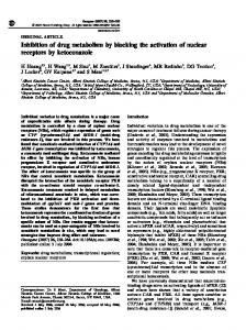
Fig. 1. Efficacy of sIFNγR gene transfer into the thigh muscle. A: Representative immunoblot against the extracellular portion of the interferon-γ receptor showing the temporal changes in serum sIFNγR protein levels after a single sIFNγR gene transfer (200 μg) into the thigh muscle of wildtype mice. Four independent experiments showed similar results. B: Representative microphotographs of immunohistostaining for sIFNγR protein (red) in atherosclerotic plaques in sIFNγR- and mock-treated ApoEKO mice at 12 weeks of age. Western-type diet was started from 8 weeks of age, and gene transfers were performed twice: at 8 and 10 weeks of age. An antibody against the mouse IgG1 Fc-fragment was used to detect the sIFNγR protein conjugated with the mouse IgG1 Fc-fragment. Similar results were obtained in five independent experiments.
with ice-cold PBS. Thereafter, the heart and the aorta were immediately excised and subjected to the histological and RT-PCR experiments.
Fig. 2. Effects of sIFNγR treatment on atherosclerotic plaque formation. A: Representative photographs showing oil red-O–stained atherosclerotic plaques (yellow-red) in the ascending aorta and the aortic arch at 12 weeks of age. B: Pooled data on the effects of sIFNγR treatment on the en face plaque area in the ascending aorta and the aortic arch at 12 weeks of age. Bar: 1 × SD (n = 5). *p< 0.05 vs. mock plasmid treatment.
En Face Plaque Area Immediately after the mice were killed, the aorta (n = 5 per group) was excised and fixed in 10% buffered formalin for quantification of the en face plaque area, as described previously (11). Briefly, after the adventitial tissue was carefully removed, the aorta was opened longitudinally, stained with oil red-O (Sigma, St. Louis, USA), and pinned on a black wax surface. En face images were obtained by a stereomicroscope (SZX12, Olympus, Tokyo, Japan) equipped with a digital camera (Dxm1200, Nikon, Tokyo, Japan) and analyzed using
262
Hypertens Res Vol. 30, No. 3 (2007)
Fig. 3. Effects of sIFNγR treatment on the structural features of the plaques. Representative microphotographs of the oil red-O (lipid, red)–, MOMA-2 (monocytes/macrophages, red)–, Azan (fibrous tissue, blue)-, and αSMA (smooth muscle cells, red)– stained cross sections of atherosclerotic plaques in the aortic root of sIFNγR- and mock-treated apoEKO mice. Five independent experiments showed similar results.
Koga et al: Blocking Interferon-γ for Atherosclerosis Prevention
263
using FastPrep homogenizer (ThermoSavant, Holbrook, USA), the total RNA was extracted and reverse-transcribed by using first-strand reaction mix beads (Amersham Biosciences) (13, 14). Aliquots of the RT products were amplified using KOD-plus (Toyobo, Osaka, Japan) and commercially available primer pairs for interferon-γ, IL-1β, MCP-1, VCAM-1 and GAPDH (Applied Biosystems, Foster City, USA) according to the manufacturer’s instructions. The RT-PCR products were electrophoresed on a 2% agarose gel stained with SYBR Gold (Invitrogen).
Serum Lipids and C-Reactive Protein Analysis Fig. 4. Representative photographs of the electrophoresis of RT-PCR products showing the effects of sIFNγR treatment on the mRNA expressions of interferon-γ (IFNγ), interleukin-1β (IL-1β), monocyte chemoattractant protein-1 (MCP-1) and vascular cell adhesion molecule-1 (VCAM-1) at 12 weeks of age. Five independent experiments showed similar results. WT, wild-type mice.
Adobe Photoshop version 7.0 and Scion Image software. The percentage of the luminal surface area stained by oil red-O was determined (11).
Histology and Immunohistostaining After the mouse was sacrificed and perfused with ice-cold PBS, the heart and the ascending aorta (n = 5 per group) were removed en bloc and snap-frozen in OCT compound (Sakura FineTech, Tokyo, Japan) for histological and immunohistochemical analysis. Atherosclerotic plaques were investigated at five independent section sets, each separated by 120 μm, of serial cryostat sections (6 μm thick) of the aortic root, as described previously (12). Immunohistostaining was performed using primary antibodies against mouse IgG1 Fcfragment (MP Biomedicals, Solon, USA), interferon-γ (Chemicon, Temecula, USA), interleukin-1β (IL-1β, Santa Cruz Biotechnology), monocyte chemoattractant protein-1 (MCP-1, Santa Cruz Biotechnology), and vascular cell adhesion molecule-1 (VCAM-1, BD Pharmingen, San Diego, USA) and a commercially available detection system (DAKO Cytomation, Kyoto, Japan). The serial sections were subjected to oil red-O staining, Mallory-Azan staining, and immunohistostaining using MOMA-2 (Serotec, Oxford, UK) and an anti–α-smooth muscle actin (αSMA) antibody (DAKO) to determine lipid-rich core, fibrotic tissue, monocytes/macrophages, and smooth muscle cells (SMCs), respectively.
Serum total cholesterol, high-density lipoprotein (HDL) cholesterol and high-sensitivity C-reactive protein (hsCRP) were measured by a commercially available laboratory (SRL, Fukuoka, Japan).
Peritonitis Model for In Vivo Monocyte Transmigration Assay To examine the effects of interferon-γ function blocking on in vivo monocyte transmigration, we employed an additional inflammation model, as described previously (15). Three days after sIFNγR or mock gene transfection (n = 6 per group), ApoEKO mice received an injection of 2 ml of 4% oyster glycogen medium (Sigma) into the peritoneal cavity. Six hours after the injection, the abdominal wall was cut and the peritoneal cavity was opened with special care to avoid blood contamination. The peritoneal cavity was washed with 2 ml of warm heparinized saline; then peritoneal fluid was collected and centrifuged at 400 g for 10 min. After the pellets were resuspended in PBS (1 ml), the number of total leukocytes was counted using an automatic analyzer. Also, the resuspended solutions were subjected to Giemsa staining and the percentage of monocytes was obtained in the leukocytes (> 500 leucocytes were counted). The monocytes were calculated by multiplying the total leukocyte count by the percentage of monocytes.
Statistical Analysis Each quantitative analysis of plaque area and monocyte count was performed by a single observer blinded to the experimental protocol. Data are expressed as means ± SD. Differences between the groups were determined using unpaired Student’s t-test. Probabilities of less than 0.05 were considered statistically significant.
Results RNA Extraction and RT-PCR Analysis The aorta (n = 5 per group) was rapidly snap-frozen in dry ice/ acetone and stored at − 80°C until use. After the frozen samples were homogenized in TRIzol (Invitrogen, Tokyo, Japan)
Efficiency of sIFNγR Gene Transfer In wild-type mice, we evaluated the temporal changes in serum sIFNγR protein levels after a single gene transfer of
264
Hypertens Res Vol. 30, No. 3 (2007)
sIFNγR plasmid (200 μg). Western blot analysis showed that sIFNγR protein was secreted into the serum, peaking at 3–7 days after gene transfer and remaining elevated for at least 2 weeks (Fig. 1A). The minimum dose of the injected sIFNγR plasmid to induce the maximum elevation of serum sIFNγR levels was 200 μg (data not shown). Accordingly, throughout the following experiments using apoEKO mice, gene transfers of 200 μg of sIFNγR plasmid were performed twice: first on the day of the starting Western-type diet (8 weeks of age) and then 2 weeks later. Next, we determined whether or not the overexpressed sIFNγR protein was present in the atherosclerotic plaques in sIFNγR-treated apoEKO mice. After the 4-week sIFNγR treatment, immunohistostaining demonstrated immunoreactivity against the IgG1 Fc-fragment–conjugated sIFNγR protein in the plaques and the media of the aortic root (Fig. 1B). In contrast, in the mock-treated apoEKO, sIFNγR protein was not detected in the plaques, the media or the adventitia.
Effects of 4-Week sIFNγR Treatment on Baseline Characteristics of apoEKO Mice All mice transfected with sIFNγR or mock plasmid appeared healthy and survived during the observation period. As shown in Table 1, sIFNγR treatment for 4 weeks had no significant effects on body weight, blood pressure, total and HDL cholesterol levels or serum hsCRP in apoEKO mice. Also, there were no significant differences in the peripheral blood leukocyte count and monocyte count between sIFNγR- and mocktreated mice (data not shown).
Effects of sIFNγR Treatment on Atherosclerotic Plaque Formation At 12 weeks of age, mock-treated apoEKO mice showed oil red-O–stained atherosclerotic plaques from the ascending aorta to the aortic arch (Fig. 2A). sIFNγR treatment remarkably prevented the aortic plaque formation, resulting in an approximately 60% reduction in en face plaque area (Fig. 2B). The cross-section at the aortic root was evaluated to determine the structural features of the atherosclerotic lesions (Fig. 3). In mock-treated apoEKO mice, the plaques in the aortic root consisted of a lipid-rich core and massive macrophage accumulation with a very thin fibrous cap and few SMCs. In sIFNγR-treated mice, the formation of the lipid- and macrophage-rich plaques was remarkably inhibited at the aortic root. And, sIFNγR treatment increased the fibrous tissue and SMCs in the luminal surface of the plaques (Fig. 3).
Effects on Inflammatory Changes in the Aortic Wall The induction of inflammatory cytokines (i.e. IL-1β), chemokines (i.e. MCP-1) and adhesion molecules (i.e.
VCAM-1) has been shown to play a key role in plaque formation in apoEKO mice (16). There was no significant expression of interferon-γ, IL-1β, MCP-1 or VCAM-1 in the aorta of wild-type mice (Figs. 4 and 5). In the mock-treated apoEKO mice, the expression of these inflammation-related molecules was remarkably upregulated in the aortic wall at 12 weeks of age (Fig. 4). Immunoreactive interferon-γ, IL-1β and MCP-1 were observed in the atherosclerotic plaques, whereas VCAM-1 was expressed in both the plaques and the medial SMCs (Fig. 5). sIFNγR treatment prevented the induction of interferon-γ, IL-1β, MCP-1 and VCAM-1 at the mRNA and protein levels (Figs. 4 and 5).
Effects on In Vivo Monocyte Transmigration in Peritonitis Model We examined whether sIFNγR reduced monocyte infiltration in another model of inflammation. Intraperitoneal administration of oyster glycogen induces peritonitis, which is an established inflammation model in mice (15). In this model, peritonitis was associated with a transmigration of monocytes in the peritoneal cavity of the mock-treated apoEKO mice (Fig. 6). sIFNγR treatment significantly reduced the monocyte transmigration.
Discussion In the present study, we have demonstrated for the first time that postnatal blocking of interferon-γ function prevents atherosclerotic plaque formation in apoEKO mice by overexpressing the interferon-γ inhibitor protein, sIFNγR. Moreover, sIFNγR treatment attenuated the inflammatory changes in the aortic wall. Taken together, these findings suggested that interferon-γ plays a crucial role in the development of early atherosclerotic lesions by activating inflammation. We used apoEKO mice to investigate the role of interferonγ in the development of atherosclerotic lesions, because the production of interferon-γ was documented in infiltrated lymphocytes within the atherosclerotic lesions in this model (17). In the present study, intrinsic interferon-γ activity was postnatally inhibited in apoEKO mice by overexpressing sIFNγR. We overexpressed sIFNγR protein fused to the IgG1 Fc fragment as the inhibitor instead of the truncated sIFNγR alone, because the fusion molecule secreted as homodimers has a much longer half-life than truncated sIFNγR (18, 19) and because the dimeric sIFNγR fusion protein exhibits higher binding activity for the ligand than single-chain receptors (20). In fact, a single sIFNγR gene transfer into the thigh muscle significantly elevated the serum sIFNγR protein levels for at least 2 weeks (Fig. 1A). And, the overexpressed sIFNγR protein was detected in the aortic atherosclerotic lesions in the sIFNγR-treated apoEKO mice (Fig. 1B). Moreover, sIFNγR treatment inhibited monocyte transmigration in the peritonitis model (Fig. 6). These results, together with those of previous studies (4, 5), suggested that the overexpressed sIFNγR pro-
Koga et al: Blocking Interferon-γ for Atherosclerosis Prevention
265
Fig. 5. Representative immunohistostainings showing the effects of sIFNγR treatment on the protein expressions (red) of interferon-γ (IFNγ), interleukin-1β (IL-1β), monocyte chemoattractant protein-1 (MCP-1) and vascular cell adhesion molecule-1 (VCAM-1) at 12 weeks of age. Similar results were obtained in five independent experiments. WT, wild-type mice.
266
Hypertens Res Vol. 30, No. 3 (2007)
Fig. 6. Effects of sIFNγR treatment on in vivo monocyte transmigration in peritonitis model. In apoEKO mice, experimental peritonitis was induced by injecting oyster glycogen 3 days after sIFNγR- or mock-gene transfection. Six hours later, peritoneal fluid was collected and the monocytes were counted. Bar: 1 × SD (n= 6). *p< 0.05 vs. mock plasmid treatment.
teins were secreted into the systemic circulation and in turn inhibited interferon-γ–induced inflammation in the remote organs. The most important finding of this study was that sIFNγR treatment abolished atherosclerotic plaque formation in the pre-atherosclerotic stage of apoEKO mice (Fig. 2). This was in line with the earlier study demonstrating that apoE/interferon-γ receptor α-chain double-KO mice had smaller atherosclerotic lesions than apoEKO mice (17). Our observations not only support the notion that interferon-γ plays a substantial role in atherosclerotic plaque formation, but also raise the possibility that blocking the interferon-γ–mediated signaling would be a new target for the prevention of atherosclerosis. It is interesting to note that sIFNγR treatment altered the structural features of the plaque in addition to reducing the plaque area. In mock-treated apoEKO mice, the plaques had a large lipid-rich core and massive macrophage accumulation accompanied by few SMCs and a very thin fibrous cap (Fig. 3), indicating markedly low plaque stability. sIFNγR treatment improved the histological stability of the plaques not only by preventing lipid and macrophage accumulation but also by increasing SMCs and fibrotic tissue. Several mechanisms were considered to explain the effects of sIFNγR. Serum cholesterol and hsCRP levels were not altered by sIFNγR treatment (Table 1). Accordingly, it is plausible that the observed effects are independent of systemic inflammation and cholesterol levels. As shown in Figs. 4 and 5, sIFNγR treatment suppressed the upregulation of IL1β, MCP-1 and VCAM-1 expression in the aortic wall, especially in the plaques. Interferon-γ is the major activator of infiltrated macrophages (2, 21). The activated macrophages are transformed into lipid-laden foam cells. Also, they secrete various kinds of inflammatory cytokines (e.g. IL-1β), which in turn leads to the production of MCP-1 and VCAM-1 in the vascular wall cells, such as endothelial cells, SMCs and mac-
rophages themselves (2, 21–23). Subsequently, these changes elicit further interferon-γ induction and inflammatory cell infiltration. This suggested that the inhibition of interferon-γ function by sIFNγR treatment abolished the interferon-γ– induced vicious cycle of plaque inflammation and macrophage recruitment, and subsequently prevented plaque formation (Figs. 4 and 5). In addition, interferon-γ has been shown to suppress SMC proliferation and collagen production in vitro (24–26). As shown in Fig. 3, sIFNγR treatment increased SMCs and fibrous tissue deposition in the plaques. In contrast, a recent study has shown that neointima formation after vascular injury is attenuated in interferon-γ receptor αchain knockout mice (27), suggesting that interferon-γ might promote SMC proliferation during neointima formation. The diversity in the roles of interferon-γ in SMC proliferation in vivo may be explained by the differences in the underlying pathophysiology, the disease stage, the SMC phenotype, the interactions of SMCs to the surrounding cells and the presence of other factors between the atherosclerotic lesions and the neointimal lesions after vascular injury (3). Our study suggested that interferon-γ inhibited SMC proliferation and collagen deposition in the plaques of this model. Further investigations are warranted to explore this issue. The present study has clearly demonstrated that sIFNγR is potentially useful for the prevention and treatment of atherosclerosis. However, there are several limitations to its clinical application. First, although no apparent side effects were observed during the period of this study, careful observation needs to be done over longer periods. In this regard, the combination of sIFNγR treatment with a local delivery technique to the target lesions, e.g. drug-eluting stent, may be desirable to avoid the possible side effects related to immunosuppression caused by systemic inhibition of the interferon-γ function. Second, the effects of sIFNγR treatment on established plaques should be determined. Based on the results of this study, we are now performing a series of experiments to see if sIFNγR treatment would regress and stabilize the existing, established plaques in the advanced stage of atherosclerosis in apoEKO mice. In conclusion, interferon-γ promotes atherosclerotic plaque formation by activating inflammation in the arterial wall. Moreover, inhibition of the interferon-γ–mediated pathway may be a new target for the prevention and treatment of atherosclerosis.
Acknowledgements We appreciate the skillful technical assistance of Kaoru Moriyama, Yayoi Yoshida, Reiko Fujiyoshi and Kimiko Kimura.
References 1. 2.
Ross R: Atherosclerosis: an inflammatory disease. New Engl J Med 1999; 340: 115–126. Young YL, Libby P, Schonbeck U: Cytokines in the patho-
Koga et al: Blocking Interferon-γ for Atherosclerosis Prevention
3. 4.
5.
6.
7.
8.
9.
10.
11.
12.
13.
14.
genesis of atherosclerosis. Thromb Haemost 2002; 88: 554– 567. Harvey EJ, Ramji DP: Interferon-γ and atherosclerosis: proor anti-atherogenic? Cardiovasc Res 2005; 67: 11–20. Piccirillo CA, Prud’homme GJ: Prevention of experimental allergic encephalomyelitis by intramuscular gene transfer with cytokine-encoding plasmid vectors. Hum Gene Ther 1999; 10: 1915–1922. Lawson BR, Prud’homme GJ, Chang Y, et al: Treatment of murine lupus with cDNA encoding IFN-gammaR/Fc. J Clin Invest 2000; 106: 207–215. Tahara N, Kai H, Niiyama H, et al: Repeated gene transfers of naked prostacyclin synthase plasmid into skeletal muscles attenuate monocrotaline-induced pulmonary hypertension and prolong survival in rats. Hum Gene Ther 2004; 15: 1270–1278. Danko I, Fritz JD, Jiao S, Hogan K, Latendresse JS, Wolff JA: Pharmacological enhancement of in vivo foreign gene expression in muscle. Gene Ther 1994; 1: 114–121. Niiyama H, Kai H, Yamamoto T, et al: Roles of endogenous monocyte chemoattractant protein-1 in ischemiainduced neovascularization. J Am Coll Cardiol 2004; 44: 661–666. Molloy CJ, Taylor DS, Weber H: Angiotensin II stimulation of rapid protein tyrosin phosphorylation and protein kinase activation in rat smooth muscle cells. J Biol Chem 1993; 268: 7338–7345. Kai H, Griendling KK, Lassegue B, Ollerenshaw JD, Runge MS, Alexander RW: Agonist-induced phosphorylation of the vascular type 1 angiotensin II receptors. Hypertension 1994; 24: 523–527. Inoue S, Egashira K, Ni W, et al: Anti-monocyte chemoattractant protein-1 gene therapy limits progression and destabilization of established atherosclerosis in apolipoprotein E−knockout mice. Circulation 2002; 106: 2700–2706. Ni W, Egashira K, Kitamoto S, et al: New anti-monocyte chemoattractant protein-1 gene therapy attenuates atherosclerosis in apolipoprotein E−knockout mice. Circulation 2001; 103: 2096–2101. Kuwahara F, Kai H, Tokuda K, et al: Transforming growth factor-β function blocking prevents myocardial fibrosis and diastolic dysfunction in pressure-overloaded rats. Circulation 2002; 106: 130–135. Kai H, Mori T, Tokuda K, et al: Pressure overload−induced transient oxidative stress mediates perivascular inflamma-
15.
16.
17.
18.
19.
20.
21.
22. 23. 24.
25.
26.
27.
267
tion and cardiac fibrosis through angiotensin II. Hypertens Res 2006; 29: 711–718. Vaporciyan AA, DeLisser HM, Yan HC, et al: Involvement of platelet-endothelial cell adhesion molecule-1 in neutrophil recruitment in vivo. Science 1993; 262: 1580–1582. Yoshii T, Iwai M, Li Z, et al: Regression of atherosclerosis by amlodipine via anti-inflammatory and anti-oxidative stress actions. Hypertens Res 2006; 29: 457–466. Gupta S, Pablo AM, Jiang X, Wang N, Tall AR, Schindler C: IFN-gamma potentiates atherosclerosis in ApoE knockout mice. J Clin Invest 1997; 99: 2752–2761. Kurschner C, Ozmen L, Garotta G, Dembic Z: IFN-gamma receptor-Ig fusion proteins. Half-life, immunogenicity, and in vivo activity. J Immunol 1992; 149: 4096–4100. Ozmen L, Gribaudo G, Fountoulakis M, Gentz R, Landolfo S, Garotta G: Mouse soluble IFN gamma receptor as IFN gamma inhibitor. Distribution, antigenicity, and activity after injection in mice. J Immunol 1993; 150: 2698–2705. Kurschner C, Garotta G, Dembic Z: Construction, purification, and characterization of new interferon gamma (IFN gamma) inhibitor proteins. Three IFN gamma receptor− immunoglobulin hybrid molecules. J Biol Chem 1992; 267: 9354–9360. Schroder K, Hertzog PJ, Ravasi T, Hume DA: Interferongamma: an overview of signals, mechanisms and functions. J Leukoc Biol 2004; 75: 163–189. Lusis AJ: Atherosclerosis. Nature 2000; 407: 233–241. Charo IF, Taubaman MB: Chemokines in the pathogenesis of vascular diseases. Circ Res 2004; 95: 858–866. Amento EP EN, Palmer H, Libby P: Cytokines and growth factors positively and negatively regulate interstitial collagen gene expression in human vascular smooth muscle cells. Arterioscler Thromb 1991; 11: 1223–1230. Yuan W, Yufit T, Li L, Mori Y, Chen SJ, Varga J: Negative modulation of alpha1(I) procollagen gene expression in human skin fibroblasts: transcriptional inhibition by interferon-gamma. J Cell Physiol 1999; 179: 97–108. Sibinga NE, Wang H, Perrella MA, et al: Interferongamma−mediated inhibition of cyclin A gene transcription is independent of individual cis-acting elements in the cyclin A promoter. J Biol Chem 1999; 274: 12139–12146. Zohlnhofer D, Richter T, Neumann F, et al: Transcriptome analysis reveals a role of interferon-gamma in human neointima formation. Mol Cell 2001; 7: 1059–1069.