Diabetologia (2004) 47:987–997 DOI 10.1007/s00125-004-1404-9
Potent activation of multiple signalling pathways by C-peptide in opossum kidney proximal tubular cells N. M. Al-Rasheed1, 3 · F. Meakin1 · E. L. Royal1 · A. J. Lewington1, 2 · J. Brown2 · G. B. Willars1 · N. J. Brunskill1, 2 1 Department
of Cell Physiology and Pharmacology, Faculty of Medicine and Biological Sciences, University of Leicester, Leicester, United Kingdom 2 Department of Nephrology, Faculty of Medicine and Biological Sciences, University of Leicester, Leicester, UK 3 Department of Pharmacology, King Saud University, Riyadh, Saudi Arabia
Abstract Aims/hypothesis. Proinsulin C-peptide is generally believed to be inert without any appreciable biological functions. However, it has been shown to modulate a variety of cellular processes important in the pathophysiology of diabetic complications. We therefore investigated the ability of C-peptide to stimulate intracellular signalling pathways in kidney proximal tubular cells, the altered activation of which may possibly be related to the development of diabetic nephropathy. Methods. Extracellular signal-regulated kinase (ERK) and Akt phosphorylation were evaluated by western blotting. ERK activity was measured by in vitro kinase assay. Intracellular Ca2+ was evaluated by confocal imaging. The membrane and cytosol-associated fractions of protein kinase C (PKC) isoforms were evaluated by western blotting. Proliferation was assessed by thymidine incorporation assay. Results. Using the opossum proximal tubular kidney cell line as a model, we demonstrated that at high picomolar to low nanomolar concentrations, C-peptide
Received: 30 October 2003 / Accepted: 26 January 2004 Published online: 26 May 2004 © Springer-Verlag 2004
stimulates extracellular signal-regulated mitogen-activated kinase (3.3±0.1-fold over basal at 3 minutes) and phosphatidylinositol 3-kinase (4.1±0.05-fold over basal at 5 minutes). ERK activation was attenuated by pre-treatment with a PKC inhibitor and abolished by pertussis toxin. Elevations of intracellular [Ca2+] are seen in response to 5 nmol/l C-peptide with consequent activation of PKC-α. Pre-treatment with pertussis toxin abolished PKC-α. C-peptide is also a functional mitogen in this cell type, stimulating significantly increased cell proliferation. Proliferation was attenuated by wortmannin and pertussis toxin pretreatments. None of these effects is reproduced by scrambled C-peptide. Conclusions/interpretation. This study provides evidence that C-peptide, within physiological concentration ranges, stimulates many signalling pathways in opossum kidney cells. Keywords Calcium · C-peptide · Diabetic nephropathy · Kidney · MAP kinase · Mitogen · PI 3-kinase · PKC
Introduction
N. J. Brunskill (✉) Department of Cell Physiology and Pharmacology, Faculty of Medicine and Biological Sciences, University of Leicester, Medical Sciences Building, University Road, Leicester, LE1 9HN, United Kingdom E-mail:
[email protected] Tel.: +44-116-2588043, Fax: +44-116-2525045
Proinsulin connecting peptide (C-peptide) is an enzymatic cleavage product derived from proinsulin during the biosynthesis of insulin [1]. Insulin and C-peptide are stored together within the secretory granules of pancreatic beta cells, being eventually released simultaneously and in equimolar amounts into the portal circulation [2]. C-peptide forms a linker between the
Abbreviations: Akt/PKB, protein kinase B · DTT, dithiothreitol · ERK, extracellular signal-regulated kinase · GPCR, G-protein coupled receptor · MAPK, mitogen-activated protein kinase · OK, opossum
kidney · PI3-K, phosphoinositide 3-kinase · PKC, protein kinase C · PMA, phorbol myristate acetate · PMSF, phenylmethansulfonylfluoride · PTC, proximal tubular cells · PTX, pertussis toxin
988
A and B chains of insulin, facilitating correct folding and thus the establishment of disulfide bonds between adjacent cysteine residues [3]. Early after the discovery of C-peptide, workers attempted to delineate a role for C-peptide in the regulation of glucose homeostasis [4, 5, 6]. No such role could be found, encouraging the traditional belief that C-peptide is a biologically inert molecule. Accumulating evidence, however, has begun to challenge this view by suggesting that C-peptide plays a role in experimental and clinical diabetes. The infusion of C-peptide into patients with Type 1 diabetes over the short term (hours), in addition to the administration of insulin, ameliorates a variety of associated renal functional abnormalities [7]. Similarly, the more prolonged administration of C-peptide (months) is associated not only with improved renal function, but also enhanced autonomic and sensory nerve function [8, 9, 10, 11]. Characteristic myocardial perfusion and limb blood flow changes observed in patients with Type 1 diabetes are also reversed by C-peptide [11, 12]. No effects of C-peptide infusion on any of these parameters have ever been demonstrated in healthy non-diabetic control subjects [8, 9, 10, 11]. At the cellular level various effects of C-peptide have now been described. Activation of Na+/K+ATPase occurs in numerous nephron segments in response to C-peptide stimulation, together with increased intracellular Ca2+ concentrations ([Ca2+]i) in proximal tubular cells [13, 14, 15, 16]. Recently Kitamura et al. [17] demonstrated C-peptide-mediated stimulation of a kinase cascade in Swiss 3T3 fibroblasts involving mitogen-activated protein kinases (MAPK), protein kinase C (PKC) and phosphoinositide-3-kinase (PI3-K). Specific cell membrane binding sites for C-peptide have recently been identified in renal tubular cells, skin fibroblasts and saphenous vein endothelial cells. Abolition of this binding by pertussis toxin (PTX) was observed, suggesting the involvement of a G-protein coupled receptor (GPCR). The calculated association rate constant (Kass) of this receptor was about 3 nmol/l, which suggests that under normal physiological conditions it is probably fully occupied, possibly explaining the absence of C-peptide effects in normal subjects [18, 19]. Diabetic nephropathy remains a serious complication of diabetes, and is characterised initially by renal growth. Hypertrophy of glomerular and tubular cells is prominent, together with thickening of basement membranes, but hyperplasia does not occur [20, 21, 22]. Later, matrix components accumulate in the glomerulus and tubulo-interstitium with progressive sclerosis. Tubular atrophy with almost complete loss of proximal tubular cells (PTC) is evident, a clear sign of major disturbances in growth and survival of this cell type. Interestingly, the pathogenesis of nephropathy does not seem to be substantially different in Type 1 and Type 2 diabetes [23, 24, 25].
N. M. Al-Rasheed et al.:
Much attention has been given to signal transduction pathways activated in response to the persistent homeostatic disturbances in diabetes that may initiate and/or propagate diabetic renal pathophysiology. Activation of PKC in the kidney occurs within days of the onset of experimental diabetes [26], and this may in turn stimulate phospholipase A2, leading to the release of membrane-bound arachidonate [26, 27]. Another consequence of renal PKC activation is stimulation of MAPK [28]. The activation of MAPK in glomeruli obtained from rats with streptozotocin-induced diabetes and in mesangial cells grown in high glucose concentrations is mediated by PKC and contributes crucially to cell hypertrophy [29]. The precise role of C-peptide, or the lack thereof, in the modulation of these signalling pathways in diabetes and in the pathogenesis of diabetic nephropathy remains uncertain. Therefore, in view of our long-standing interest in renal tubular disease and the body of literature supporting a physiological role for C-peptide, we examined signalling and functional roles for C-peptide in the kidney. Our primary aim was to determine whether C-peptide has the capacity to signal in PTC, focusing especially on pathways involved in cell growth and the potential development of diabetic nephropathy. We used opossum kidney (OK) cells, an immortalised kidney PTC cell line [30] that retains many characteristics of the proximal tubule [31]. Many typical proximal tubular functions such as parathyroid hormone responses and endocytic uptake of proteins have been investigated using this cell type [32, 33].
Materials and methods Materials. Human 31 amino acid (aa) C-peptide and a 31 aa scrambled C-peptide were generously provided by Dr J. Wahren (Karolinska Institute, Stockholm, Sweden). These peptides were prepared as 250 µmol/l stock solutions in 50 mmol/l glacial acetic acid, and stored at −20 °C. All tissue culture media and plasticware were from Invitrogen Life Technologies (Paisley, UK) and general laboratory chemicals were from Sigma (Poole, UK) unless otherwise stated. Wortmannin, PTX and Ro-31-8220 were from Calbiochem (Nottingham, UK). The extracellular signal-regulated MAP kinase (ERK) substrate, a peptide (RRELVEPLTPSGEAPNQALLR) derived from the cytoplasmic tail of the epidermal growth factor receptor, was synthesised by the Protein and Nucleic Acid Chemistry Laboratory (University of Leicester, UK). Rabbit polyclonal anti-ERK-1 and antiphospho-ERK1/2 were obtained from Santa Cruz Biotechnology (Santa Cruz, Calif., USA). Sheep monoclonal anti-Akt1/PKBα and anti-phospo-Akt1/PKBα were from Upstate Biotechnology (Milton Keynes, UK). Anti-PKC isoforms were from Transduction Laboratories (San Diego, Calif., USA). Protein A sepharose CL-4B was from Pharmacia Biotech (Uppsala, Sweden). We obtained γ-[32P] ATP from Perkin Elmer Life Sciences (Cambridge, UK) and [3H]-thymidine from Amersham Biosciences (Chalfont St. Giles, Buckinghamshire, UK). Cell culture. Opossum kidney cells were obtained from Dr J. Caverzasio (University Hospital, Geneva, Switzerland) and used between passages 72-87. Cells were maintained in
Potent activation of multiple signalling pathways by C-peptide
989
Dulbecco’s modified Eagle’s media-Ham’s mix (DMEM-F12), supplemented with 10% FCS, 2 mmol/l L-glutamine, 100 U/ml penicillin, 100 µg/ml streptomycin and 10 mmol/l HEPES. In all experiments cells were serum-starved overnight before being subject to agonist stimulation to ensure as far as possible that cells were not exposed to mitogenic agents or C-peptide derived from FCS.
3-AM, Molecular Probes, Cambridge, UK). This was done at room temperature for 45 to 90 min. Cover slips were then mounted in a chamber on a heated stage of an Olympus 1x 70-SIF inverted microscope. The chamber was perfused with Krebs buffer (bath temperature 37 °C), with or without C-peptide or scrambled C-peptide, at 5 ml per minute. The intracellular fluorescence, as an index of [Ca2+]i, was determined by using an Ultraview confocal imaging system (Perkin Elmer). Cells were excited with a krypton/argon laser at 488 nm and emitted light was collected above 510 nm with a narrow-band pass filter.
Determination of ERK and Akt activation by western blotting of phospho-ERK and phospho-Akt. Cells growing in 6-well plates were stimulated with various concentrations of C-peptide or scrambled peptide in serum-free DMEM-F12 for various times. After stimulation, cells were rinsed with ice-cold PBS and then lysed in Laemmli buffer (60 mmol/l Tris, pH 6.8, 10% glycerol, 2% SDS, 100 mmol/l dithiothreitol [DTT] and 0.01% bromophenol blue). Samples were separated by SDS-PAGE and then transferred to nitrocellulose membranes. Membranes were blocked with a 5% (w/v) solution of dried milk powder in TTBS (20 mmol/l Tris base, 100 mmol/l NaCl and 0.05% Tween 20, pH 7.5) and immunoblotted using antisera against phosphorylated ERK or phosphorylated Akt. To check the uniformity of loading, blots were stripped and re-probed with antisera to total (phospho- and non-phospho ERK-1) ERK or total Akt. In all cases, primary antibodies were visualised using peroxidase-conjugated secondary antibodies and enhanced chemiluminescence (Amersham). Immunoblots were quantitated using Scion Image analysis software (version 4.0.2; Scion, Frederick, Md., USA). Determination of ERK activity by in vitro kinase assay. ERK activity in OK cells after treatment with test agents was measured using an in vitro kinase assay, in essence as previously described [34]. Briefly, confluent monolayers of OK cells were grown in 6-well plates. After stimulation, the cells were washed once with ice-cold PBS and then solubilised with icecold lysis buffer (20 mmol/l Tris-HCl, pH 8.0, 0.5% Nonidet P-40, 250 mmol/l NaCl, 3 mmol/l EDTA, 3 mmol/l EGTA, 1 mmol/l phenylmethansulfonylfluoride [PMSF], 2 mmol/l Na3VO4 and 1 mmol/l DTT). The lysates were clarified by 10 minutes of centrifugation at 14000 g at 4 °C. Next rabbit polyclonal anti-ERK-1 antiserum (stock concentration 200 µg/ml) was added to give a final concentration of 2 µg/ml, and incubated for 120 min on ice. Immune complexes were incubated for 30 min at 4 °C with 100 µl of a 15% (v/v) slurry of protein A-sepharose. Immunoprecipitates were then washed twice in 200 µl lysis buffer and twice in 200 µl kinase buffer (20 mmol/l HEPES, pH 7.2, 20 mmol/l β-glycerophosphate, 10 mmol/l MgCl2, 1 mmol/l DTT and 50 µmol/l Na3VO4). An aliquot of 40 µl kinase buffer containing 20 µmol/l γ[32P]-ATP (92.5 kBq/nmol) and 200 µmol/l of synthetic ERK peptide substrate was added to immunoprecipitates to initiate the kinase reaction. The reactions were terminated after 20 minutes by adding 10 µl of 25% (w/v) trichloroacetic acid. The mixtures were then centrifuged at 14000 g for 2 min and 40 µl of the supernatant spotted on to squares of P81 cation exchange paper (Whatman, Maidstone, UK). Papers were washed three times for 5 min in 0.5% (v/v) orthophosphoric acid, rinsed once with acetone and air-dried, after which radioactivity was determined by liquid scintillation counting. Confocal imaging of [Ca2+]i. OK cells were plated on to sterile 25-mm diameter poly-D-lysine-coated glass cover slips and allowed to adhere for 24 h. Cells were incubated in Krebs buffer (10 mmol/l HEPES, 4.2 nmol/l NaHCO3, 11.7 mmol/l D-glucose, 1.18 mmol/l MgSO4·7H2O, 1.18 mmol/l KH2PO4, 4.69 mmol/l KCl, 118 mmol/l NaCl, 1.29 mmol/l CaCl2·2H2O, pH 7.4) containing 6 µmol/l fluo-3-acetoxymethyl ester (fluo-
Determination of PKC activation by translocation assay. The cytosol to membrane translocation of PKCα was determined by cell fractionation and western blotting as an index of activation as previously described [35]. Cells were stimulated and then lysed in 200 µl ice-cold lysis buffer (20 mmol/l Tris-HCl, 5 mmol/l EGTA, 2 mmol/l EDTA, 1 mmol/l DTT, 0.5 mmol/l PMSF, 10 µmol/l iodoacetamide, pH 7.4) and incubated on ice for 30 min. Samples were then centrifuged (20800 g, 15 min, 4 °C) to separate cytosolic and membrane fractions. A 100 µl aliquot of supernatant (cytosolic fraction) was removed. The remaining supernatant was aspirated and the membrane pellet re-suspended in 200 µl volume of solubilisation buffer (9.2 mmol/l Tris, 0.92 mmol/l EGTA, 150 mmol/l NaCl, 0.1% [v/v] SDS, 1% [v/v] ethylphenylpolyethylene glycol [Nonidet P-40], 0.5% [w/v] deoxycholate, 0.5 mmol/l PMSF, 10 µmol/l benzamidine hydrochloride and 5 µmol/l iodoacetamide, pH 7.4). An equal volume of sample buffer (100 mmol/l Tris, 2% SDS, 0.1% bromophenol blue, 10% [v/v] glycerol, 200 mmol/l DTT) was added to the membrane and cytosolic fractions. Proteins were separated by SDS-PAGE, transferred to nitrocellulose and immunoblotted for PKCα, ε and ι as described above, using anti PKCα, ε and ι at 0.1, 0.5 and 0.5 µg/ml respectively. The nitrocellulose was subsequently stained using Ponceau-s (0.2% Ponceau-S and 3% TCA) to ensure equivalent protein loading and transfer. Incorporation of [3H]-thymidine. Proliferation of OK cells in response to C-peptide was assessed by [3H]-thymidine incorporation. Cells were plated in 24-well plates and grown to approximately 80% confluence. Serum-starved cells were exposed to various concentrations of C-peptide, 10% fetal calf serum or serum-free media as a control. After 22 h of incubation at 37 °C, the culture medium was replaced with serumfree medium containing 37 kBq of [3H]-thymidine. After a further 2 h of incubation, cells were washed with ice-cold PBS and fixed at 4° C in 50% methanol and 10% glacial acetic acid for 30 min, and then in 10% trichloroacetic acid for 10 min. Cells were then solubilised overnight in 100 mmol/l NaCl containing 0.1% SDS, after which [3H]-thymidine incorporation was determined by standard liquid scintillation counting. Data presentation and analysis. Data are presented as means ± SEM. Statistical analysis was by unpaired Student’s t test for comparison between two experimental groups. For multiple comparisons, one-way ANOVA with Tukey’s correction was used. Differences were considered significant at a p value of less than 0.05.
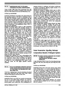
Results Effect of C-peptide on activation of ERK-1 and -2. Incubation of OK cells with C-peptide resulted in a time- and a concentration-dependent increase in phos-
990
Fig. 1. Stimulation of ERK phosphorylation by C-peptide but not scrambled C-peptide in opossum kidney cells. a. Time course of activation. Cells were treated with 5 nmol/l C-peptide for the indicated times and total extracellular signal-regulated kinase (ERK) or phosphorylated ERK were detected by immunoblotting. The left panel shows a representative blot. Phosphorylated bands were quantified and values expressed graphically (right panel). **p