PRECISION HIGH-BANDWIDTH OUT-OF-PLANE ACCELEROMETER AS CONTACT MICROPHONE FOR BODY-WORN AUSCULTATION DEVICES Pranav Gupta*, Yaesuk Jeong, Jaehoo Choi, Mark Faingold, Anosh Daruwalla, and Farrokh Ayazi Georgia Institute of Technology, Atlanta, Georgia, USA
sensitive to vibrations from its contact surface. Its small size allows it to replace bulky stethoscopes with ergonomic wearable auscultation systems that can precisely measure cardiopulmonary sounds, chest wall motion, ballistocardiogram (BCG) signal as well as body motion of the user simultaneously. This opens new gateways in telemedicine and remote health monitoring. Moreover, such an integrated solution significantly reduces fabrication cost of wearable technology, making it more accessible and affordable to the masses. In this paper, a low noise, wide bandwidth out-of-plane accelerometer with nano-gaps (270nm) capacitive electrodes implemented using HARPSS+ process [5] is fabricated and characterized as a contact microphone. The performance of the accelerometer contact microphone (ACM) is compared with a commercial piezoelectric contact microphone. Further, the paper addresses the use of this contact microphone as a body-worn auscultation device by mounting it on the chest and recording sounds produced in the thoracic cavity. The recorded signals are filtered and processed to extract the heart sound and BCG signal from the recorded data. Similarly, lung sound and chest wall motion are extracted using data processing techniques, demonstrating the possibility of accurately capturing multiple types of acoustic and vibrational data from the body at the same time.
ABSTRACT
This paper reports on the implementation and characterization of wafer-level-packaged accelerometer contact microphones having wide bandwidth (f res >10 kHz) and low-noise (10 kHz) and low noise ( 20Hz) of the recorded signal correspond to the cardiopulmonary sounds. The filtering algorithm shown in Fig. 7 is used to separate the low frequency component of chest wall motion and the high frequency component of the sound signal. The filtering is performed using high-order Butterworth filters on the basic criteria of audible (> 20Hz) and inaudible frequencies (< 20Hz). A wavelet denoising technique is used on the high frequency components to reduce noise and extract signal features [13]. The resultant waveforms are shown in Fig. 8, corresponding to extracted cardiopulmonary signals. There are two major cardiac sounds; S1 and S2, which occur due to closing of the atrioventricular valves (Mitral and Tricuspid) and closing of the semilunar valves (Aortic and Pulmonary), respectively. The period from S1 to S2 is known as Systole, whereas the S2 to S1 period is known as Diastole [14], as shown in Fig. 8. The recorded signal, known as phonocardiogram (PCG), is characterized by a ‘lub-dub’ sound, and provides a solid foundation for preliminary diagnosis, based on the frequency content and amplitude of the signal, of any cardiac disease. Another feature captured along with the heart sounds is the ballistocardiogram (BCG) signal, defined as the micro-movements of the body due to shift in center of mass with the pumping of blood at every heartbeat [15]. As this signal lies in the frequency range of 0-20Hz, it is typically captured using a precision micro-g accelerometer. The dual nature of the proposed contact microphone enables the detection of the BCG signal, while measuring cardiac sounds. As shown in Fig. 8, the BCG signal is characterized by the H, I, J, K, L wave, forming a ‘W’ pattern within the waveform. The importance of capturing this waveform is marked by its unobtrusive nature and ability to detect early onset of several diseases such as acute myocardial disease, asymptotic coronary artery disease and congestive heart failure [16]. Auscultation of the lungs provides vital information regarding their physiology such as obstructions in airway or presence of liquid in the organ. The qualities of breath sounds modify as air passes through the lungs. The pitch and duration of recorded lung sounds differ with respect to location of the senor. The presence of adventitious breath sounds such as crackles or wheezes usually indicate disease [2]. To accurately identify the timing of abnormal breath sounds within a respiratory cycle, the inspiration and expiration can be monitored by tracking the movement of the chest wall. Normal “vesicular” breath sound recorded by our accelerometer contact microphone is shown in Fig. 8. The ACM demonstrates high-fidelity cardiopulmonary auscultation sensing capability as well as high sensitivity towards motion artifacts. Characteristic features of heart sounds, BCG signal and lung sound are easily detected using the ACM.
Figure 5: Audio Reconstruction Test showing time domain signals of original and recorded signals MATLAB program, the captured data is reconstructed into an audio file format for playback. Fig. 5 shows the time domain signals for the original audio clip along with the recorded data. A high degree of similarity is observed between the recorded sounds and original audio clip. High frequency components (>2 kHz) are not picked up by the ACM due to its operation in air. However, on playback, the ACM demonstrates high quality signal specifically at lower frequencies compared to the Knowles microphone which exhibits noticeable distortion. This behavior can be attributed to the ACM’s inherent ability to capture high quality signals at low frequencies, including the inaudible range (0-20 Hz), unlike the piezoelectric contact microphones.
BODY-WORN AUSCULTATION DEVICES
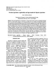
Sensor Placement Several auscultation locations for cardiopulmonary sounds are available in the intercostal spaces (ICS) (i.e. the space between two consecutive ribs) on the chest. Cardiopulmonary sounds, which lie between 20 to 2500 Hz frequency range [12], are recorded by mounting the sensor left of the sternum in the 5th intercostal space, shown in Fig. 6. The PCB is held in place using a chest strap to provide firm contact between the board and skin. Such configuration is essential to pick-up high-fidelity audio signals. Cardiopulmonary Sound Sensing The senor board is interfaced with NI-9220 DAQ to collect the data and a MATLAB program is used to filter the recorded signals which can be further stored in audio file format for playback. Unlike piezoelectric microphones, the ACM can measure accelerations
Figure 6: Sensor placement on the body for heart and lung sound acquisition
32
Figure 8: Recorded heart (left) and lung(right) sounds when the subject is sitting idle and breathing heavily respectively. The spectrogram shows the frequency content of the sound signal and its variation with heartbeats and respiratory cycle.
CONCLUSION
[5]
The growing demand for remote health monitoring and wearable technologies will necessitate the integration of sensors into smaller form-factor with increased functionality and precision performance. The presented accelerometer contact microphone, whose functionality and feasibility are validated by an audio reconstruction test followed by an on-body auscultation test to capture heart and lung sounds, along with simultaneous recording of BCG signals and chest wall motions, represents as a major step towards this goal. By integrating the functionality of a microphone and an accelerometer in a single MEMS device, the cost of manufacturing wearable devices can be reduced significantly. Using this device, a physician can observe heart and lung sounds remotely with more accuracy and precision than a conventional stethoscope, while having access to low-frequency chest motions and body activities for extracting additional information. Additional degrees of freedom of motion sensing can be added to the same die, e.g. a tri-axial wide-bandwidth micro-g accelerometer can be implemented using the same process platform technology.
[6] [7] [8]
[9]
[10] [11] [12]
ACKNOWLEDGEMENT
This work was supported by a grant from the Georgia Tech Institute for Electronics and Nanotechnology (IEN). The fabrication of the WLP MEMS accelerometers were supported by the DARPA MTO Single-Chip Timing and Inertial Measurement Unit (TIMU) program under contract # N66001-11-C-4176.
[13] [14]
REFERENCES [1]
[2] [3] [4]
S. Swarup and A. N. Makaryus, "Digital stethoscope: technology update," Medical Devices (Auckland, N.Z.), vol. 11, pp. 29-36, M. C. Boyars and B. Karnath, "Pulmonary Auscultation," Hospital Physician, pp. 22-26, 2002. S. Leng, R. S. Tan, K. T. C. Chai, C. Wang, D. Ghista, and L. Zhong, "The electronic stethoscope," BioMedical Engineering OnLine, vol. 14, p. 66, July 10 2015. Y. Seo, D. Corona, and N. A. Hall, "On the theoretical maximum achievable signal-to-noise ratio (SNR) of piezoelectric microphones," Sensors and Actuators A: Physical, vol. 264, pp. 341-346,
[15]
[16]
H. Wen, et al., "A high-performance single-chip timing and inertial measurement unit with robust mode-matched gyroscopes," presented at the 31st International Conference on Micro Electro Mechanical Systems, Belfast, 2018. J. P. Guyer, Fundamentals of Acoustics, 2013. Invensense : AN-1112 Microphone Specifications Explained. https://www.invensense.com/wpcontent/uploads/2015/02/AN-1112-v1.1.pdf Y. Jeong, A. Daruwalla, H. Wen, and F. Ayazi, "An out-ofplane "hinge-shaped" nano-gap accelerometer with high sensitivity and wide bandwidth," in 2017 19th International Conference on Solid-State Sensors, Actuators and Microsystems (TRANSDUCERS), 2017, pp. 2131-2134. F. Ayazi and K. Najafi, "High aspect-ratio combined poly and single-crystal silicon (HARPSS) MEMS technology," Journal of Microelectromechanical Systems, vol. 9, pp. 288294, 2000. Irvine Sensors, MS3110 Datasheet. Knowles, BU-23173 Datasheet, 2007. S. Reichert, R. Gass, C. Brandt, and E. Andrès, "Analysis of Respiratory Sounds: State of the Art," Clinical Medicine. Circulatory, Respiratory and Pulmonary Medicine, vol. 2, pp. 45-58, S. R. Messer, J. Agzarian, and D. Abbott, "Optimal wavelet denoising for phonocardiograms," Microelectronics Journal, vol. 32, pp. 931-941, Z. Syed, et al. "A Framework for the Analysis of Acoustical Cardiac Signals," IEEE Transactions on Biomedical Engineering, vol. 54, pp. 651-662, 2007. L. Giovangrandi, O. T. Inan, R. M. Wiard, M. Etemadi, and G. T. A. Kovacs, "Ballistocardiography – A Method Worth Revisiting," Conference proceedings : IEEE Engineering in Medicine and Biology Society. Annual Conference, vol. 2011, pp. 4279-4282, 2011. E. Pinheiro, O. Postolache, and P. Girão, "Theory and Developments in an Unobtrusive Cardiovascular System Representation: Ballistocardiography," The Open Biomedical Engineering Journal, vol. 4, pp. 201-216
CONTACT
*P. Gupta, tel: +1-404-907-7467;
[email protected]
33