tumor antigens to CD4 T lymphocytes by murine melanoma cells transfected with major histocompatibility complex class II genes. Peter W. Chen, Stephen.
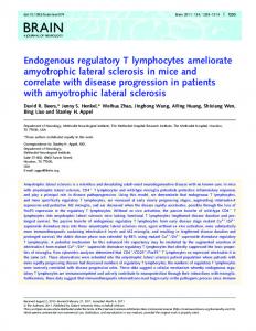
Presentation of endogenous tumor antigens to CD4 T lymphocytes by murine melanoma cells transfected with major histocompatibility complex class II genes Peter
W. Chen,
Department
Abstract:
In
a
Stephen
of Immunology,
previous
E. Ullrich,
The
study,
we
University
and
Honnavara
of Texas
demonstrated
M.D
that
N. Ananthaswamy
Anderson
Cancer
Center,
Houston,
that
Akbearing
K1735 transfectants expressing either Kk or Ak antigens alone produced tumors in syngeneic mice, whereas transfectants that expressed both antigens were rejected. In this study, we investigated whether Ki 735 transfectants
hole and
limpet stimulate
expressing molecules can present endogenous tumor antigens to CD4 T lymphocytes in the absence of normal accessory cells. Our results indicate that K1735 transfectants expressing and A” molecules presented antigen to both CD4 and CD8 T lymphocytes, whereas K1735 transfectants expressing only the Ak or the Kk antigen preferentially stimulated either CD4 or CD8 T cells. Analogous to endogenous antigens, K1735 transfectants expressing A” molecules also presented exogenous hen egg lysozyme (HEL) to HEL-specific 3A9 hybridomas in the absence of normal accessory cells. These results demonstrate that Ki 735 murine melanoma cells expressing molecules can function as antigen-presenting cells and that the generation of an effective antitumor immune response by K1735 melanoma cells expressing Kk and Ak antigens is due to their ability to present endogenous tumor antigens to both helper and cytotoxic T cells. J. Leukoc. Biol. 56: 469-474; 1994.
APCs. Ostrand-Rosenberg et al. [20] have demonstrated that transfection of Sal sarcoma cells with class II genes resulted in the rejection of these tumors in syngeneic mice. In a previous study, we transfected a K1735 melanoma cell
Key Words: antigen-presenting complex #{149} melanoma - gene transfer
cells
.
major
tigens.
These
that
that
expressing
lysed
by preformed
This
histocompatibility
by
of
molecules
addition,
activated
B cells
can
tion.
Interestingly,
MHC Alexander primary
class
also
cells, which augment the immune cytokines [5], recognize peptide antiof MHC class II molecules [3, 4], lymphocytes (CTLs), which kill [6], recognize peptide antigens in the I molecules. In general, professional for initiation oftumor-specific imcells
[14],
function
some [17]
macrophages
and
induce
transformed
as APCs
II molecules
et al. melanoma
or
have lesions
[15],
nonlymphoid
can
also
act
shown that that express
[7-13].
In
or malignant
[16]
T cell proliferacells
as APCs.
expressing
Studies
cell lines from human leukocyte
some
key-
can
IAk
(HEL) these an-
nonlymphoid
antigens
and/or
Kk
antigen,
CTLs, was
K1735
to
[1-4]. response
dendritic
exogenous
egg lysozome specific for
express
H2Kk
only
cells
also
function
either
MHC
genes
and
although
were
supported
(III)
cells
in the
professional
CD8 text
as
class
I or
determined
with
recognized
unable
to stimulate
by
finding
the
the
and
Th
that
interleukin-2
cells.
transfec-
(IL-2)
by
the
T cells
that
can
MHC class To prove formally can
arm
of
the
recognize
transfectants tumor
2,2’-azino-bis
salt;
mercaptoethanol;
BCS,
ELISA, enzyme-linked cell sorter; GAM,
gene
antigen
Eagle’s
medium;
bovine
goat
SDS,
DR;
serum;
immunosorbent anti-mouse;
murine
sodium
Reprint
requests: The
University
combe
Blvd.,
Box
Peter
W.
Received
Journal
Honnavara
178,
April
of Leukocyte
present 5,
20,
1994;
hemocyanin;
Biology
HLA-DR,
human
MEM,
minimal
complex; saline;
Roswell
Park
mAb,
rMuIL-2, Memorial
recomInstitute;
toxoid.
Ananthaswamy,
Department
Anderson
TX
Cancer
of
Center,
Immu-
1515
Hol-
77030. The
Stanford
accepted
2f3-
lymphocyte;
limpet
tetanus
address:
School,
T
FACS, fluorescence-activated hen egg lysozyme; HEPES,
M.D.
Houston,
23-ME,
cytotoxic
histocompatibility
N.
of Texas
Chen’s
Medical
TT,
Ak
Th
assay; HEL,
RPMI,
sulfate;
con-
to
cell;
CTL,
phosphate-buffered
interleukin-2;
dodecyl
nology,
Harvard
major
PBS,
in the expressing
acid; keyhole
MHC,
to CD4 bypassgenerate
antigens
antigen-presenting calf
KLH,
antibody;
binant
response
(3-ethylbenzothiazoline-6-sulfonic
APC,
N-2-hydroxyethylpiperazine-N’-2-ethanesulfonic leukocyte
immune
antigens
endogenous
ABTS,
diammonium
tumor
I antigens. that K1735
present
Abbreviations: acid)
helper
endogenous tumor antigens directly context of MHC class II antigens (thus APCs), as well as their ability to
of
monoclonal
early an-
tigen (HLA) DR can process and present soluble tetanus toxoid (TT) antigen to autologous and H LA-DR-matched allogeneic TT-itnmune helper T cell clones. In addition, Malissen et al. [181 and Norcross et al. [19] demonstrated
stimulate
presenting
ing
either
II
constitutively
conclusion
tion
Tumor-specific immunity mediated by T lymphocytes is initiated by recognition of the peptide antigen in the context of major histocompatibility complex (MHC) molecules present on the surface of antigen-presenting cells (APCs)
are
that
class
with
cells
T cells
munity
not
present
their tumorigenicity and immunogenicity [21]. K1735 transfectants expressing either or Ak molecules alone produced tumors in normal C3H mice, whereas most transfectants that expressed both molecules were rejected in normal C3H mice but produced tumors in nude mice. These results suggested that K1735 transfectants expressing Kk and Ak molecules were able to induce both the helper and the cytotoxic arms of the immune response, whereas K1735
ability
helper (Th) by releasing gens in the context whereas cytotoxic T antigen-specific targets context of MHC class APCs that are responsible
did
could
suggest
MHC
II molecules
cells
(KLH) or hen T cell hybridomas
results
express
line
L
hemocyanin helper
resulted in abrogation oftumorigenicity and induction of antitumor immunity. Based on these results, we hypothesize that the rejection of K1735 transfectants expressing Kk and Ak antigens seen in normal syngeneic hosts is due to their
INTRODUCTION
T
mouse
Texas
Schepens
Street, May
Volume
31,
Eye
Boston,
Research
MA
Institute,
02114.
1994.
56,
October
1994
469
cells
in
the
absence
of
known
accessory
cells,
we
purified
CD4 T cells from immunized mice and then determined whether these cells proliferate when cocultured with K1735 transfectants expressing molecules. In addition, we investigated whether K1735 transfectants expressing A’ molecules can present exogenous antigens to an antigenspecific T cell hybridoma.
MATERIALS
AND METHODS
the plates and gentle pipeting. The resulting cells were subjected to two more cycles of plating on platic to deplete macrophages. The lymphocytes were then passed through a nylon wool column twice and nonadherent cells were collected.
CD4
and
CD8),
Cell
lines
K1735
is a murine
melanoma
induced
in
a mammary
tumor
C3H/HeNCr mouse by initiation with radiation and promotion with repeated croton oil The derivation of K1735 transfectants ex-
L22
pressing
K
been
(1735-6),
described
(1735-il), previously
or both
[21].
(1735-1)
Tumor
ceils
were
antigens grown
minimal Eagle’s medium (MEM; Gibco Laboratories) supplemented with 10% bovine calf serum (BCS; Hyclone Laboratories), 2% vitamin solution, 1.0 mM sodium pyruvate, 0.1 mM nonessential amino acids, 2.0 mM glutamine, 100 U/ml penicillin, and 100 g/ml streptomycin. 3A9 is a murine T cell hybridoma that is activated to produce IL-2 when the nominal antigen HEL is presented by professional APCs [23]. This hybridoma responds to HEL amino acid residues 46-61 [24, 25] presented by IAk_bearing APCs. 3A9 cells were grown in RPMI 1640 supplemented with 10% BCS, 1% vitamin solution, 1.0 mM sodium pyruvate, 0.1 mM nonessential amino acids, 5 x l0 M 2/3-mercaptoethanol (2/3-ME), 0.5 M N-2-hydroxyethylpiperazine-N-2-ethanesulfonic acid (HEPES) buffer, 100 U/ml penicillin, and 100 tg/ml streptomycin. All cell lines were tested for and found free of Mycoplasma and specific pathogenic viruses. in
Indirect
immunofluorescence
The K1735 cell line and its various transfectants were analyzed for expression of and A’ antigens using monoclonal antibodies (mAbs) 16-1-uN and 10-3.6.2, respectively [26]. Single-cell suspensions (2 x 10⁶ cells) were washed with PBS + (phosphate-buffered saline containing 1% goat serum, 0.1% bovine serum albumin, and 0.02% sodium
azide)
of
16-1-uN
ter for
two 30
and or
washes
mm
isothiocyanate
on
ice
for
hybridoma
with with
(Organon + , the cells
1%
goat
Teknica Corp.). were suspended and
fluorescence-activated Coulter Electronics).
cell sorter As a control,
mAb
either
200
supernatants.
tl Af-
PBS+, the cells were incubated on 100 sl of 1:500 diluted fluorescein
paraformaldehyde
FITC-GAM
1 h with
culture
(FITC)-conjugated
antibody with PBS ing
incubated 10-3.6.2
alone
to
anti-mouse
(GAM)
After two more in 1 ml of PBS
washes contain-
then
(FACS; cells
analyzed
ice
the
by
background
fluorescence.
Purification C3H 1735
RPMI RPMI cytes
mice
IH
of CD4
and
T cells
were immunized by subcutaneous transfectants (1 x 10⁶ cells/mouse)
1640 medium. 1640 medium
Journal
in
injection serum-free
of
As a control, mice were injected with alone. Two weeks later, splenic lympho-
from unimmunized vested separately and (2 x 10⁶ cells/dish) and 37#{176}Cfor 2 h. Nonadherent
470
CD8
and immunized mice were harplated on 100-mm plastic dishes incubated in RPMI 1640 medium at cells were collected by swirling
of Leukocyte
Biology
Volume
56, October
positively
selected
by passing
The
absence
of macrophages
T cell population (Ml/70.15.l1.5HL)
FITC-conjugated
goat
was
anti-rat
mAb
Lymphocyte Purified munized 1640
proliferation
CD4 mice medium
and CD8 (1 x l0 in
B cells
assay T cells cells/well)
96-well
and
ascertained by staining and anti-B220 mAb. (Organon Teknica We found that T cells contained 90-96% T cells. Similarly, T contained 84-99% T cells. In addition, did not react with mAb.
from unimmunized were cocultured
microtiter
plates
with
and imin RPMI varying
num-
bers (1 x l0 to 5 x l0 cells) of y-irradiated (10,000 R) K1735 parent or l735NI, i735PII, or i735III transfectants. After incubation at 37#{176}Cfor 3 days, [3H]thymidine (1 tCi/well) was added, and 16 h later incorporation of radioactivity was determined using a Wallac Betaplate Reader (Wallac Inc., Gaithersburg, MD). As controls, T cells were cultured in the absence of ‘y-irradiated tumor cells to determine background radioisotope uptake. These values were subtracted from the values obtained with tumor and T cell cocultures.
of exogenous
HEL
to 3A9
hybridoma
by
K1735 transfectants Parental K1735 or K1735 transfectants (1 x i0 cells/well) were plated in 24-well plates in complete RPM! 1640 medium in the absence or presence of 100 tg/ml HEL (Pierce). After 24 h of incubation at 37#{176}C,the medium was removed and the cells were washed 3 times with PBS. 3A9 cells in complete RPM! medium were then added to each well (1 x 10⁶ cells/well) and incubated for 2 days at 37#{176}C. Culture rpm,
and
supernatants analyzed
were the
for
collected, presence
centrifuged of IL-2.
at
1200
IL-2 assay
EPICS Profile; were treated with
determine
were
Corp.) was used a secondary antibody. eluted from anti-CD4 mAb columns CD4 T cells and less than 0.5% CD8 cells eluted from anti-CD8 columns CD8 T cells and less than 0.8% CD4 enriched CD4 and CD8 T cells anti-Mac-i mAb or with anti-B220
Presentation
assay
T cells
respectively.
in the purified with anti-Mac-i
virus-negative ultraviolet treatment has
CD8
the enriched T cells over anti-CD4 or anti-CD8 mAb-coated columns (Accurate Chemical and Scientific Corp., New York) according to the manufacturer’s instructions. The purity of CD4 and CD8 T cell subsets was determined by FACS analysis using mAbs GK1.5 (anti-CD4) and 53.672 (anti-
1994
Microtiter (96-well) plates were coated anti-IL-2 mAb (2 sg/ml) (PharMingen) (0.1 M NaHCO3, pH 8.2) and incubated Each well was washed three times with taming 0.05% Tween 20 and dried at Two hundred microliters of 10% BCS in each well and incubated for 2 h at room were washed three times with 200 l of dried culture
hundred microliters of the to each well, and the plates were incubated overnight at 4#{176}C.Plates were washed four times with 200 jl of PBS-Tween 20 and dried at room temperature. One hundred microliters of biotinylated anti-IL-2 (2
tg/ml)
at
room temperature. supernatant was
with 50 j.tl of in coating buffer overnight at 4#{176}C. 200 ,sl of PBS conroom temperature. PBS was added to temperature. Plates PBS-Tween 20 and
(PharMingen)
and incubated for 45 washed six times with
One
added
in
mm
10%
PBS
was
added
at room temperature. 200 tl of PBS-Tween-20
to
each
well
Plates were and dried at
room temperature. One-hundred fifty microliters of 0.1 tg/ml avidin peroxidase (Pierce) was added to each well and incubated for 2 h at room temperature. Plates were washed eight times temperature.
with
200 One
l
of PBS-Tween 20 and hundred microliters
bis(3-ethylbenzothiazoline-6-sulfonic (ABTS)-peroxidase (Pierce) cubated for 30 mm at room 1% sodium dodecyl sulfate stop the reaction and the MR5000 enzyme-linked plate reader (Dynatech) at dard curve using various binant murine IL-2 (rMuIL-2; generated ture
and
used
100
0
E
dried at room of 2,2’ -azino-
80
acid) diammonium salt was added to each well and intemperature. Fifty microliters of (SDS) was added to each well to plates were read on a Dynatech immunosorbent assay (ELISA) a wavelength of 405 nm. A stanknown concentrations of recomCollaborative Research) was
to determine
the
levels
of IL-2
in the
60
G) C.)
40
0 C 20
C-)
cul-
supernatant.
0
01 0
RESULTS Expression of Kk and Ak antigens K1735 transfectants
and tumorigenicity
Fig.
The
K1735 in this study
transfectants (1735-6, 1735-11, and 1735-1) used express high levels ofeither K’, Ak or both antigens, respectively (Fig. 1). In contrast, the parental Kl735 murine melanoma cell line does not constitutively express either MHC class I or class II antigens. In addition, all cell lines with the exception of 1735-i, which expresses both Kk and Ak antigens, produce tumors in normal C3H mice following injection of 1 x l0 cells/mouse (Fig. 2). Although the 1735-1 cell line is rejected in normal C3H mice even at a dose of 1 x 10⁶ cells/mouse, this cell line produces tumor in athymic nude mice at a dose of 1 x lO cells/mouse (data not shown), suggesting that the rejection of 1735-i transfecin fact
( P11)
normal that
C3H MHC
transfectants,
to postulate and present us
mice (PII)
were
that MHC endogenous
is immunologically transfectants,
rejected (IIP) tumor
mediated. not MHC
but
in normal
C3H
mice
led
cells may function as APCs antigens directly to Th cells.
4
Weeks
of 2. Tumorigenicity
tigens.
tants The
2
Tumor
mal
C3H
weeks.
cells mice.
(0)
Presentation
CD8 We
(1
x
expressing
were
growth
(#{149}) 1735-6
Injection
transfectants
105/mouse)
Tumor
K1735;
After
of K1735
was
injected
(A
of endogenous
)
at
1735-Il
tumor
Kk and/or
subcutaneously
measured
(I’IIi;
6
weekly
(III’);
intervals (A)
antigens
1735-I
12
(I’ll’).
to CD4
and
investigated
whether
cells
from
immunized
mice
Ki735
cells,
K1735
can
transfectants
present
endogenous
expressing
tumor
Kk
antigens
and/or CD8 T cells in the absence of in vitro and cause their proliferation. The results shown in Figure 3A and B reveal that lethally irradiated 1735-1 (IIP) transfectants caused the proliferation of both CD4 and CD8 T lymphocytes from immunized but not from unimmunized mice (data not shown). However, 1735-11 (III) transfectants caused the proliferation of CD4 T cells but not CD8 T cells from immunized mice only (Fig. 3A). Conversely, 1735-6 (III) transfectants caused the proliferation ofCD8 T cells but not CD4 T cells which
antigens,
did
only
do
not
not
(Fig.
3B).
express
cause
As expected,
detectable
the
proliferation
parental of K’ or of either T cell levels
subset.
‘I)
K1735
Presentation of HEL antigen to 3A9 T cell h’bridoma by K1735 transfectants expressing Kk and/or A molecules
0 ‘4-
0 I.-
(Ilr)
1735-6
a)
E
z
(Iii’)
1735-11
a)
Because transfected antigens
several studies have shown that nonlymphoid cells with MHC class II genes can present exogenous to T helper cells in the absence of normal accessory
cells
19,
[18,
27-29k,
we
investigated
tants
expressing A antigens tigens to Tb cells. To test
> 1735-1
a)
1735-il
(lif’)
(III)
whether
can this,
transfectants
K1735
transfec-
also present exogenous we pulsed 1735-1 (III)
with
HEL
and
then
and 3A9
Log Fluorescence Fig.
1. Surface
Single-cell
(10-3.6.2) staining
mAbs, with
cycles
expression
suspensions
as described secondary
of fluorescent
antigen-positive
of Kk and were
stained
Ak antigens with
in Materials FIIC-GAM
intensity.
and mAb
The
on
antiKk
number
K1735
Methods.
alone.
transfectants.
(16-1-1IN)
panel
(PII)
antiAk
and
represents
four
the HEL produced
represents
per-
positive
Control
Abscissa
in each
and
1735-11 (PIP) transfectants cells to produce IL-2.
represents
cells.
transfectants
Chen
et al.
A”-bearing
1735-6
(PIP)
transfectants
antigen to 3A9 IL-2 only when transfectants, (Fig.
K1735
pulsed However,
but
Th cells. stimulated not with
with the
were
anand
determined
whether these cells can stimulate 3A9 cells to produce The results shown in Figure 4 reveal that both 1735-i
cent
norfor
T cells by K1735 transfectants
and/or Ak antigens to purified CD4 known accessory
A’ (I)
log
Ak aninto
IL-2. (PIP)
HEL stimulated parental K1735
unable
to present
As expected, 3A9 cells with HEU-pulsed A”KUH-pulsed A”-positive
4).
melanoma
cells
function
as APCs
471
A
60000
3A9
0 50000
C
KLH HEL
K1735(V11)
.2
a,
40000
0
U
.
-I
1735-11
(I -
Il
___________________________ I
-
0
U
30000
C
20000
a, 0
C
1735-6(llI)
(I
1735-1
)
II
I
l0000i
I
0
10
0 1000
10000
100000
Number
1000000
of Stimulator
Fig. 4. Presentation pressing
Cells
Ak
HEL
or KLH
cells
for
presence
IQ.
of HEL
antigen
Tumor
cells
antigens.
for 24 h, washed
2
days
of
IL-2.
at
30
20
lL-2
Release to 3A9 (I
with
x
PBS,
37#{176}C. Culture
cells
l0)
40
50
60
(Units/mi) by Kl735
were
and
incubated
supernatants
transfectants
pulsed
with
100
with
were
exg/ml
1 x 10⁶ 3A9
analyzed
for
the
B
C-) C
stimulate CD8 cells that the context of MHC class strate that Ki735 melanoma
0
I :
K”
and
Ak
molecules
well as CD8 other known
pressing CD4 cells, cause
10000
E
only
0 1000
±
C”
10000
Number Fig. Kk
(I
x
3. Presentation and/or Akbearing l0)
1735-1
from
irradiated dine
Kl735
stimulator k)r
16 h,
tumor
were
cells
for
incorporation
mice
of
and
at
CD4
from
or
CD8
mice
immunized
with
various
numbers
37#{176}C. After
pulsing
radioactivity
(B) CD8 ‘I’ cells. (#{149}) 1735-I (A) K1735 parent.
cells. (I11);
to T lymphocytes
Purified
cocultered
3 days
Cells
antigens
transfectants. C3H
transfectants
1000000
of Stimulator
c)f endogenous
unimmunized
(1I1)
100000
was
(I’II);
(0)
with
determined. 1735-11
by T
cells with of
-y-
the
cause
tumor antigens in Our results demonexpressing both proliferation of CD4 as mice in the absence of K1735 transfectants ex-
the
from immunized cells, whereas A”
or
the
Kk
antigen
stimulated
either
or CD8 T cells. In contrast, parental Ki735 tumor which lack expression of K” and A” molecules, did not the proliferation of either CD4 or CD8 T cells.
In addition I-
can
T cells accessory
can recognize I antigens. transfectants
gens
to
CD4
these
transfectants
to the presentation ofendogenous T cells by A”-bearing K1735 can
present
exogenous
tumor antitransfectants, HEL
antigen
to
3A9 hybridomas in the absence of known accessory cells. The 3A9 cell line is a murine helper T cell hybridoma that specifically produces cytokines when HEL antigen is processed and presented by professional APCs. This hybridoma is reactive to HEL amino acid residues 46-61 [24, 25]. Because our A”-bearing K1735 transfectants pulsed with
[3Hthymi(A)
(Ill’);
CD4
(A)
T
1735-6
No mAb Since efficiently
1735-1 (III) and 1735-Il presented HEL antigen to
(IIP) 3A9
>
transfectants cells, we inves-
Anti-isotype
tigated context shown blocked
whether the antigenic peptides were presented in the of MHC class I or class II molecules. The results in Figure 5 reveal that anti-As’ mAb completely the ability of HEL-pulsed 1735-1 (PIP) and 1735-1
(IIP)
cells
whereas
to
stimulate
antiKk
and
3A9
anti-isotype
hybridoma
matched
to
produce
mAb
had
C
< ,
.E
AntiAk
IL-2,
.2
no effect.
Anti-K
DISCUSSION 0
In
this
K1735 normal
study
we tested
transfectants syngeneic
helper dogenous text of
arm
472
Journal
the
expressing hosts is due
hypothesis
that
K” and Ak to their ability
of the immune response tumor antigens directly to CD4 MHC class II antigens as well
of Leukocyte
Biology
the
rejection
56,
October
IL-2
of
antigens seen in to stimulate the
by presenting enT cells in the conas their ability to
Volume
10
1994
Fig.
5.
mAb.
(10-3.6.2),
then
described
30
Release
40
50
analyzed before.
or
for
anti-isotype-matched
their
ability
(IgG2a)
to present
60
(Units/mI)
Blocking of APC function of Ak-bearing transfectants 1735-I (III) cells were incubated with 50 sg/ml anti-K
anti-Ak
and
20
HEL
mAb
antigen
for
by anti-Ak (l6-l-llN), 1 h at
to 3A9
37#{176}C
cells
as
HEL caused the proliferation of 3A9 cells, it is reasonable to assume that our Akbearing tumor cells can indeed process HEL and present the reactive antigenic peptides 46-61 to
ACKNOWLEDGMENTS This
work
3A9
can
Cancer
cells
in
a manner
cells. In any transfectants antigens to reports that
case, can CD4 MHC
dogenous ternatively,
exogens because
and
similar
to
that
of
normal
accessory
our demonstration that Akbearing Ki735 present both endogenous and exogenous T cells is in agreement with previous class II molecules can present both en-
rejection
response
failure class
could be I expression
nize
tumor
molecules,
MHC
to cells
in
insufficient
class
in syngeneic
attributed on these
antigens
to CD4 T cells have demonstrated
Institutes
ported Institutes
We
supported
Society by
a
of
Dr.
and
(to
S.E.U.).
Health
Emil
IM-598
H.N.A.)
predoctoral Health under
of
thank
by grants
(to
for
association
with
may
or
result
(Fig.
of
MHC recog-
class
I
expression
escape
2. Schwartz, with gene Annu. Rev.
2). This
MHC
lack
in their
cells
expressing
transfected
MHC
class
II molecules
cells
tumors the
were
that
invariant
are
rejected
chain.
transfected
with
finding
that
and A” molecules CD4 and CD8
Ki735
Surprisingly,
the
be achieved class I and
augmenting
invariant
efficient forms on stimulation of the immune
T cells under
in our
sytem
exhibit
Sal
MHC
IP
they
be-
gene, suggest class
of both
activity.
supplying
us with
the
3A9
the
major
in association complex.
Miles, C., Grey, ovalbumin pepUSA 83, 3968-3971.
Sci.
Miles,
C.,
Grey,
histocompatibility
capacity
ofla
K., of
immunogenic
Feldman,
J.D.
(1983)
Young,
J.W.,
R.M.
lymphocytes
J.,
Melief,
decreases
C.J.M.
or
(1987)
by
cytomajor
the
J.
Inaba, Steinman,
K.,
cells
vivo.
in
Shevach, phages
Metlay, J.P., R.M. (1990) Int.
Rev.
EM., in antigen
macrophage response.
Ziegler,
Unanue,
J. ER. event
presentation
pig
regulation Med 138,
Exp. (1981)
T-
response.
Witmer-Pack, M., antigen presenting
197-206.
S.A. (1973) by guinea
in the
with
ofCD4
T lymphocyte MT., cells as
6,
Immunol.
Rosenthal, recognition
Role of the the immune K.,
Crowley, Dendritic
Function of T lymphocytes.
of genetic 1213-1229.
Identification
required
for
to T lymphocytes.j
ofa
I-region
macroII.
control
127,
Immunol.
gen
presenting
cells:
131,
munol. Issekutz,
anti-
1869-1875.
T.,
Chu,
L.H.,
gen-beaning alloantigen fashion
E.,
Geha,
Kim,
(1983)
for
B cell
B cells
antiasProc.
as anti-
activation.].
MA.,
presentation
R.S.
(1982)
Antigen
proliferation induced cells. J. Immunol. 129,
K.,
Green,
tumor
in a major to antigen-reactive
advanced,
H.M.
requirement
T cell
B cell
Alexander, tigen
the
175-178. R.W., Grey,
is
Im-
109-114.
by human B cells: virus B lymphoblastoid Glimcher,
79,
Sci. USA T., Chestnut,
of
macrophage
restricted
Zeigler, K., Unanue, ER. (1982) Decrease in macrophage gen catabolism caused by ammonia and chloroquine sociated with inhibition of antigen presentation to T cells. NatI. Acad Kakiuchi,
Exp.
Stimulation
requirement
in the induction ofcytotoxic Immunol. 18, 219-223.
activa-
J.
cells.
(1988)
obviates
Direct
dendritic
cells
Eur.
17.
restricted
role of polymorphic T cell restriction-specificity,
Steinman,
cytotoxic
cells
helper
16.
the biological determining
Inhibition
by a monoclonal
Adv. Immunol. 27, 51-178. M. (1985) Properties of purified T cell subresponses of purified T cell subsets. J. Exp. Med.
CD8
dendritic
15.
peptides.
responsiveness.
166, 182-194. C.J., Boes,
Med.
11.
(1987)
(MHC)
2068-2088.
tion
10.
H.M.
complex
to bind
J.S.,
studies on antigens
I. In vitro
8. Inaba,
MHC
lines
I., can
Paul,
present
(1982)
protein
J.,
by
melanoma
cell
lines
early,
disease.
j
but
not
biologically
B.,
Steinmetz,
Ia
anti-
antigen
and
complex-restricted Med 155, 445-459.
Bennicelli,
Guerry,
Epstein-Barr
1446-1450.
WE.
histocompatability T cells. J. Exp. human
presentation by
D. (1989)
Defective cultured Immunol.
anfrom
142,
4070-4078.
18.
Malissen, Hood,
et al.
Acad
SM.,
Rosenberg,
and
162,
14.
is
recognition histocompatibility
(1985)
J., Schaefer,
Sprent, sets.
K”
investigation.
G., Unanue, ER. to Ia histocompatibility
1353-1358.
S.C.,
function 7.
gen
helper could
Chen
and
235,
NaIl.
Colon,
between
toxic T cells: transplantation
for
This
Proc.
A.,
antigen-processing
tumor cells, thereby stimulation of CD4 then secrete cytokines [41-43] and augment CTLs. In addition T cells may be cytoevidence that CD4 T do not know whether
cytotoxic
sup-
the National T32-CA-09598.
ofinterleukin synthesis and T cell proliferation anti-Ia antibody. j Immunol. 130, 1236-1240. 6. Zinkernagel, R.M., Doherty, P.C. (1979) MHC
12.
to both accessory
of immunotherapy of both the response. This
relation
5. Gilman,
13.
both
Sette,
restriction
that the IP tumors
antigens of known
expression
Na-
was
Smith, C., Freed, J.H., between a “processed”
Ia molecules. S.,
Science
expres-
expressing
II molecules on autologous transforming the cells into APCs for cells. The activated CD4 Th cells can such as IL-2, interferon-rny, and IL-7 the proliferation of tumor-specific CD8 to secreting cytokines, activated CD4 toxic to tumor cells. Although there is cells can kill tumor cells [44-48], we currently
chain
transfectants
the
lack
when
can present endogenous T cells in the absence more depend arm
CD4
mice
These results chain on Sal MHC functioning.
cells implies that melanoma may and the cytotoxic
by
in normal
and
The
to act as et al.
came highly tumorigenic. presence ofthe invariant prevents the APCs from The
tide
9. Boog,
APCs [19, 27-29]. In addition, Ostrand[38, 39] demonstrated that an immune response against Sal mouse sarcoma depends on the presence of the cytoplasmic domain of MHC class II molecules, which suggests that an unknown intracellular signaling event is required for tumor-specific immunity. However, Clements et al. [40] demonstrated that Sal MHC
class II sion of
Chen
from Grant
RH. (1985) T-lymphocyte products of the major Immunol. 3, 237-261.
S., Colon, SM., (1986) Interaction
4. Buus,
CTL
(
are able Rosenberg
3. Buus, H.M.
of
from
recognition and destruction. The fact that Ki735 transfectants expressing A” molecules caused the proliferation of only CD4 but not CD8 T cells led us to conclude that antigen presentation by Akbearing K1735 transfectants is MHC class II restricted. This conclusion is further supported by the finding that presentation ofthe HEL antigen to 3A9 cells by 10735 transfectants expressing both Kk and A” molecules was completely blocked by anti-As’ mAb but not by anti-Ks’ mAb Fig. 5). These results are in agreement with previous reports showing that Ia-bearing L cells could present antigens to T cell hybridomas in an antigen-specific, MHCrestricted manner [18, 19, 27-29]. Several investigators have demonstrated that mouse and
human
Amenthe
[30-35]. that
the lack of sufficient (Fig. 1). Since CTLs
expression
I antigens
hosts
the from
Peter
fellowship Training
Unanue
from
AR40824
Alhybnidoma. A’#{176} APCs can stimulate autorea#{231}tive T cell clones [36, 37], it is REFERENCES quite possible that K1735 transfectants expressing A” molecules can stimulate autoreactive T cell clones to produce 1. Babbett, B.P., Allen, P.M., Matsueda, IL-2, thus providing help for MHC class I restricted CTLs. Binding of immunogenic peptide Although 1735-il (PIP) transfectants can present enmolecules. Nature 317, 359-361. dogenous antigens to CD4 T cells, they are unable to induce
a tumor
antigens others
tional
was
A’-bearing
U.
(1983)
K1735
Expression
melanoma
M.,
McMillen,
of
IAk
cells
class
function
M., II
genes
Pierres, in
as APCs
mouse
M., U
473
cells after 19. Norcross, RN.
DNA-mediated MA., Bentley,
(1984)
Membrane
gene transfer. Nature 305, D.M., Marguiles, D.H., Ia
expression
and
Ostrand-Rosenberg,
S.,
Thakur,
with
Med Clements,
A.,
class II major 160, 1316-1337. V.K.
(1990)
Re-
jection of mouse sarcoma cell after transfection of MHC class II genes. j Immunol. 144, 4068-4071. 21. Chen, P.W., Ananthaswamy, H.N. (1993) Rejection of K1735 munine
22.
23.
melanoma
in
MHC
class I antigens
munol.
151,
Knipke,
in
Cancer
Inst.
Allen,
P.M.,
and
hosts
either
requires
class
expression
II antigens
or IL-2.J.
Im-
(1979)
the
Speculations
development 63, 541-545. Unanue,
on
of
the
role
malignant
ofultraviolet
j
Allen,
(1984)
P.M.,
Unanue,
ER.
macrophages: identification two T cell hybridomas. Proc. 25.
Allen, P.M., Matsueda, Specificity of the T cell
generated 26.
munol. Ozato,
Differential
NatI.
requirements
Mayer,
N.,
(1984)
Processing
of the
determinant
Natl.
G.R., receptor:
by the same 135, 368-373. K.,
27.
Sachs,
for
tional 28.
29.
30.
31. 32.
33.
RN.,
Norcross,
expression
Nature 306, Raboudin-Combe,
of
Sci.
the (1980)
to mouse
MA.,
of
IAk
by
by
murine
(1985) are
molecule.
H-2
and D.H.
class
j
Im-
cell
lines
Ia antigens. (1984)
II
from
Func-
MHC
gene.
Journal
42.
C.,
Mach,
B. (1983)
Expression
those
derived
from
exogenous
of HLA-DR
antigen.
j
Nabavi,
N.,
Mosmann,
of Leukocyte
Biology
Volume
56,
October
G.J., commajor
1994
EM. T
Ghogawala,
T.R.,
(1982)
cell
Production
J.
hybnidomas.
Myer,
A.,
properties.
blood.]
Exp.
44. Nagarkatti, M., zation of tumor lymphokine
144, Rotteveel,
RU.
oflymphokine
43. Alderson, MR., leukin 7 enhances duces lymphokine
45.
Z.,
Coffman,
patterns
tional
Immunol.
Shevach,
of autoreacExp.
156,
Med.
Griffith,
I.J.,
Wade,
W.F.,
Chen, Z.Z., McKean, D.J., Glimcher, U.H. (1989) Antigen presentation abgrogated in cells expressing truncated Ia molecules. j Immunol. 142, 1444-1447. Ostrand-Rosenberg, S., Roby, CA., Clements, V.K., Cole, GA. (1991) Tumor-specific immunity can be enhanced by transfection of tumor cells with syngeneic MHC-class-II genes or allogeneic MHC-class-I genes. mt. j Cancer 6, 61-68. Ostrand-Rosenberg, S., Roby, CA., Clements, V.K. (1992) Abrogation of tumorigenicity by MHC class II antigen expression requires the cytoplasmic domain of the class II molecule. j Immunol. 147, 2419-2422. Clements, V.K., Baskar, S., Armstrong, T.D., OstrandRosenberg, S. (1992) Invariant chain alters the malignant phenotype of MHC class IP tumor cells. J. Immunol. 149, 2391-2396. Gilman, S.C., Rosenberg, J.S., Feldmam, J.D. (1983) Inhibition of interleukin synthesis and T cell proliferation by a monoclonal anti-Ia antibody. j Immunol. 130, 1236-1240. different
190-194.
147, 3306-3313. 34. Adorini, U., Moreno, J., Mombourg, F., Hammerling, Guery, J.C., Valli, A., Fuchs, S. (1991) Exogenous peptides pete for the presentation of endogenous antigens to
474
40.
41.
antigens at the surface of mouse U cells cotransfected with cloned human genes. Nature 303, 670-674. Malissen, B., Peele-Price, M., Governman, M., McMillen, M., White, J., Kappler, J., Marrack, P. (1984) Gene transfer of H2 class II genes: antigen presentation by mouse fibroblast and hamster B cell lines. Cell 36, 319-327. Nuchtern, J.G., Biddison, WE., Klausner, RD. (1990) Class II MHC molecules can use the endogenous pathway of antigen presentation. Nature 343, 74-76. Weiss, S., Bogen, B. (1991) MHC class Il-restricted presentation of intracellular antigen. Cell 64, 767-776. Brooks, A., Hartley, S., Kjer-Nielsen, U., Perera, J., Goodnow, CC., Basten, A., McCluskey, J. (1991) Class Il-restricted presentation of an endogenously derived immunodominant Tcell determinant of hen egg lysozyme. Proc. Nati. Acad Sci. USA 88, 3290-3294. Moreno,J., Vignali, D.A.A., Nadimi, F, Fuchs, S., Adorini, U., Hammerling, G.J. (1991) Processing of an endogenous protein can generate MHC class Il-restricted T cell determinants distinct
39.
2489-2492.
Hybridoma
Marguiles,
a transfected
lysozyme
recognized
81,
USA
T cell
E., Unanue, ER. different determinants
and D.H.
monoclonal antibodies Immunol. 124, 533-540.
Germain,
Acad. Haber, two
peptide
secreting
J.
37.
38. ER.
L.H.,
I region-restricted
640-645.
radi-
melanoma.
Glimcher, tive
of
antigen processing by macrophages for lysozyme-specific hybridomas. j Immunol. 132, 1077-1079. 24.
36.
244-255.
ML.
ation
syngeneic
histocompatibility complex class Il-restricted T cells. j Exp. 174, 945-948. 35. Calin-Laurens, V., Forquet, F., Lombard-Platet, S., Bertolino, P., Chretien, I., Trescol-Biemont, M.-C., Gerlier, D., Rabourdin-Combe, C. (1992) High efficiency of endogenous antigen presentation by MHC class II molecules. ml. Immunol. 4, 1113-1121. Med.
antigen-presentating
accessory cell function of U cells transfected histocompatibility complex genes. j Exp. 20.
440-443. Germain,
(1989)
TH1
and
TH2
lead to different funcAnnu. Rev. Immunol. 7, 145-173. Sassenfeld, H.M., Widmer, MB. (1990) Intercytolytic T lymphocyte generation and inactivated killer cells from human peripheral
172,
Med.
577-587.
Clary, S.A., Nagarkatti, P.S. (1990) Characteriinfiltrating CD44 T cells as Thi cells based
secretion
and
functional
j
properties.
on
Immunol.
4898-4905. ET.M.,
Kokkelink,
I., van
Lier,
RAW.,
Kuenen,
Meager, A., Miedema, F., Lucas, C.J. (1988) Clonal functionally distinct human CD4 T cell subsets. j 168, 1659-1673. 46. Erb, P., Gorgg, D., Troxler, M., Kennedy, M., Fluri,
CD4
cells:
secretion
T cell mediated
killing
of MHC
presenting cells: I. Characterization in vivo or in vitro activated CD4
class
II
positive
B.,
analysis Exp.
of Med.
M.
(1990) antigen
of target cell recognition killer T cells.]. Immunol.
by
144,
790-795.
Ozdemirli, M., El-Khatib, M., Bastiani, U., Akdeniz, H., Kuchroo, V., Ju, S.-T. (1992) The cytotoxic process ofCD4 ThI clones. j Immunol. 149, 1889-1895. 48. Ruijs, T.C., Uouste, K., Brown, E.A., Antel, J.P. (1993) Lysis of human glial cells by major histocompatibility complex-unrestricted CD4 cytotoxic lymphocytes.]. Neuroimmunol. 42, 105-111.
47.