究包括 103 位在特里布文教学医院工作的显微镜工作者。 .... equivalent power) were classified as emmetropic, greater than. + 0. ... Near point of convergence.
国际眼科杂志摇
2018 年 7 月摇 第 18 卷摇 第 7 期摇 摇 http: / / ies. ijo. cn
电话:029鄄82245172摇 摇 85263940摇 摇
电子信箱:IJO. 2000@ 163. com
·Original article·
Prevalence of myopia and binocular vision dysfunctions in microscopists Rajeshwori Ngakhushi1 , Raju Kaiti2,3 , Sanjeev Bhattarai4 , Gulshan Bdr Shrestha4 Reiyukai Eiko Masunaga Eye Hospital, Banepa 45210, Nepal 2 Department of Ophthalmology, Dhulikhel Hospital, Kathmandu University, Kavrepalanchowk 45200, Nepal 3 Drishti Eye Care System, Kalanki 44620, Nepal 4 Department of Ophthalmology, B. P. Koirala Lions Centre for Ophthalmic Studies, Institute of Medicine, Tribhuvan University, Kathmandu 44613, Nepal Correspondence to: Raju Kaiti. Department of Ophthalmology, Dhulikhel Hospital, Kathmandu University, Kavrepalanchowk 45200, Nepal. rajukaiti@ gmail. com Received: 2018-01-25摇 摇 Accepted: 2018-04-27 1
显微镜工作者近视和双眼视力障碍的患病率分 析
Rajeshwori Ngakhushi1 , Raju Kaiti2,3 , Sanjeev Bhattarai4 , Gulshan Bdr Shrestha4 ( 作者单位:1 45210 尼泊尔,巴内帕,Reiyukai Eiko Masunaga 眼科 医院;2 45200 尼泊尔,Kavrepalanchowk,加德满都大学,图利凯尔 医院,眼科;3 44620 尼泊尔,卡兰基,Drishti 眼护系统;4 44613 尼 泊尔,加德满都,特里布文大学医学院,B. P. Koirala Lions 眼科 研究中心) 通讯作者:Raju Kaiti. rajukaiti@ gmail. com
摘要 目的:测定临床显微镜工作者屈光和双眼视力状态。 方法:这是一项以医院为基础的观察性和横断面研究。 研 究包括 103 位在特里布文教学医院工作的显微镜工作者。 受试者均行全面的眼部检查,包括静态检影,动态检影和 视轴评估。 收集受试者显微镜下视觉状态信息。 结果: 该 组 显 微 镜 工 作 者 屈 光 不 正 患 病 率 为 69. 90% 。 68. 93% 受试者近视,平均近视误差为-1. 58 依1. 89 D。 研 究发现 61. 20% 受试者汇聚功能不全。 调节不足与调节 功能不全的发病率分别为 41. 30% 和 40. 06% 。 研究人群 的融合性转向也有所降低。 结论:研究发现,临床显微镜工作者屈光不正尤其是近视 的患病率增加。 其中大多数有转斜和调节不足。 大多数 受试者视疲劳症状与其显微镜工作有关,这可能会影响他 们的工作效率。 关键词:显微镜工作者;屈光不正;显微镜近视;视轴矫正; 视疲劳 引用:Ngakhushi R, Kaiti R, Bhattarai S, Shrestha GB. 显微镜工
作者近视和双眼视力障碍的患病率分析. 国际眼科杂志 2018; 18(7) :1180-1183 1180
Abstract
誗 AIM: To determine the refractive and binocular vision
status in clinical microscopists. 誗METHODS: It was an observational and cross sectional hospital based study. One hundred and three microscopists working at Tribhuvan University Teaching Hospital were recruited in the study. All subjects had a comprehensive eye examination including static retinoscopy, dynamic retinoscopy and orthoptic evaluation. Information about their visual symptoms associated with microscopy was also collected. 誗 RESULTS: The prevalence of refractive error in this group of microscopists was 69. 90% . Majority of the subjects were myopic (68. 93% of total subjects) with the mean myopic error of - 1. 58 依 1. 89 D. Convergence insufficiency was found in 61. 20% of the study population. Prevalence of accommodative insufficiency and infacility were 41. 30% and 40. 06% respectively. Fusional vergence was also reduced in this study population. The outcomes of this study were expected to increase the awareness about the refractive and binocular vision anomalies among this population. 誗 CONCLUSION: There was found to be increased prevalence of refractive error in clinical microscopists, especially myopia. Majority of them had vergence and accommodative anomalies. Most of the subjects reported asthenopic symptoms associated with their microscopy work, which may affect their work efficiency. KEYWORDS: microscopists; refractive error; 誗 instrumental myopia; orthoptic; asthenopic
DOI:10. 3980 / j. issn. 1672-5123. 2018. 7. 03
Citation: Ngakhushi R, Kaiti R, Bhattarai S, Shrestha GB.
Prevalence of myopia and binocular vision dysfunctions in microscopists. Guoji Yanke Zazhi(Int Eye Sci) 2018;18(7):1180-1183
INTRODUCTION efractive error is a condition where an unfocused image is formed on the retina. Microscopy work, which involves prolong near focus can lead to refractive error, oculomotor imbalance and asthenopic symptoms. According to a study, among 50 clinical microscopists, 60% of the subjects reported refractive errors [1] . Heavy near work is the most important factor for higher incidence of myopia, poor convergence and exophoria [2] . Near work is primary, environmental based factor in the aetiology and progression of myopia [3] . Majority of people whose myopia progressed were law students [4] ,
R
Int Eye Sci, Vol. 18, No. 7, Jul. 2018摇 摇
Tel:029鄄82245172摇
cadets in the air force academy [5] and microscopists [6] . According to a study by Fritzsche et al [7] on 163 pathologists, 89% suffered from ametropia. Myopia was the most common refractive error affecting 75. 50% of the cases. Instrumental myopia is the over accommodation that occurs when looking through optical instruments, for example binoculars, telescopes, phoropter, auto refractor and microscopes, even though these devices render the image at optical infinity [8-10] . This over accommodation can create an imbalance between the accommodative and vergence system which potentially lead to myopia progression [11] . Sustained and chronic accommodation can lead to vitreous chamber elongation and myopia due to scleral stretching [12] . Ninety four percent of subjects mentioned different kinds of asthenopic symptoms [1] . The association between prolonged use of microscope and visual problems has been recognized for decades. However awareness about these problems is still ignored. In this study, we sought to determine the presence of refractive and binocular vision anomalies in a group of Nepalese microscopists. We wanted to assess asthenopic and visual symptoms associated with the near work and provide awareness about their ocular health. SUBJECTS AND METHODS A cross sectional hospital based study was conducted at B. P. Koirala Lions Centre for Ophthalmic Studies from November 2014 to October 2015. A total of 103 subjects (53 female and 50 male ) were enrolled from Pathology and Microbiology laboratories at Tribhuvan University Teaching Hospital. A verbal consent was taken from each subject for participation after explaining the objectives of the study, examination procedures and assuring that information collected was for research purpose only and their privacy will be maintained. The study was conducted in accordance with the Declaration of Helsinki. Static retinoscopy was performed on all subjects using Heine BETA 200 retinoscope. We followed the same criteria for refractive error classification by Adams and McBrien. Refractive errors - 0. 25 DS to + 0. 75 DS ( spherical equivalent power) were classified as emmetropic, greater than + 0. 75 DS as hyperopic and less than - 0. 25 DS as myopic [13] . Dynamic retinoscopy was performed on non presbyopic subjects by monocular estimated method ( MEM) over distance correction in place. Both distant ( 6 m) and near ( 0. 4 m ) heterophoria were measured by using prism bar. Near point of convergence ( NPC) was assessed with the help of royal air force ( RAF) rule. NPC of less than 7. 6 cm was considered as reduced convergence [14] . Monocular and binocular amplitude of accommodation ( AA ) was assessed on non - presbyopic subjects with the help of RAF rule. AA is considered as reduced when it was less than age normal expected value. The value of negative relative accommodation ( NRA ) was measured by adjusting the phoropter at 40 cm with refractive correction in place. Subject was asked to fixate N6 letters, plus power was added in 0. 25 DS steps until first sustained
http: / / ies. ijo. cn
85263940摇 摇 Email:IJO. 2000@163. com
Table 1摇 Normal value of positive fusional vergence ( PFV) for distance and near Blur
Break
Recovery
Distance
Near
7-11 pd
14-20 pd
8-12 pd
7-15 pd
15-23 pd
18-24 pd
Table 2 摇 Normal value of negative fusional vergence ( NFV) for distance and near Blur
Break
Recovery
Distance
Near
Not applicable
11-15 pd
3-5 pd
10-16 pd
5-9 pd
19-23 pd
blur was reported. The value of + 1. 50 D to + 2. 50 D was considered normal [15] . The value of positive relative accommodation ( PRA ) was measured by adjusting the phoropter at 40 cm with refractive correction in place. Subject was asked to fixate N6 letters, minus power was added in 0. 25 D steps until first sustained blur was reported. The PRA value of -1. 25 D to -3. 50 D was considered normal [15] . Accommodative facility was assessed monocularly and binocularly with flipper lens of + / - 2. 00 D and recorded as number of cycle per minute ( cpm) . Value equal to or less than 6 cpm and 3 cpm were considered as abnormal for monocular and binocular flipper test respectively [15] . Horizontal vergence ranges at distance (6 m) and near ( 40 cm) were measured with prism bar. Prism bar was placed base out ( BO) to measure positive fusional vergence ( PFV) and base in ( BI ) to measure negative fusional vergence ( NFV) . Normal value of positive fusional vergence ( Table 1 ) and negative fusional vergence ( Table 2 ) are as follows [16] . Stereopsis was measured with help of Titmus vectographic plate ( Stereo fly test with wirt rings) and Polaroid glasses in seconds of arc. Information about subject蒺s age, refractive correction, work history, working hours and symptoms associated with microscopy work was collected via a specially prepared questionnaire. All the clinical findings were entered in the standard study Performa. The results were depicted in the form of diagrams and tables by using computer data analysis software ( SPSS 20. 0 ) . Data was subjected to statistical analysis including descriptive statistics, frequency analysis, paired t - test and bivariate Pearson correlations. RESULTS The mean age of the subjects was 29. 56 依8. 82 ( range from: 19-59) y. Their working duration ranges between 3mo to 40y with the mean value of 7. 84y. They had been using microscope 1 to 10h per day with an average working hours of 2. 59依2. 01h per day. The prevalence of refractive error in this group of microscopists was found to be 69. 90 % ( n = 103) . Myopia constituted about 98. 61% ( n = 71) of the refractive error and 1181
国际眼科杂志摇
2018 年 7 月摇 第 18 卷摇 第 7 期摇 摇 http: / / ies. ijo. cn
电话:029鄄82245172摇 摇 85263940摇 摇
电子信箱:IJO. 2000@ 163. com Table 4摇 Distribution of accommodative anomalies Accommodative anomalies
Higher lag of accommodation Lead of accommodation
Accommodation insufficiency RE Accommodation insufficiency LE Accommodation excess
Accommodative infacility
n (92)
Prevalence
4
4. 35%
28 38 32 1
30
RE: Right eye; LE: Left eye. Table 5摇 Prevalence of ocular symptoms Symptoms
Table 3摇 Distribution of binocular vision anomalies Binocular vision anomalies
n (103)
Prevalence
Reduced PFV at distance
66
64. 1%
Convergence insufficiency Reduced PFV at near
Reduced NFV at distance Reduced NFV at near Gross stereopsis
63 38 3
92 11
61. 2% 36. 9% 2. 9%
89. 3%
10. 68%
PFV: Positrve fusional vergence; NFV: Negative fusional vergence.
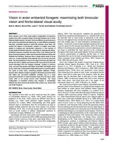
remaining 1. 39% ( n = 1 ) was hyperopia. Out of 103 subjects, 23 had astigmatism of greater than -0. 75 D. The mean myopic error was - 1. 58 依 1. 89 D in OD and -1. 69依1. 99 D in OS. The mean spherical equivalent of RE and LE were found to be statistically similar ( P = 0. 373) . In term of eye ( n = 206) , 63. 11 % of the eyes were myopic ( Figure 1) . Among 71 myopic subjects, 23 reported onset of myopia before entering this profession whereas 48 developed myopia afterward. There was no correlation between years spent working as microscopist and refractive error ( r = 0. 157, P = 0. 19) and similarly no correlation between working hours and refractive error ( r = -0. 13, P = 0. 19) . Majority of subjects had orthophoria (85. 40% ) at distance and exophoria ( 50. 50% ) at near. None of them had any constant manifest deviation. The mean amount of deviation at distance was 0. 51 pd exophoria ( XP) ( range from:14 Pd base in to 20 Pd base out ) and near was 2. 91 Pd of XP ( range 48 pd base in to 16 pd base out) . The mean value for near point of convergence was 9. 45 依4. 47 ( range from: 6 to 42) cm. Of the 103 subjects, 63 had receded near point of convergence. Subjects having reduced PFV at distance and near were 66 and 38 respectively. Subjects having reduced NFV at distance and near were 3 ( 2. 90% ) and 92 ( 89郾 30% ) respectively. Majority of them had normal stereopsis 92 ( 89. 32 % ) and 11 ( 10. 68% ) had gross stereopsis present ( Table 3) . 1182
41. 30% 34. 78% 1. 08%
40. 06%
Prevalence
Eyestrain / eyeball pain
70 (67. 6% )
Blurred near vision
16 (15. 5% )
Blurred distance vision
Figure 1 摇 Distribution of refractive errors in term of eye ( n =206) .
30. 43%
Nausea
Headache Dry eyes
Dizziness
Double vision
37 (35. 9% ) 16 (15. 5% ) 65 (63. 1% ) 54 (52. 4% ) 23 (22. 3% ) 10 (9. 71% )
Among non - presbyopic group ( n = 92 ) , subjects having reduced AA in OD and OS were 38 and 32 respectively. Accommodative excess was found in one subject. Mean lag of accommodation in non - presbyopic subjects was 0. 73 依 0. 37 ( range - 0. 50 to 1. 75 ) D. Subjects having high lag of accommodation were 30. 43% while 4. 35 % had lead of accommodation. Subjects having low PRA were 19 (18郾 40% ) and 12 ( 11. 70% ) had high PRA. Twelve subjects had low NRA and 21 subjects had high NRA. The mean value of accommodative facility, monocularly and binocularly were 8. 35 依 2. 84 cpm and 7. 51 依 2. 68 cpm respectively. The accommodative facility of OD and OS were found to be statistically similar ( t = 1. 21, P = 0. 229, paired t-test) . Out of 92 non - presbyopic subjects, 8 ( 18. 7% ) subjects failed to perform flipper test and 22 (21. 36% ) had accommodative infacility ( Table 4) . Majority of subjects reported symptom of eyestrain (67. 60% ) whereas least reported double vision (9. 71% ) ( Table 5 ) . Due to the imbalance in accommodation and convergence system after prolonged near work they often reported symptoms of headache, dizziness, blurred near or distance vision, nausea, double vision, difficulties to focus at distance after microscope use etc. DISCUSSION The prevalence of refractive error in this group of Nepalese microscopists (69. 90% ) was found to be greater than that of general population (10. 8% ) [17] . A study done on Nepalese student showed lesser prevalence of refractive error (8. 58% ) than microscopists. In this group of Nepalese students, myopia constituted about 44. 79% of refractive error, which is lesser than Nepalese Microscopists ( 98. 61% of refractive error) [18] . This study provides further evidence that prolonged near work and visual environment can have a major impact on refractive state of eye irrespective of age. The prevalence of
Int Eye Sci, Vol. 18, No. 7, Jul. 2018摇 摇
Tel:029鄄82245172摇
refractive error in this group of microscopist found to be less than study done by Fritzsche et al [7] (69. 9% cf. 89% ) . This may be due to the fact that their study was based on an online questionnaire survey and there is high probability that ametropic subjects with ocular problems may had participate. The prevalence of myopia in our study is comparatively similar to that of study done by Adams and McBrien in 1989 [13] . In our study the prevalence of myopia was 68. 93% and mean spherical equivalent was - 1. 50 D, which was found to be lower than Chinese microscopists (87% and - 4. 45 D) [19] . Since Chinese population are known to have higher prevalence of myopia [20] . Majority of subjects in this study had higher lag of accommodation. Thus hyperopic retinal defocus caused by this higher lag is believed to play a role in myopia development and progression [21-23] . Many subjects were also found to have accommodation insufficiency and infacility. Spending long time on microscope can also lead to problem of shifting focus from near to distance or vice versa. It was speculated that accommodative facility might be a good predictor of future myopic progression [24] . In this study population, fusional vergence range was found to be reduced which may be due to stress on convergence and accommodative system. Most of them have reduced NFV at near ( 89. 30% ) which may be due to over accommodative converge at near. Accommodative and vergence anomalies may lead to different kind of signs and symptoms which lower productivity and impaired quality of life. Symptoms associated with accommodative and vergence anomalies include eyestrain, blurred vision, headache, nausea, dizziness, diplopia and loss of concentration during a task performance. These symptoms tend to worsen by the end of day and related to use of eyes. Asthenopia related to near work could be eliminated with proper lens correction or vision therapy. This study recommended the need for increasing awareness about ocular problems and binocular vision anomalies related to microscopy work. There was found to be increased prevalence of refractive error in clinical microscopists, especially myopia. Majority of them had vergence and accommodative anomalies. Most of the subjects reported asthenopic symptoms associated with their microscopy work, which may affect their work efficiency.
REFERENCES 1 Jain G, Shetty P. Occupational concerns associated with regular use of microscope. Int J Occup Med Environ Health 2014;27(4) :591-598 2 Risovic DJ, Misailovic KR, Eric-Marinkovic JM, Kosanovic-Jakovic NG, Milenkovic SM, Petrovic LZ. Refractive errors and binocular dysfunctions in a population of university students. Eur J Ophthalmol 2008;18(1):1-6 3 Ciuffreda KJ, Vasudevan B. Nearwork - induced transient myopia ( NITM) and permanent myopia-is there a link? Ophthalmol Physiol Opt
http: / / ies. ijo. cn
85263940摇 摇 Email:IJO. 2000@163. com
2008;28(2) :103-114 4 Zadnik K, Mutti DO. Refractive error changes in law students. Am J Optom Physiol Opt 1987;64(7) :558-561 5 O蒺Neal MR, Connon TR. Refractive error change at the United States Air Force Academy - class of 1985. Am J Optom Physiol Opt 1987;64 (5) :344-354 6 McBrien NA, Adams DW. A longitudinal investigation of adult-onset and adult-progression of myopia in an occupational group: refractive and biometric findings. Invest Ophthalmol Vis Sci 1997;38(2):321-333 7 Fritzsche FR, Ramach C, Soldini D, Caduff R, Tinguely M, Cassoly E, Moch H, Stewart A. Occupational health risks of pathologists-results from a nationwide online questionnaire in Switzerland. BMC Public Health 2012;12(1) 8 Schober HAW, Dehler HKR. Accommodation during observations with optical instruments. J Opt Soc Am 1970;60(1) :103 9 Richards OW. Instrument myopia - microscopy. Am J Optom Physiol Opt 1976;53(10) :658-663 10 Kotulak JC, Morse SE. Relationship among accommodation, focus, and resolution with optical instruments. J Opt Soc Am A Opt Image Sci Vis 1994;11(1) :71-79 11 Birnbaum MH. Nearpoint visual stress: clinical implications. J Am Optom Assoc 1985;56(6) :480-490 12 Wallman J, Gotrlieb MD, Rajaram V, Fugate - Wentzek LA. Local retinal regions control local eye growth and myopia. Science 1987;237 (4810) :73-77 13 Adams DW, McBrien NA. Prevalence of myopia and myopic progression in a population of clinical microscopist. Optom Vis Sci 1992; 69(6) :467-473 14 Duane A. A new classification of motor anomalies of the eye based upon physiological principles, together with their symptoms, diagnosis and treatment. Ann Ophthalmo Otolarngol 1896:72 15 Garcia A. Evaluating relative accommodations in general binocular dysfunctions. Optom Vis Sci 2002;79(12):779-787 16 Saladin J. Phorometry and stereopsis. Borish蒺s clinical refraction 2006;899-960 17 Shrestha SP, Bhat KS, Binu VS, Barthakur R, Natarajan M, Subba SH. Pattern of refractive errors among the Nepalese population: a retrospective study. Nepal J Ophthalmol 2010;2(2):87-96 18 Shrestha GS, Sujakhu D, Joshi P. Refractive error among school children in Jhapa, Nepal. J Optom 2011;4(2) :49-55 19 Ting PW, Lam CS, Edwards MH, Schmid KL. Prevalence of myopia in a group of Hong Kong microscopists. Optom Vis Sci 2004;81 ( 2 ): 88-93 20 Lam CS, Goh WS, Tang YK, Tsui KK, Wong WC, Man TC. Changes in refractive trends and optical components of Hong Kong Chinese aged over 40 years. Ophthalmic Physiol Opt 1994;14(4):383 388 21 Gwiazda J, Thorn F, Bauer J, Held R. Myopic children show insufficient accommodative response to blur. Invest Ophthalmol Vis Sci 1993; 34(3) :690-694 22 Rosenfield M, Gilmartin B. Disparity - induced accommodation in late-onset myopia. Ophthalmic Physiol Opt 1988;8(3) :353-355 23 McBrien NA, Millodot M. The effect of refractive error on the accommodative response gradient. Ophthalmic Physiol Opt 1986;6 (2 ) : 145-149 24 Allen PM, O'Leary DJ. Accommodation functions: co - dependency and relationship to refractive error. Vision Res 2006;46(4) :491-505
1183