SUMMARY. A novel approach to the production of a human glucagon in E. coli is described. The 29 amino acids of human glucagon and pentapeptide linker ...
BIOTECHiVOLC)GY TECHNIQUES Volume 10 No. 9 September (1996) pp.669-672 Received as revised 21 July 1996
PRODUCTION OF RECOMBINANT HUMAN GLUCAGON IN ESCHERZCHZA COLZ BY A NOVEL FUSION PROTEIN APPROACH Dae-Young Kim, Nam-Kyu Shin, Seung-Gu Chang and Hang-Cheol Shin* Protein Engineering Laboratory, Hanhyo Institute of Technology San 6, Daeya-Dong, Shiheung-Shi, Kyungki-Do, Republic of Korea SUMMARY A novel approach to the production of a human glucagon in E. coli is described. The 29 amino acids of human glucagon and pentapeptide linker containing enzyme processing site were fused at the amino terminus to a 57 residue N-terminal portion of the human tumor necrosis factor-alpha (hTNF-a). The fusion protein was expressed in the E. coli cytoplasm at levels up to 30% of the total cell protein. Precipitation of the fusion protein near its isoelectric point, specific enterokinase cleavage at the linker site and subsequent HPLC purification makes this approach suitable for the production of glucagon as well as other relatively small peptides with therapeutic interests. INTRODUCTION Glucagon is a 29-residue peptide hormone that is produced by the alpha-cells of the islets of Langerhans in the pancreas. Glucagon regulates the blood glucose level in concert with insulin by stimulating hepatic glycogenolysis and gluconeogenesis (Unger & Orci, 198 l), thus maintaining fuel homeostasis. Glucagon has several therapeutic applications, including emergency treatment of hypoglycaemia, pretreatment in radiographic and endoscopic examination of the digestive tract. Recombinant DNA technology has been widely used for the production of biologically important proteins or peptides in bacterial strains. However, the direct expression of relatively small peptides is generally inefficient, mainly due to the proteolytic degradation of the foreign product by the host cells (Gottesmann, 1989). In order to increase the yield of foreign products, such products have been produced as fusion proteins in which the polypeptide product is fused to a larger protein which is known to accumulate in the host organism. In many cases, the proteins, which are used to form a fusion protein with desired polypeptides, are large in comparison with a typical polypeptide product. The desired polypeptide therefore represents only a small percentage of the fusion protein produced by a tranformed host organism, and the efficiency of the process is low. In order to devise a simple and efficient way of producing small peptides, such as glucagon, we developed a new fusion protein approach using human tumor necrosis factor-alpha (hTNF-a). We have found that the N-terminal portion of hTNF-a is suitable for the fusion protein approach. In this study, a 57 residue N-terminal fragment of hTNF-a was tailored to serve as a fusion partner for the human glucagon. MATERIALS Plasmids,
bacterial
strains
AND METHODS
and growth 669
condition:
E.coli BL21 (DE3) carries the
T7 RNA polymerase gene under lacUV5 control. Transformation was carried out as described by Hanahan ( 1985). Transformed E.coli cells wcrc grown in LB medium (1% Bacto-tryptone, 0.5% yeast extract, 1% sodium chloride) at 37°C. DNA Manipulation and Plasmid Construction: E. coli BL21 (DE3) was used for the propagation of the plsmids as well as for the cloning procedures. Standard DNA techniques were used. Restriction endonucleases, T4 DNA ligase, Vent DNA polymerase from NEB were used as described in the manufacturer’s instructions. PCR primers were synthesized using AI31 DNA synthesizer. A 174bp DNA fragment encoding the N-terminal 57 residues of hTNF-a was amplified by PCR, digested with restriction endonucleases NdeI and BamHI and inserted downstream of the T7 promoter of the expression plasmid PET-3a (Studier et al., 1990) which was linearized with NdeI and BamHI. The resulting plasmid was termed pT1. The expression plasmid pTlG for the fusion protein, was constructed by inserting the gene encoding the glucagon and the N-terminal pentapeptide linker into the plasmid pT1. SDS-PAGE, Enzyme Cleavage and HPLC Purification: iT.coli cells were harvested and suspended in 50mM Tris/Acetatc buffer (pH 6.0). After cell disruption by sonication, soluble and insoluble fractions were separated by centrifugation at 5000 g for 10min. The total cell lysate, soluble and insoluble fractions were analyzed by SDS-PAGE, followed by staining with Coomassie blue. The pellet was washed twice with 50mM Tris/Acetate buffer (pH 6.0) dissolved in IM urea, 5mM CaCl?, 50mlM Tris-HCI (pH 8.0) and then treated with enterokinase for 24h at 37°C. The released glucagon from the fusion protein was purified using HPLC. The authentic glucagon was purchased from Sigma and used as a standard for analysis. RESULTS
AND DISCUSSION
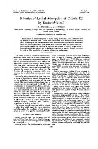
TNF-(I is a 157-residue cytokine, which exhibits a wide range of biological activities, including antitumor activity (Aggarwal, 1987). This protein was stably expressed in E. coli with high expression level (Pennica et al., 1984; 1985). This protein does not contain any methionine residue, thus making it suitable as a fusion partner for fusion system cleavable by CNBr method. Since human glucagon contains one methionine, CNBr method cannot be used in this case. Therefore, in our study, highly specific enterokinase cleavage site (Asp-Asp-Asp-Asp-Lys) was incorporated. To increase the proportion of glucagon relative to the TNF-a moiety in the fusion protein, fusion proteins of different sizes with deletions at the carboxy terminus of TN&a moiety were produced and tested for high expression. The smallest amino terminal fragment of TN&a that fulfilled these criteria was 57 residues (data not shown). For expression of the human glucagon, the truncated TNF-a gene encoding the N-terminal 57 residues was placed under the control of the T7 promoter in the expression plasmid PET-3a (Studier et al., 1990). Subsequently, a synthetic DNA fragment encoding the human glucagon and the pentapeptide linker Asp-Asp-Asp-Asp-Lys was ligated in the same reading frame to the truncated gene TNF-a (Fig. 1). The resulting expression plasmid for the fusion protein (9 1 residues) was used to transform E. coli BL2 1 (DE3) cells harboring a chromosomal copy of the bacteriophage T7 RNA polymerase (Studier er al., 1990). After 24h culture of transformed E. cofi cells, expression level and partition of the fusion protein between soluble and insoluble parts were tested by analysing the total cell lysate, soluble and insoluble fractions with SDS-PAGE. About 80% of the fusion protein was present in the soluble fraction. Since the isoelectric point (PI) of the fusion protein is around 5, we have tested optimum pH where the fusion protein can be selectively precipitated. At pH 6.0, the fusion protein was almost selectively precipitated (Fig. 2). About 20% of the fusion protein was retained in the soluble fraction. Since the
670
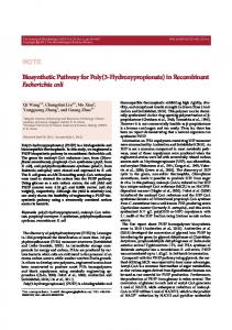
A Fig. 1. (A) Map of theE.coZi expression vector for the T 1G fusion protein. (B) Nucleotide sequence encoding the TlG fusion protein. The restriction sites used for cloning are underlined.The enterokinase recognition site DDDDK is boxed and the cleavage position is arrowed.
Jj
Tl(l-57) 10 VVANPQAEGQ MPSDKPVAH cat ATG CCG AGT GAC AAG CCT GTA GCC CAT GTT GTA GCA AAC CCT CAA GCT GAG GGG CA( NdeI 20 30 LQWLNRR A N A L L ANGVELR CTC CAG TGG CTG AAC CGC CGG GCC AAT GCC CTC CTG GCC AAT GGC GTG GAG CTG AGA 50 40 DNQLVVPIE GLFLI Y SRGS GAT AAC CAG CTG GTG GTG CCA ATA GAG GGC CTG ‘I-IX CTC ATC TAC TCC CGG GGA TCC RamHI I 70 60 * Glucagon(63-9 1) ID D D D K1 H S Q G T F T S D Y S K Y L D GAC GAC GAC GAC AAA CAC TCT CAG GGT ACT ITC ACT TCT GAC TAC TCT AAA TAC CTG GAC 90 80 VQWLMNT** SRRAQDF TCT CGT CGT GCT CAG GAC TIC GTT CAG TGG CTG ATG AAC ACT TAG TAA gctt Hind111
MTS
I Fig.2. SDS-PAGE analysis of the TlG preparation. M : molecular weight marker T : total cell lysate S : soluble fraction after cell disruption I : insoluble fraction after cell disruption
36.5 31 21.5 14.4 6
4-TlG
I L-
u”. II
I I I I I I I I I I I I I I I I I I I I I I I ln m In m u-l t rt r’, M c-4
I
I m k-4
I
I
I
I
I In c4
I
l
I
l
l Q) t-3
Fig.3. HPLC chromatograms of crude enterokinase digested fusion protein(A) and the glucagon after reverse phase purification(B). The purity of the recombinant glucagon from one step C 18 semi-preparative column was above 99% as judged by analytical column.
671
size of the fusion protein is small with no disulfide bridges, the precipitate was easily solubilized with Tris-HCI buffer, pH 8.Ocontaining 1M urea. After addition of enterokinase into the solution at 1:4700 ratio (w/w, enzyme/peptide), enzyme cleavage reaction was carried out by incubating the solution for 24 h at 37°C. The digested peptide mixture was separated by Vydac Cl8 reverse-phase HPLC chromatography (Fig. 3). The major peak which was eluted at the same position as that of authentic glucagon was collected and the purity was judged by analytical RP-HPLC. The autheticity was confirmed by tryptic digestion, electron spray mass spectrometry and amino acid analysis. The tryptic digestion of the peptide sample and standard glucagon yielded an identical HPLC elution profile. The peptide sample gave the expected electron spray mass spectrum within the error range (expected: 3482.8, found: 3480.93k1.95). Amino acid composition also agreed well with the theoretical values.
1Cell disruption (50mM Tris/Acetate(pH 6.0)) 1 +
Fig 4. Flow diagram of human glucagon production from T 1G fusion protein
In conclusion, the glucagon was produced by a novel fusion protein approach and purified to homogeneity by one step HPLC purification. The production yield was approximately 18 mg from lg dry cell weight. This is equivalent to approximately 720 mg of glucagon production from 1L culture when 40g dry cells/L of high cell-density fermentation is performed. This approach is suitable for the production of relatively small peptides with therapeutic interests by the simple procedure and high productivity (Fig. 4). ACKNOWLEDGMENT We thank Drs. Jong-Suhl Kim and Baik-Lin Seong for helpful discussions. This work was supported by Hanil Synthetic Fiber Co. REFERENCES Aggarwal, B.B. (1987) Drugs of the Future 891-897. Gottesmann, S. (1989) Annu. Rev. Genet. 2 3, 163-198 Hanahan, D. (1985) In: DNA Cloning 1 (Ed. D.M. Glover), pp. 109-135, IRS Press Pennica, D., Nedwin, G.E., Hayflick, J.S., Seeburg, P.H., Derynck, R., Palladino, M.A., Kohr, W.J., Aggarwal, B.B. and Goeddel, D.V. (1984) Nature 3 12,724-729 Pennica, D., Hayflick, J.S., Bringman, T.S., Palladino, M.A. and Goeddel, D.V. (1985) Proc. Natl. Acad. Sci. U.S.A. 82, 6060-6064 Studier, W.F., Rosenberg, A.H., Dunn, J.J. and Dubendorff, J.W. (1990) Methods in Enzymol. 185, 60-89 Unger, R.H. and Orci, L. (1981) New Engl. 1. Med. 304, 1518-1524
672