system for simultaneous separation of CD3 and CD15 cells and for CD3 isoaltion in comparison to MACS magnetic isolation. Methods: pluriSelect cascade ...
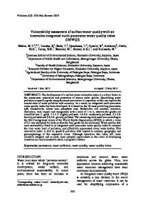
Quality testing of an innovative cascade separation system for multiple cell separation Arkadiusz Pierzchalski1,3, Aleksandra Moszczynska1,3, Bernd Albrecht3, Jan-Michael Heinrich2, Attila Tarnok1,3 1
Translation Centre for Regenerative Medicine, University of Leipzig, Leipzig, Germany pluriSelect GmbH, Leipzig, Germany 3 Department of Paediatric Cardiology, Cardiac Centre GmbH, Leipzig, Germany 2
Abstract Isolation of different cell types from mixed samples in one separation step by FACS is feasible but expensive and cheaper but still challenging by magnetic separation. An innovative bead-based cascade-system (pluriSelect GmbH, Leipzig, Germany) relies on simultaneous physical separation of different cell types. It is based on antibody-mediated binding of cells to beads of different size and isolation with sieves of different mesh-size. We validated pluriSelect system for single parameter (CD3) and simultaneous separation of CD3 and CD15 cells from EDTA blood-samples. Results were compared with those obtained by MACS (Miltenyi-Biotech) magnetic separation (CD3 separation). pluriSelect separation was done in whole blood, MACS on Ficoll gradient isolated leukocytes, according to the manufacturer’s protocols. Isolated and residual cells were immunophenotyped (7-color 8-antibody panel (CD3;CD16/56;CD4;CD8;CD14;CD19;CD45;HLADR) on a CyFlowML flow cytometer (Partec GmbH). Cell count (Coulter), purity, yield and viability (7-AAD exclusion) were determined. There were no significant differences between both systems regarding purity (92-98%), yield (50-60%) and viability (92-98%) of isolated cells. PluriSelect separation was slightly faster than MACS (1.15 h versus 1.5h). Moreover, no pre-enrichment steps were necessary. In conclusion, pluriSelect is a fast, simple and gentle system for efficient simultaneous cell separation of two cell subpopulation directly from whole blood and can provide a simple alternative to FACS. The isolated cells can be used for further research applications. Introduction In multicellular organisms, all the cells are identical in their DNA but the proteins vary tremendously. Therefore, it is useful to separate cells that are phenotypically different from each other for their deeper characterization. The separation of cells that differ in size, shape (morphology), color or other physical characteristics requires good knowledge of the available technologies and the features of the cell mixture. In general the separation methods can be divided into 4 groups. 1. Physical - centrifugationbased techniques - The oldest and crudest method, this technique only requires the resuspension of the cells in an appropriate buffer and the knowledge of their approximate composition and density or size. By setting the centrifuge to spin at various speeds, cells of different masses and densities are pelleted accordingly, with the densest cells pelleting first and at comparatively low centrifugation speeds, while the smallest, lightest cells require much faster speeds. 2. Biochemical separation – by perturbing a biochemical process that is required by the cell for its growth and/or survival. Biochemical separations include blocking DNA synthesis and serum deprivation. It results in the inhibition of key metabolic pathways and, as such, arrest a cell in particular stages of the cell cycle. 3. Antibody-based fluorescence cellsorting techniques - this method requires more extensive understanding of the cell's biology.
This approach was accompanied by a rapid instrument development called Fluorescenceactivated cell sorter (FACS) which is a specialized type of flow cytometry. The first cell sorter was invented by Mack Fulwyler in 1965 (Fulwyler 1965) 1. The technique was expanded by Len Herzenberg (Hulett et al. 1969, Bonner et al.1972) 2,3. It provides a method for sorting a heterogeneous mixture of cells into two or more containers, one cell at a time, based upon the specific light scattering and fluorescent characteristics of each cell. It provides fast, objective and quantitative recording of fluorescent signals from individual cells as well as physical separation of cells of particular interest. 4. Antibody-based magnetic cell sorting this method is basically a combination of antibody tagging of the desired cells with a tiny magnet (Molday1977) 4. The cells are then applied to a magnetic column that is capable of retaining these tagged cells when a magnetic field is generated within it. Although positive selection is in general the fastest and the most efficient way to isolate a particular cell subset with high purity and yield, negative selection is needed when the cells of desire have to be “untouched” for subsequent analyses (Abst 1989, Miltenyi 1990,Qren 2005, Grutzkau 2010) 5 8 – . A sequential magnetic sorting strategy can be used, if different cell populations have to be isolated from a single sample. This approach is however time consuming and laborious. pluriSelect has introduced an innovative, bead-based cascade-system which relies on simultaneous physical separation of different cell types. It is based on antibody-mediated binding of cells to beads of different size and isolation with sieves of different mesh-size. Thus by relatively easy to handle system one can separate a few different cells of interest upon one separation. In this paper we present for the first time validation of pluriSelect system for simultaneous separation of CD3 and CD15 cells and for CD3 isoaltion in comparison to MACS magnetic isolation.
Methods: pluriSelect cascade isolation The overall analysis workflow for both cell separation systems for CD3 isolation is shown in the Figure 1 and the analysis workflow for CD3/CD15 isolation is shown in Figure 2. CD3 cells or CD3/CD15 cells were separated from 4 ml or 1 ml whole EDTA-blood respectively by pluriSelect cascade isolation. Briefly, the blood cells were incubated with antibody-coated beads (pluriBead) in a mixing container at room temperature for 30 minutes on a horizontal pluriPlix roller. Next cells were placed on a sieve cascade (CD3/CD15) with bigger mesh-size on lower mesh-size. The cells were washed thoroughly with ca. 45 ml wash buffer (PBS w/o Ca2+ w/o Mg2+ pH 7.4 with 2mM EDTA and 0.5% BSA) to remove erythrocytes and unbound cells and to forward the smaller beads onto the low size mesh sieve. Each separate sieve was attached via connector ring to a sterile 50 ml centrifuge tube The Luer-Lock was closed. 2 ml detachment buffer was applied. The sample was swirled and incubated for 10 minutes at RT. Cells were separated from the beads by careful pipetting, The Luer-Lock was opened and the detached cells ran into the centrifuge tube underneath the sieve. Sieves were washed with additional 8 ml wash buffer. Cells were centrifuged 10 minutes at 250g and transferred and resuspended in 1 ml wash buffer for further analysis. MACS isolation CD3 cells were isolated via MACS microbeads system by means of miniMACS column. PBMC from 4 ml EDTA-blood was isolated by density gradient centrifugation using Biocoll (AG Biochrom, Berlin Germany). PBMC were washed twice in wash buffer (PBS w/o Ca2+ w/o Mg2+ pH 7.4 with 2mM EDTA and 0.5% BSA) and subjected to further magnetic labelling. Cell number was determined, cells were resuspended in 80 µl of buffer and 20 µl of CD3 MicroBeads was added. After 15 minutes incubation in a refrigerator (2-8oC) cells were washed in 1.5 ml wash buffer and centrifuged at 300g for 10 minutes. Cells were resuspended
in 500 µl wash buffer. Magnetic separation was done on the rinsed MS column. Cell suspension was applied onto the column. The column was 3 times washed. The wash through was collected. The column was removed from the separator and placed on an Epp tube. CD3 positive cells were eluted with 1 ml wash buffer and then subjected for the further analysis.
Figure 1. Workplan summary. The original blood sample was sorted into two subsets by
dual-layer pluriSelect separation system and was subjected to further analysis. The analysis included cell counting, viability assessment and immunophenotyping of the original blood, the isolated samples and the residual cells in the wash-through. One isolation by Miltenyi MACS system were conducted. Briefly - cells isolated on Ficoll gradient were subjected to isolation and the isolated and residual cells were counted, and viability and immunophenotype were assessed.
Figure 2. Workplan summary. The original blood sample was sorted into two subsets by
dual-layer pluriSelect separation system and was subjected to further analysis. The analysis
included cell counting, viability assessment and immunophenotyping of the original blood, the isolated samples and the residual cells in the wash-through. Immunophenotyping As shown in the Fig. 1 there were all together 6 different cell fractions for immunophenotyping. Immunophenotyping was done in a 1 tube (7 colors, 9 markers staining) with the following markers: CD3-FITC ;CD16/56-PE;CD4-PE-Cy7;CD8-APC;CD14-APCH7;CD19-APC;CD45-PacBlue;HLADR-PerCP. 50 µl sample was stained for 1 h at RT in dark and then the erythrocytes were lysed and the sample was fixed in Versalyse lysing solution for 8 minutes. The samples were analyzed on CyFlow ML Partec FCM (Partec GmbH, Germany) with use of the 3 lasers – blue 488 nm; red 645 nm; violet 405 nm with the following filter settings: FL1 (FITC) – 527/30nm BP; FL2 (PE) – 590/30nm BP; FL3 (PerCP) – 682/30nm BP or FL3 (PE-Cy7) – 680nm LP; FL4 (APC) - 675/20nm BP; FL5 (APC-H7) – 748nm LP; FL6 (PacBlue) – 455/30nm BP. Analysis was done as shown in Fig. 3.
Figure 3. FCM immunophenotyping analysis scheme. The gating strategy for percentual cell estimation. The amount of the following parameters was estimated - T cells in leukocytes and in lymphocytes. Thelper (Th), Tcytotoxic (Tc), B cells in leukocytes and in lymphocytes and NK cells in non-CD3+ lymphocytes (non T cells). Cell count estimation The cell counts from the whole blood and residual pluriSelect blood samples were estimated on Sysmex KX-21 hematology analyser. The count for all the positively isolated cells and Ficoll gradient isolated samples and MACS residual samples was estimated on Coulter Counter (Z2 Coulter Coulter, USA). The cell counts of leukocytes served next as basis for yield and cell loss as well as cellular contamination estimation.
Cell viability estimation CD3 positively selected cells were stained with CD3 FITC and CD45APC Abs for 30 minutes and then costained with Propidium Iodide PI (5 µg/ml) for 5 minutes (Fig.4B). The cells were analysed by CyFow ML Partec FCM. Whole blood sample was prestained with anti-CD3 FITC Abs for 30 minutes which was followed by PI staining (5 µg/ml) see Fig. 4A. Samples others than positively isolated cells were stained as for immunophenotyping which was followed by 7-AAD staining (5 µg/ml).
Count
CD45 APC
CD3+ cells
PI
CD3 FITC
CD3 FITC
CD3 FITC
Count
PI
Count
PluriSelect
CD45 APC CD45 APC
MACS
pluriSelect
Figure 4A: Cell viability test of T-cells in whole blood (Staining A). Figure displays the measurement and gating strategy to determine the % of PI positive T-lymphocytes (cells in region RN2) in whole blood.
PI
Figure 4B: Cell viability test of isolated CD3+ cells. Displayed is the measurement and gating strategy to determine the % of PI positive T-lymphocytes in isolated CD3+ samples (pluriSelect and MACS isolation).
Analysis software For cell subpopulations cell number estimation a database has been established and software has been developed. The software facilitated the statistical sample analysis and was design to reduce time for cytometric data analysis. The software interface is shown in Figure 5.
Figure 5. Software for cytometric data analysis. The parameters being analysed are listed in 2 right columns.
Results 1. pluriSelect and MACS CD3 positive selection 1.1. Cell count/yield/purity estimation of CD3 positively isolated cells As shown in the table 1 we determined the CD3 cell count in all the samples. The number of cells allowed us to determine the yield which was better in MACS isolated CD3+ sample 40.1% vs 57.1% of total cells. In case of pluriSelect system from overall slightly above 60% of total CD3+ cells 21.8% of them were in residual sample. There was much lower CD3+ cell count in MACS residual sample (7.2%). Overall cell purity of CD3+ cells was very high in cells isolated by both systems with 98.4% and 96.5% purity for pluriSelect and MACS, respectively. We also calculated the overall yield of T helper and T cytotoxic cells to see if there is any bias as to T cell subpopulations selection. Overall there was a significantly higher cell loss (initial cell count – cells in the CD3+ and CD3- sample) of CD3+4+ cells than of CD3+CD8+ cells both for pluriSelect (50% vs. 30%) and MACS (30% vs 5%). Cell loss between the two methods and for both cell types was not significant.
pluriSelect
CD3 yield (%)
A. Whole Blood 3.5 (2.7-6.1) 19.2 (9.7-37.4) 100
CD3+4+ count 6 (x10 ) CD3+8+ count 6 (x10 ) CD3+4+ yield
2.61 (1.7-3.88) 0.986 (0.295-2.34) 100
CD3+8+ yield
100
CD4/CD8-ratio
2.84 (1.23-7.95)
Total T-cell 6 count (x10 ) CD3% FCM
MACS
B. CD3+
D. CD3-
E. CD3+
F. CD3-
1.6 * (0.51-2.3) 98.4 (95.6-99.3) 40.1* (14.8-63.8) 0.97* (0.21-1.66) 0.48 (0.234-1.206) 39.5* (11.7-62.4) 51.4* (18.5-79.9) 2.01* (0.78-6.2)
0.86* (0.14-1.88) 4.71* (1.15-11) 21.8* (4.5-34.0) 0.55* (0.085-0.96) 0.209 (0.039-0.735) 15.9* (3.25-27.4) 26.51 (6.34-54.5) 1.32* (0.26-5.48)
2.64 (1.1-3.26) 96.5 (93,62-98,13) 57.1 (31.1-80.3) 1.88 (0.98-2.82) 0.625 (0.24 -2.15) 65.8 (52.9-100.7) 78.22 (46.88-102.1) 2.31 (0.89-7.66)
0.25 (0.14-0.75) 8.56 (3,73-26.1) 7.2 (3.32-18.5) 0.103 (0.022-0.17) 0.15 (0.068-0494) 3.63 (0.7-6.48) 21.75 (4.46-44.55) 0.72 (0.09-1.62)
Table 1: Absolute CD3 cell counts, yield and purity. Table shows summary of absolute CD3+ Coulter Counter cell number estimation and FCM corrected values. Data show median and range in parenthesis. * differences between values obtained by MACS and pluriSelect (CD3+ vs CD3+ or CD3- vs CD3-) were regarded statistically significant if p 90%) very few contaminating cells were present in samples B and C. This is the reason why we obtained such a discrepancy between different samples. Viability of for isolated CD3+ cells was very high. It is remarkable that the viability of cells in the residual samples (D) were substantially higher then in the isolated fraction and only slightly below the initial sample. Viability of isolated neutrophils was even higher than for CD3+, all except two samples yielded viabilities >95%. Viabilities in the residual sample were similar and slightly below the initial sample. We do not find the reason for lower cell viability of CD3+ cells. The difference in cell viability between CD3+ and CD15+ cells suggests that the contact of CD3+ cells with beads and perhaps with sieves may render these cells more susceptible but still the viability was very high. If we look closer at comparison between pluriSelect with MACS the purity and recovery are equivalent for both systems. In both systems we got a selective lost of CD4 cells. In pluriSelect the recovery of negatively selected cells is substantially better and the viability of CD3 cells is slightly reduced. Moreover the pluriSelect preparation time was 15 minutes shorter than MACS separation.
Summing up pluriSelct separation system seems to be a versatile, easy to handle and reliable cell separation system suitable for a wide range of applications. References [1] Fulwyler, M.J., “Electronic separation of biological cells by volume,” Science (New York, N.Y.) 150(3698), 910-911 (1965). [2] Hulett, H.R., Bonner, W.A., Barrett, J., and Herzenberg, L.A., “Cell sorting: automated separation of mammalian cells as a function of intracellular fluorescence,” Science (New York, N.Y.) 166(3906), 747-749 (1969). [3] Bonner, W.A., Hulett, H.R., Sweet, R.G., and Herzenberg, L.A., “Fluorescence activated cell sorting,” The Review of Scientific Instruments 43(3), 404-409 (1972). [4] Molday, R.S., Yen, S.P., and Rembaum, A., “Application of magnetic microspheres in labelling and separation of cells,” Nature 268(5619), 437-438 (1977). [5] Abts, H., Emmerich, M., Miltenyi, S., Radbruch, A., and Tesch, H., “CD20 positive human B lymphocytes separated with the magnetic cell sorter (MACS) can be induced to proliferation and antibody secretion in vitro,” Journal of Immunological Methods 125(1-2), 19-28 (1989). [6] Miltenyi, S., Müller, W., Weichel, W., and Radbruch, A., “High gradient magnetic cell separation with MACS,” Cytometry 11(2), 231-238 (1990). [7] Øren, A., Husebø, C., Iversen, A.-C., and Austgulen, R., “A comparative study of immunomagnetic methods used for separation of human natural killer cells from peripheral blood,” Journal of Immunological Methods 303(1-2), 1-10 (2005). [8] Grützkau, A., and Radbruch, A., “Small but mighty: how the MACS-technology based on nanosized superparamagnetic particles has helped to analyze the immune system within the last 20 years,” Cytometry. Part A: The Journal of the International Society for Analytical Cytology 77(7), 643-647 (2010). [9] Willasch, A., Eing, S., Weber, G., Kuçi, S., Schneider, G., Soerensen, J., Jarisch, A., Rettinger, E., Koehl, U., et al., “Enrichment of cell subpopulations applying automated MACS technique: purity, recovery and applicability for PCR-based chimerism analysis,” Bone Marrow Transplantation 45(1), 181-189 (2010).