J Infect Chemother (2009) 15:92–98 DOI 10.1007/s10156-009-0670-3
© Japanese Society of Chemotherapy and The Japanese Association for Infectious Diseases 2009
ORIGINAL ARTICLE Naoko Chiba · Somay Y. Murayama · Miyuki Morozumi Eiichi Nakayama · Takafumi Okada · Satoshi Iwata Keisuke Sunakawa · Kimiko Ubukata
Rapid detection of eight causative pathogens for the diagnosis of bacterial meningitis by real-time PCR
Received: October 10, 2008 / Accepted: January 28, 2009
Abstract We aimed to detect causative pathogens in cerebrospinal fluid (CSF) collected from patients diagnosed with bacterial meningitis by real-time polymerase chain reaction (PCR). In addition to Streptococcus pneumoniae, Haemophilus influenzae, and Mycoplasma pneumoniae described previously, five other pathogens, Neisseria meningitidis, Escherichia coli, Streptococcus agalactiae, Staphylococcus aureus, and Listeria monocytogenes, were targeted, based on a large-scale surveillance in Japan. Results in CSF from neonates and children (n = 150), and from adults (n = 18) analyzed by real-time PCR with molecular beacon probes were compared with those of conventional culturing. The total time from DNA extraction from CSF to PCR analysis was 1.5 h. The limit of detection for these pathogens ranged from 5 copies to 28 copies per tube. Nonspecific positive reactions were not recognized for 37 microorganisms in clinical isolates as a negative control. The pathogens were detected in 72.0% of the samples by real-time PCR, but in only 48.2% by culture, although the microorganisms were completely concordant. With the real-time PCR, the detection rate of H. influenzae from CSF was high, at 45.2%, followed by S. pneumoniae (21.4%), S. agalactiae (2.4%), E. coli (1.8%), L. monocytogenes (0.6%), and M. pneumoniae (0.6%). The detection rate with PCR was
N. Chiba · S. Y. Murayama · M. Morozumi · E. Nakayama · K. Ubukata (*) Laboratory of Molecular Epidemiology for Infectious Agents, Graduate School of Infection Control Sciences, Kitasato University, 5-9-1 Shirokane, Minato-ku, Tokyo 108-8641, Japan Tel. +81-3-5791-6385; Fax +81-3-5791-6386 e-mail:
[email protected] E. Nakayama Department of Pediatrics, Tokyo Metropolitan Cancer and Infectious Diseases Center, Komagome Hospital, Tokyo, Japan T. Okada · S. Iwata Department of Pediatrics, National Hospital Organization Tokyo Medical Center, Tokyo, Japan K. Sunakawa Laboratory of Infectious Disease, Graduate School of Infection Control Sciences, Kitasato University, Tokyo, Japan
significantly better than that with cultures in patients with antibiotic administration (χ2 = 18.3182; P = 0.0000). In conclusion, detection with real-time PCR is useful for rapidly identifying the causative pathogens of meningitis and for examining the clinical course of chemotherapy. Key words Real-time PCR · Bacterial meningitis · cerebrospinal fluid(CSF) · Neonate · Adult
Introduction Bacterial meningitis is a serious and sometimes fatal infection in both children and adults. The main causative pathogens are S. pneumoniae, Haemophilus influenzae type b (Hib), and Neisseria meningitidis.1 The incidence rate and causative pathogens of meningitis vary in various countries due to different social backgrounds. These are heavily affected by: (i) the availability of vaccination against Hib and S. pneumoniae, (ii) the availability of a medical insurance system, and (iii) the hygienic and sanitary conditions of each country. In addition to the introduction of the Hib vaccine in 1987,2 developed countries in Europe, as well as United States, implemented vaccination with a 7-valent pneumococcal conjugate vaccine (7PCV) against pneumococci in 2000–2001.3,4 In these countries, the number of meningitis cases due to Hib has decreased dramatically5,6 and the number of cases of invasive pneumococcal disease has been decreasing gradually.4,7–10 In Japan, on the other hand, the incidence rate of bacterial meningitis is estimated to be between 10 and 13 per 100 000 in children aged less than 5 years.11 According to the 2005 and 2006 large-scale surveillance carried out by Sunakawa et al.,12 55% of these cases were caused by Hib and 19.5% by S. pneumoniae. For meningitis in neonates and infants aged 3 months or less, Escherichia coli (2.5%) and S. agalactiae (7.7%) were the dominant pathogens. Among these causative pathogens, the resistance of Hib and S. pneumoniae to therapeutic antibiotics has rapidly
93
increased from about 2000 and has became a topic of controversy in the clinic.13–16 β-Lactamase-nonproducing ampicillin-resistance (BLNAR) Hib accounts for 40% of these cases, and 35% of S. pneumoniae cases were penicillin-resistant S. pneumoniae (PRSP) in 2007. The resistance mechanism in BLNAR originated from some mutations of the ftsI gene, encoding penicillin-binding protein 3, that mediate septal peptidoglycan synthesis.17 In 2007, Hib vaccination was finally approved by the government in Japan, but approval has not yet been granted for 7PCV. Considering this situation, it is desirable to create rapid detection methods for causative pathogens in patients diagnosed with meningitis, to allow for the proper selection of chemotherapeutic agents. Multiplex real-time PCR for simultaneously detecting S. pneumoniae, Hib, and N. meningitidis was previously reported by Corless et al.18 In addition to these pathogens, a single identification system for S. agalactiae19 and Mycobacterium tuberculosis20 has been described, but a detection system that covers bacterial meningitis in neonates to adults has not been developed yet. In the present study, we aimed to develop a real-time PCR that could simultaneously detect eight pathogens; namely, in addition to S. pneumoniae, H. influenzae, and N. meningitides, E. coli, S. agalactiae, and Staphylococcus aureus, which are the major causative pathogens in neonatal meningitis; and Listeria monocytogenes and Mycoplasma pneumoniae, which are rarely the causative pathogens. We report an identification system using real-time PCR with pathogen-specific molecular beacon (MB) probes and primers for eight meningitis pathogens; we also describe the results when applied to cerebrospinal fluid (CSF) assay, together with the results of conventional culturing.
Methods Clinical samples A total of 168 CSF samples collected from patients who were diagnosed with bacterial meningitis, based on clinical symptoms, CSF findings, and blood examination testing, were sent to our laboratory for bacterial identification from doctors belonging to medical institutions throughout Japan from January 2005 to December 2007. These samples were transported under frozen conditions at −20°C within 24 h of collection. For CSF collection and examination from patients, informed consent was obtained by the doctors in attendance from the parents or the responsible family members. Bacterial culture and DNA extraction Upon arrival at our laboratory, the CSF samples were thawed and immediately centrifuged at 10 000 rpm for 10 min at 4°C. From a total 150 μL of sediment, 10 μL of each sample was inoculated onto sheep blood agar and chocolate II agar
(Nippon Becton Dickinson, Tokyo, Japan). These plates were then incubated overnight at 37°C in an atmosphere of 5% CO2. On the following day, if bacterial growth was observed on the plates, the colonies were identified by the standard methods21 and also their antibiotic susceptibilities were measured.22 DNA extraction from 100 μL of the sediment was immediately carried out by using an EXTRAGEN II kit (Tosoh, Tokyo, Japan).23 Finally, the harvested DNA pellet was resuspended in 40 μL of DNase- and RNase-free H2O. The time required for the DNA extraction process was within 15 min.
Real-time PCR for bacterial detection The following eight bacterial pathogens were subjected to the real-time PCR analyses: E. coli, L. monocytogenes, N. meningitidis, S. agalactiae, S. aureus, S. pneumoniae, H. influenzae, and M. pneumoniae. Oligonucleotide primers and MB probes were designed using Beacon Designer 2.0 Software (Premier Biosoft International, Palo Alto, CA, USA). The primers, MB probes, target genes, and amplicon sizes (bp) for the eight pathogens are shown in Table 1. The eight pathogens were grouped in pairs and they were analyzed simultaneously with four tubes. Their combinations were as follows: S. pneumoniae (a) and H. influenzae (b) in tube A, E. coli (a) and S. agalactiae (b) in tube B, N. meningitidis (a) and L. monocytogenes (b) in tube C, and M. pneumoniae (a) and S. aureus (b) in tube D. The MB probes for detecting pathogens marked (a) were labeled with fluorescent dye, 6-carboxy-2′,4,4′,5′,7,7′-hexachlorofluorescein (HEX), at the 5′-terminal, whereas those marked (b) were labeled with 6-carboxyfluorescein (FAM). All MB probes were labeled with black hole quencher 1 (BHQ-1) at the 3′-terminal. The PCR reaction mixture consisted of: (i) 25 μL of 2×Real-time PCR Master Mix (Toyobo, Tokyo, Japan), (ii) 0.2 μM of each primer, and (iii) 0.3 μM of each MB probe, and the final volume of the mixture was adjusted to 50 μL by the addition of DNase- and RNAase-free H2O. Four reaction mixtures were pipetted into four wells of six-tube strip, and two of the remaining wells were used as positive and negative controls. The strip was filled with reaction reagents and stored at −30°C until used. The frozen PCR reagent, when it was used for assays, was thawed on ice and 2 μL of each DNA sample from CSF was added to each well. After that, real-time PCR was performed immediately with Stratagene Mx3000P (Stratagene, La Jolla, CA, USA). The PCR conditions were as follows: an initial DNA denaturation step of 95°C for 30 s, followed by 40 cycles of 95°C for 15 s, 50°C for 30 s and 75°C for 20 s, and at 75°C for 30 s, successively. S. pneumoniae chromosomal DNA was used in each assay as a positive control. The time required for the whole process from DNA extraction to the end of the real-time PCR operation was 1.5 h.
S. pneumoniae Sense primer Reverse primer Probe H. influenzae Sense primer Reverse primer Probe E. coli Sense primer Reverse primer Probe S. agalactiae Sense primer Reverse primer Probe N. meningitidis Sense primer Reverse primer Probe L. monocytogenes Sense primer Reverse primer Probe M. pneumoniae Sense primer Reverse primer Probe S. aureus Sense primer Reverse primer Probe
A
a
Stem oligonucleotides are underlined
D
D
C
C
B
B
A
Species, primer, and probe
Tube (paired)
5′-TACATGTCGTTAAACCTGGTG-3′ 5′-TACAGTTGTACCGATGAATGG-3′ FAM-CGCGATCCAAGAACTTGTTGTTGATAAGAAGCAACCGATCGCG-BHQ1
5′-GTAATACTTTAGAGGCGAACG-3′ 5′-TACTTCTCAGCATAGCTACAC-3′ HEX-CGCGATACCAACTAGCTGATATGGCGCAATCGCG-BHQ1
5′-CGCTTTTGAAAGATGGTTTCG-3′ 5′-CTTCCAGTTTCCAATGACCC-3′ FAM-CGCGATCGCGGCGTTGCTCCGTCAGACTTGATCGCG-BHQ1
5′-CATATCGGAACGTACCGAGT-3′ 5′-GCCGCTGATATTAGCAACAG-3′ HEX-CGCGATCCTATTCGAGCGGCCGATATCGATCGCG-BHQ1
5′-AGGAATACCAGGCGATGAAC-3′ 5′-AGGCCCTACGATAAATCGAG-3′ FAM-CGCGATCATTTGGCTAGTTATGAAGTCCCTTATGCGATCGCG-BHQ1
5′-GGGAGTAAAGTTAATACCTTTGC-3′ 5′-CTCAAGCTTGCCAGTATCAG-3′ HEX-CGCGATCACTCCGTGCCAGCAGCCGCGGATCGCG-BHQ1
5′-TTGACATCCTAAGAAGAGCTC-3′ 5′-TCTCCTTTGAGTTCCCGACCG-3′ FAM-CGCGATCCTGACGACAGCCATGCAGCACGATCGCG-BHQ1
5′-CAACCGTACAGAATGAAGCGG-3′ 5′-TTATTCGTGCAATACTCGTGCG-3′ HEX-CGCGATCAGGTCTCAGCATTCCAACCGCCGATCGCG-BHQ1
Primer or probea sequence
Table 1. Primers and probes for real-time PCR
spa
16S rRNA
16S rRNA
16S rRNA
dltS
16S rRNA
16S rRNA
lytA
Target gene
224
225
457
356
331
204
167
319
Amplicon size (bp)
This study
23
This study
This study
This study
This study
23
23
Reference
94
95
Sensitivity and specificity of real-time PCR The sensitivity of the present real-time PCR procedure was determined for the five pathogens: E. coli, L. monocytogenes, N. meningitidis, S. agalactiae, and S. aureus. The sensitivity for S. pneumoniae, H. influenzae, and M. pneumoniae had already been examined in our previous study.23 The procedure was performed with three strains each from the five species by tenfold serial dilutions of bacterial cells from 108 to 100/mL. The specificity of the MB probes and primers was tested with 37 Gram-positive and -negative microorganisms in clinical isolates in addition to the eight targeted bacteria. The species are listed in Table 2.
E. coli, 16 copies for L. monocytogenes, 2 copies for N. meningitidis, 28 copies for S. agalactiae, and 14 copies for S. aureus. A significant correlation was found between the tenfold diluted bacterial cell counts and the Ct values, ranging from γ = 0.9709 in L. monocytogenes to γ = 0.9989 in N. meningitidis. Although details of the results are not shown here, the sensitivities of the remaining three pathogens have previously been revealed to be two DNA copies for S. pneumoniae, ten copies for H. influenzae, and five copies for M. pneumoniae.23 The specificities of the 5-MB probe and primer sets were examined for 37 Gram-positive and -negative microorganisms selected from clinical strains as negative controls. Nonspecific positive reactions were undetectable after 40 cycles in the present real-time PCR procedure.
Results Comparisons of results between real-time PCR and bacterial culture
Sensitivity and specificity of real-time PCR The threshold cycle (Ct) value for a positive result was defined as the point at which the horizontal threshold line was crossed. The sensitivities of the real-time PCR assay for the five pathogens, E. coli, L. monocytogenes, N. meningitidis, S. agalactiae, and S. aureus, are shown in Table 3. The limits of detection per reaction tube were 2 DNA copies for
Table 2. Specificity panel: amplification-negative-control organisms Genus
Species
Streptococcus
S. dysagalactiae subsp. equisimilis, S. mitis, S. milleri, S. salibarius, S. oralis, S. mutans, S. sanguis, S. bovis Enterococcus E. faecalis, E. faecium, E. avium Staphylococcus S. epidermidis, S. haemolyticus Moraxella M. catarrhalis Haemophilus H. parainfluenzae, H. haemolyticus Pseudomonas P. aeruginosa Klebsiella K. pneumoniae, K. oxytoca Pantoea P. agglomerans Proteus P. mirabilis Serratia S. marcescens Acinetobacter A. calcoaceticus Enterobacter E. cloacae Citrobacter C. freundii Mycoplasma M. orale, M. hominis, M. salivarium Cryptococcus C. neoformans
Table 4 shows the details of the causative pathogens identified by real-time PCR and those confirmed by culturing from the CSF samples (n = 168) sent to our laboratory. Among the real-time PCR-positive cases, H. influenzae was detected at the highest incidence of 76 cases (45.2%), followed by S. pneumoniae in 36 cases (21.4%), S. agalactiae in 4 cases (2.4%), E. coli in 3 cases (1.8%), and L. monocytogenes (0.6%) and M. pneumoniae (0.6%) in 1 case each. There were no positive cases of N. meningitidis or S. aureus identified during the study periods. For bacterial culturing, H. influenzae was isolated in 48 cases (28.6%), S. pneumoniae in 27 cases (16.1%), S. agalactiae in 2 cases (1.2%), E. coli in 3 cases (1.8%), and L. monocytogenes in 1 case (0.6%). Ultimately, the causative pathogens were determined in as many as 72.0% of all samples by real-time PCR, but in only 48.2% by bacterial culturing. The microorganisms obtained by bacterial culture and by real-time PCR showed complete concordance. The sensitivity and specificity of the real-time PCR were calculated as 100% and 54.0%, respectively. However, this specificity does not reflect the true percentage, because in many cases with a negative culture an antibiotic had been prescribed before the bacterial cultivation of the CSF.
Table 3. Sensitivities for six pathogens identified by real-time PCR No. of DNA copies/50 μL of reaction tube 5
10 104 103 102 101 Correlation coefficienta a
Threshold cycle (Ct) E. coli
L. monocytogenes
N. meningitidis
S. agalactiae
S. aureus
18 21 25 28 31
21 24 28 31 >40
16 20 24 27 31
26 29 33 36 >40
26 28 31 34 >40
0.9987
0.9709
0.9989
Each value was calculated between the 10 fold diluted bacterial calls and the Ct values
0.9988
0.9783
96 Table 4. Causative pathogens identified by real-time PCR and by culture of the CSF samples (n = 168) Causative pathogen
PCR (%)
S. pneumoniae H. influenzae S. agalactiae E. coli L. monocytogenes M. pneumoniae
Culture (%)
No. of positive (%)
subtotal (%)
No. of positive (%)
subtotal (%)
36 76 4 3 1 1
121 (72.0)
27 (16.1) 48 (28.6) 2 (1.2) 3 (1.8) 1 (0.6) 0
81 (48.2)
(21.4) (45.2) (2.4) (1.8) (0.6) (0.6)
Not detected
47 (28.0)
87 (51.8)
* Sensitivity and specificity of the real-time PCR was calculated 100% and 54.0%, respectively; PCR and culture both positive (n = 81), PCR and culture both negative (n = 47), PCR negative and culture positive (n = 0), PCR positive and culture negative (n = 40) Table 5. Relationship between positive identification of pathogens in meningitis by real-time PCR and age of the patients Causative pathogen
na
Pediatrics (n = 106) ≤3 m
positive case S. pneumoniae H. influenzae S. agalactiae E. coli L. monocytogenes M. pneumoniae Subtotal negative case a
36 76 4 3 1 1 121
2 3 3 2
10
47
11
Subtotal
4–11 m
1y
2y
3y
6 34
3 14
2 7
3 9
4y 1 4
5–17 y 5 5
41
1 18
9
12
5
11
22 76 3 3 1 1 106
13
2
1
1
1
15
44
1 1
Adults (n = 15)
Subtotal
18–34 y
35–49 y
50–64 y
>65 y
1
3
6
4
1
1
1
4
14 1
6
4
15
1
1
3
Number of real-time PCR positive case
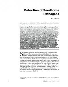
Relationship between real-time PCR-positivity and age of the patients The relationship between positive identification of pathogens by real-time PCR and the age of the meningitis patients is shown in Table 5. Among pediatric patients aged 17 years or less, a pathogen was suspected in 106 patients (70.7%) by real-time PCR. Five of the 6 patients in whom the pathogen was suspected to be either E. coli or S. agalactiae were neonates and infants aged 3 months or less. For patients aged between 4 months and 17 years, H. influenzae and S. pneumoniae were the major pathogens. Among adult meningitis patients aged 18 years or more, 15 cases (83.3%) were real-time PCR-positive, and most of them were caused by S. pneumoniae, with the exception of 1 case caused by S. agalactiae. Influence of prior antibiotic use The relationship between a history of antibiotic use prior to CSF collection and the pathogen-positive rate by realtime PCR or culturing was analyzed in 115 patients for whom a history of antibiotic use could be accurately followed up. As shown in Fig. 1, 62 patients (53.9%) had received antibiotics prior to hospital admission. Fifteen patients had received an injection and 47 patients had been treated by oral administration. In these 62 patients, the causative
pathogens were identified by culturing in only 18 patients (29.0%) and by real-time PCR in 36 patients (58.1%). In the 53 patients without a history of antibiotic administration, causative pathogens were detected by culturing in 37 patients (69.8%) and by real-time PCR in 47 patients (88.7%). Regarding the detection rate of causative pathogens, real-time PCR was significantly better than culturing both in patients with antibiotic administration (χ2 = 18.3182; P = 0.0000) and those without antibiotic administration (χ2 = 12.1338; P = 0.0005) prior to the evaluation. Of the 32 patients for whom a causative pathogen was not detected by either culturing or real-time PCR, 26 patients (81.3%) had previously received antibiotics.
Discussion In bacterial meningitis, rapid and accurate diagnosis is essential for the appropriate selection of chemotherapeutic agents to be used against the putative pathogens in a timely manner. Causative pathogens in such patients are usually estimated by Gram staining or agglutination testing of CSF upon hospitalization. We frequently encounter patients, however, in whom it is difficult to estimate the causative pathogen due to previous treatment with an antibacterial agent. Considering such a situation, studies applying real-time PCR, which is becoming more advanced, have been reported
97
Culture Antibiotic pretreated (-) (n=53)
47.2
H. influenzae
32.1
Total of detection PCR: n=47 (88.7%) Culture: n=37 (69.8%)
35.8 32.1
S. pneumoniae 5.7 5.7
Other
11.3
Not detected
Antibiotic pretreated (+) (n=62)
PCR
30.2
38.7
H. influenzae
Total of detection PCR: n=36 (58.1%) Culture: n=18 (29.0%)
21.0 12.9
S. pneumoniae
6.5 6.5
Other
1.5 41.9
Not detected
71.0
(%) 0
10
20
30
40
50
60
70
80
Fig. 1. Influence of prior antibiotics on the detection of causative pathogens by real-time PCR or culturing
for the detection of causative pathogens in meningitis.18,24–28 In particular, multiplex real-time PCR, for the identification of three bacterial species, S. pneumoniae, H. influenzae, and N. meningitidis, is noteworthy.18 This technique is beneficial for the rapid identification of a causative pathogen with high sensitivity and specificity. Distributions of causative pathogens of meningitis and their mortality rates vary significantly among countries, however, owing to different levels of infrastructure development, such as the availability of vaccination and a medical insurance system, and the hygienic and sanitary conditions in each country. According to a recent large-scale survey conducted in Japan,12 S. agalactiae and E. coli are the most dominant pathogens for meningitis in infants aged 3 months or less, and only rarely is meningitis caused by S. aureus or the Enterobacteriaceae family. In contrast, Hib (55%) and S. pneumoniae (19.5%) are reported to be the major causative pathogens in meningitis cases in children aged 4 months or more, followed by L. monocytogenes, N. meningitidis, Gram-negative bacilli, and some other bacterial species. This high dominance of Hib as a causative pathogen reflects the situation in Japan that the Hib vaccine had not been approved by the Ministry of Health, Labor and Welfare until 2007. Based on the frequencies of these meningitis pathogens, as described above, we aimed to develop a real-time PCR that could also be suitable for identifying suspected meningitis pathogens in infants. Although this real-time PCR is limited to the detection of eight causative patho-
gens, we designed it to assay two different bacterial species simultaneously in one tube to avoid decreasing the sensitivity of the species. In 98.3% of cases with a positive real-time PCR result the pathogen could be detected using two reaction tubes, tube A for S. pneumoniae and H. influenzae, and tube B for E. coli and S. agalactiae. Additionally, as described in the “Results” section, the detection rate of the real-time PCR was significantly higher, at 72.0% of all 168 CSF samples, compared with that of culturing, at 48.2%. These performance results of real-time PCR can be considered satisfactory for the detection of causative pathogens in cases diagnosed as bacterial meningitis. Although the results are not shown here, a second-stage PCR assay was performed to detect antibiotic resistance genes, using the remaining DNA samples obtained from CSF, when H. influenzae or S. pneumoniae was suspected as the causative pathogen. More specifically, the assay for H. influenzae aimed to detect the β-lactamase gene, PBP3 gene, to identify BLNAR and capsule type b.29 In cases where S. pneumoniae was suspected, the presence or absence of an abnormality in each of three genes encoding PBP1A, PBP2X, and PBP2B, which affect a decrease in βlactam susceptibility, was investigated.14 As we previously reported, the antibiotic susceptibility of causative pathogens can be estimated by the 90% minimum inhibitory concentration (MIC90) values once the resistance genes are revealed, because MIC90 is statistically calculated based on the relationship between gene mutations and antibiotic susceptibility.29,30 The time required for
98
identifying resistance genes is 3.0 h, including the initial 1.5 h for the process from receiving the samples to detecting the causative pathogen by the real-time PCR. The ability to reveal resistance genes is hugely beneficial when determining the appropriateness of an antibiotic. According to the Practice guidelines for bacterial meningitis,31 which were published in consideration of the current situation of bacterial resistance in Japan, the carbapenem antibiotic, panipenem, is recommended for PRSP meningitis, whereas the concomitant use of meropenem and either cefotaxime or ceftriaxone is preferred for Hib meningitis. In the future, diagnosis by the real-time PCR presented in this article also seems promising for the treatment of severe invasive infections in addition to meningitis. Acknowledgments This work was supported by the Japanese Ministry of Health, Labor, and Welfare (Research Project for Emerging and Re-emerging Infectious Diseases, H20-002).
References 1. World Health Organization. State of the art of new vaccines: research and development. Geneva, Switzerland: WHO; 2005. 2. Ward JI, Broome CV, Harrison LH, Shinefield H, Black S. Haemophilus influenzae type b vaccines: lessons for the future. Pediatrics 1988;81:886–93. 3. Advisory Committee on Immunization Practices. Preventing pneumococcal disease among infants and young children. Recommendations of the Advisory Committee on Immunization Practices (ACIP). MMWR Recomm Rep 2000;49:1–35. 4. Reinert RR. Pneumococcal conjugate vaccines – a European perspective. Int J Med Microbiol 2004;294:277–94. 5. Centers for Disease Control and Prevention. Progress toward eliminating Haemophilus influenzae type b disease among infants and children – United States, 1987–1997. MMWR Morb Mortal Wkly Rep 1998;47:993–8. 6. Olowokure B, Spencer NJ, Hawker JI, Blair I, Smith RL. Invasive Haemophilus influenzae disease: an ecological study of sociodemographic risk factors before and after the introduction of Hib conjugate vaccine. Eur J Epidemiol 2003;18:363–7. 7. Centers for Disease Control and Prevention. Direct and indirect effects of routine vaccination of children with 7-valent pneumococcal conjugate vaccine on incidence of invasive pneumococcal disease – United States, 1998–2003. MMWR Morb Mortal Wkly Rep 2005;54:893–7. 8. Lexau CA, Lynfield R, Danila R, Pilishvili T, Facklam R, Farley MM, et al. Changing epidemiology of invasive pneumococcal disease among older adults in the era of pediatric pneumococcal conjugate vaccine. JAMA 2005;294:2043–51. 9. Black S, France EK, Isaacman D, Bracken L, Lewis E, Hansen J, et al. Surveillance for invasive pneumococcal disease during 2000– 2005 in a population of children who received 7-valent pneumococcal conjugate vaccine. Pediatr Infect Dis J 2007;26:771–7. 10. Poehling KA, Talbot TR, Griffin MR, Craig AS, Whitney CG, Zell E, et al. Invasive pneumococcal disease among infants before and after introduction of pneumococcal conjugate vaccine. JAMA 2006;295:1668–74. 11. Kamiya H, Uehara S, Kato T, Shiraki K, Togashi T, Morishima T, et al. Childhood bacterial meningitis in Japan. Pediatr Infect Dis J 1998;17:S183–5. 12. Sunakawa K, Ubukata K, Chiba N, Hasegawa K, Nonoyama M, Iwata S, et al. Childhood bacterial meningitis trends in Japan from 2005 to 2006. Kansenshogaku Zasshi 2008;82:187–97. 13. Ubukata K. Problems associated with high prevalence of multidrug-resistant bacteria in patients with community-acquired infections. J Infect Chemother 2003;9:285–91.
14. Ubukata K, Chiba N, Hasegawa K, Kobayashi R, Iwata S, Sunakawa K. Antibiotic susceptibility in relation to penicillin-binding protein genes and serotype distribution of Streptococcus pneumoniae strains responsible for meningitis in Japan, 1999 to 2002. Antimicrob Agents Chemother 2004;48:1488–94. 15. Hasegawa K, Kobayashi R, Takada E, Ono A, Chiba N, Morozumi M, et al. High prevalence of type b beta-lactamase-non-producing ampicillin-resistant Haemophilus influenzae in meningitis: the situation in Japan where Hib vaccine has not been introduced. J Antimicrob Chemother 2006;57:1077–82. 16. Hasegawa K, Chiba N, Kobayashi R, Murayama SY, Iwata S, Sunakawa K, et al. Rapidly increasing prevalence of betalactamase-nonproducing, ampicillin-resistant Haemophilus influenzae type b in patients with meningitis. Antimicrob Agents Chemother. 2004;48:1509–14. 17. Ubukata K, Shibasaki Y, Yamamoto K, Chiba N, Hasegawa K, Takeuchi Y, et al. Association of amino acid substitutions in penicillin-binding protein 3 with beta-lactam resistance in betalactamase-negative ampicillin-resistant Haemophilus influenzae. Antimicrob Agents Chemother 2001;45:1693–9. 18. Corless CE, Guiver M, Borrow R, Edwards-Jones V, Fox AJ, Kaczmarski EB. Simultaneous detection of Neisseria meningitidis, Haemophilus influenzae, and Streptococcus pneumoniae in suspected cases of meningitis and septicemia using real-time PCR. J Clin Microbiol 2001;39:1553–8. 19. Ke D, Menard C, Picard FJ, Boissinot M, Ouellette M, Roy PH, et al. Development of conventional and real-time PCR assays for the rapid detection of group B streptococci. Clin Chem 2000;46: 324–31. 20. Takahashi T, Nakayama T. Novel technique of quantitative nested real-time PCR assay for Mycobacterium tuberculosis DNA. J Clin Microbiol 2006;44:1029–39. 21. Murray PR, Baron EJ, Jorgensen JH, Landry ML, Pfaller MA, editors. Manual of clinical microbiology. 9th ed. Washington, DC: American Society for Microbiology; 2007. 22. Clinical and Laboratory Standards Institute. Performance standards for antimicrobial susceptibility testing, 18th informational supplement. M100-S18. Wayne, PA: CLSI;, 2008. 23. Morozumi M, Nakayama E, Iwata S, Aoki Y, Hasegawa K, Kobayashi R, et al. Simultaneous detection of pathogens in clinical samples from patients with community-acquired pneumonia by real-time PCR with pathogen-specific molecular beacon probes. J Clin Microbiol 2006;44:1440–6. 24. Kearns AM, Graham C, Burdess D, Heatherington J, Freeman R. Rapid real-time PCR for determination of penicillin susceptibility in pneumococcal meningitis, including culture-negative cases. J Clin Microbiol 2002;40:682–4. 25. Bryant PA, Li HY, Zaia A, Griffith J, Hogg G, Curtis N, et al. Prospective study of a real-time PCR that is highly sensitive, specific, and clinically useful for diagnosis of meningococcal disease in children. J Clin Microbiol 2004;42:2919–25. 26. Uzuka R, Kawashima H, Hasegawa D, Ioi H, Amaha M, Kashiwagi Y, et al. Rapid diagnosis of bacterial meningitis by using multiplex PCR and real time PCR. Pediatr Int 2004;46:551–4. 27. van Haeften R, Palladino S, Kay I, Keil T, Heath C, Waterer GW. A quantitative LightCycler PCR to detect Streptococcus pneumoniae in blood and CSF. Diagn Microbiol Infect Dis 2003;47: 407–14. 28. Deutch S, Moller JK, Ostergaard L. Combined assay for 2-hour identification of Streptococcus pneumoniae and Neisseria meningitidis and concomitant detection of 16S ribosomal DNA in cerebrospinal fluid by real-time PCR. Scand J Infect Dis 2008; 5:1–8. 29. Hasegawa K, Yamamoto K, Chiba N, Kobayashi R, Nagai K, Jacobs MR, et al. Diversity of ampicillin-resistance genes in Haemophilus influenzae in Japan and the United States. Microb Drug Resist 2003;9:39–46. 30. Ubukata K, Muraki T, Igarashi A, Asahi Y, Konno M. Identification of penicillin and other beta-lactam resistance in Streptococcus pneumoniae by polymerase chain reaction. J Infect Chemother 1997;3:190–7. 31. Committee of Practice Guidelines for Bacterial Meningitis. Practice guidelines for bacterial meningitis. Tokyo: Igakusyoin; 2007.