Xu JJ, Ciuffreda KJ, Chen H, Fan Lu. Effect of retinal defocus on rapid serial visual presentation (RSVP) digit recognition. J Behav Optom 2009;20:67-69.
EFFECT OF RETINAL DEFOCUS ON RAPID SERIAL VISUAL PRESENTATION (RSVP) DIGIT RECOGNITION Jing Jing Xu, M.D.1 Kenneth J. Ciuffreda, O.D., Ph.D.1,2 Hao Chen, O.D.1 Lu Fan, O.D. M.D.1 1. Wenzhou Medical College 82 Xue Yuan Road Wenzhou, Zheijiang 325000, China 2. SUNY/ State College of Optometry Department of Vision Sciences 33 West 42nd Street New York, NY 10036
Abstract Nearly all objects in one’s visual field are defocused to some extent, and its potentially deleterious effect on visual task performance is variable. The purpose of the present study was to assess the effect of retinal defocus on rapid serial visual presentation (RSVP) single digit recognition. Subjects were 17 myopic and two emmetropic, visually-normal, young adults. Single digit, random Arabic numbers (1-9; 40 total at each level) were presented on a computer screen using an RSVP paradigm under binocular viewing conditions. Full refractive corrections at 4m and 40cm were in place. Target contrast was 50% with 20/50 font size. Defocus lenses were introduced in descending order: +2.5, 2.0, 1.5, 1.0, 0.5D, and plano. Maximum digit recognition per minute was determined under each lens condition using a modified staircase technique. There was a rapid, linear decrease in digit recognition rate with increase in retinal defocus. The effect was greater at distance than at near. Digit recognition speed was highly susceptible to retinal defocus, perhaps due to the dynamic nature of the stimulus. The results have implications related to everyday tasks in a complex and constantly Xu JJ, Ciuffreda KJ, Chen H, Fan Lu. Effect of retinal defocus on rapid serial visual presentation (RSVP) digit recognition. J Behav Optom 2009;20:67-69. Journal of Behavioral Optometry
changing dynamic visual environment, such as during driving and sports.
Key Words accommodation, depth-of-focus, digit recognition, rapid serial visual presentation, RSVP, refractive error, retinal focus
R
INTRODUCTION etinal defocus of varying magnitudes is present for virtually all objects in one’s visual field, including the most sensitive central foveal region.1,2 Despite this ubiquitous presence of retinal defocus in one’s visual world, it may have an adverse effect on human visual performance if it exceeds an individual’s “functional blur threshold.”3 For example, under a variety of conditions, such as reading4 and sports5 activities, there appears to be considerable tolerance. Up to approximately 2D of retinal defocus can be present without significant adverse consequences in task performance. In contrast, both conventional visual acuity and contrast sensitivity are subject to relatively rapid performance degradation with smaller amounts of retinal defocus (e.g., 1D).6 Furthermore, under more stringent and restrictive dynamic viewing conditions, such as reading television captions with limited viewing durations, these smaller amounts of retinal defocus (e.g., +1D) have also been found to be deleterious.7 Another such limiting and restricted viewing condition is rapid-serial visual presentation (RSVP).8 Here the temporal component of the target rapidly changes within a fixed spatial location. The importance of retinal defocus effects becomes especially critical because the RSVP paradigm has been advocated for use in reading text in low vision patients having oculomotor dysfunctions.9 However, in such patients,
the refractive error may be difficult to correct accurately due to the presence of abnormal fixational eye movements and/or visual field defects.10 These could result in increased and undesirable retinal defocus. If the retinal defocus exceeds the depthof-focus (DOF), one will experience the perception of blur.11 Thus, the purpose of the present study was to investigate the effect of spherical retinal defocus at both distance and near on RSVP in visually-normal, young adult subjects under binocular viewing conditions.
METHODS Subjects Nineteen visually-normal, young adult subjects (ages 18-30 years of age) participated in the study. There were 8 males and 11 females. The subjects were either clinical faculty or students at the Wenzhou Medical College. All were free of ocular pathology. They were comprised of 17 myopes (refractive range -0.50 to -5.62D, with a mean of -2.95D spherical equivalent) with astigmatism of less than or equal to 1.0D, and 2 emmetropes (refractive range +0.50 to -0.25D). The study protocol was approved by the Wenzhou College Medical Institutional Review Board, and written informed consent was obtained from each subject prior to their participation.
Apparatus The stimuli were displayed on the center of a laptop computer screen placed at distances of either 4m or 40cm. Stimuli consisted of 40 single-digit, 20/50 random numbers (0-9) having a contrast of 50%. Numbers were used rather than letters or words to minimize the cognitive demand, as well as to simplify the report of blur. Screen luminance was 45 cd/m.2
Procedures Subjects were placed within a chinrest/ headrest assembly which was aligned with Volume 20/2009/Number 3/Page 67
the center of the computer screen. A trial frame with full distance refractive correction was placed on the subject. Testing was performed under binocular viewing conditions at far, and then repeated at near. All testing was performed on the same day to minimize intra-subject variability. Defocusing convex lenses were then added binocularly in descending order: +2.5, 2.0, 1.5, 1.0, 0.5D, and plano. This sequence was used to maximize and maintain blur adaptation in order to be relatively constant throughout all testing. For the far test condition (4m), cycloplegia was not used, so that viewing was naturalistic. Pupil size was approximately 4-5mm and was not controlled during the far testing. For the near test condition (40cm), cycloplegia was used to control accommodative fluctuations that are prominent at this distance.12 Plus lenses were added binocularly to obtain optical conjugacy with the computer screen. One drop of 1% cyclopentolate was administered, followed by two drops, 5 and 10 minutes apart. Testing proceeded 30 minutes later to allow full cycloplegia to take place and be maintained during the one hour of near testing.13 Pupil size was approximately 7-8mm and was not controlled during the near testing. For each distance and defocus amount, the 40 random numbers were presented. The sequence was as follows: After a five minute practice session at an initial “equivalent reading rate” of 200 “words” per minute (wpm), testing commenced. Subjects were instructed to call out the test numbers as rapidly as possible. A staircase procedure was used. If the error rate did not exceed 5% (2 out of 40), the presentation rate was increased by the equivalent of 20 wpm. This continued until it exceeded 5%, at which time it was decreased by the equivalent of 10 wpm. Finally, when the error rate once again was less than 5%, the presentation rate was decreased by the equivalent of 5 wpm, which was the final designated “equivalent reading speed.” This paradigm was then repeated in descending order of lens power.
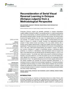
Results Figure 1 presents the distance findings. There was a rapid and linear reduction in maximum equivalent reading rate with retinal defocus. It was 190 wpm under the plano condition and reduced to 15 wpm equivalent under the 1.5D condition, and zero beyond. The largest fallVolume 20/2009/Number 3/Page 68
Figure 1: Equivalent reading rate as a function of lens defocus at distance. Plotted is the mean +/- 1 sem. The open symbols have a zero value and were not included in neither the statistical nor linear regression analysis.
Figure 2: Equivalent reading rate as a function of lens defocus at near. Plotted is the mean +/- 1 sem. The open symbols have a zero value and were not included in neither the statistical nor linear regression analysis.
off in processing rate was between 0.5 and 1.0D of defocus. A one-way ANOVA was performed, and the effect of retinal defocus was found to be significant [F(3,50)=50.776, p=