□
CASE REPORT
□
Rapidly Progressive Pulmonary Fibrosis Following the Onset of Diffuse Alveolar Hemorrhage in Sjögren’s Syndrome: An Autopsy Case Report Yusuke Tomita 1,2, Shunsuke Mori 3, Nobuyuki Arima 4, Koichiro Fukuda 1 and Hirotsugu Kohrogi 2
Abstract We report an autopsy case of a patient with Sjögren’s syndrome (SjS) who presented with rapid progressive pulmonary fibrosis following the onset of diffuse alveolar hemorrhage (DAH) without cryoglobulinemia. Despite early and aggressive immunosuppressive therapy, pulmonary fibrosis progressed and the patient succumbed to his illness. An autopsy was performed and revealed DAH and interstitial pneumonia with a fibrotic nonspecific interstitial pneumonia pattern. We could not find any previously-reported underlying causes of DAH. The findings from this case suggest that DAH can occur as a pulmonary manifestation of SjS as well as other connective tissue diseases or vasculitis. Key words: Sjögren’s syndrome, diffuse alveolar hemorrhage, nonspecific interstitial pneumonia (Intern Med 51: 295-299, 2012) (DOI: 10.2169/internalmedicine.51.6288)
Introduction Diffuse alveolar hemorrhage (DAH) is a rare yet serious and frequently life-threatening complication of a variety of conditions. Most cases of DAH are caused by capillaritis associated with systemic autoimmune diseases such as antineutrophil cytoplasmic antibody-associated vasculitis, antiglomerular basement membrane disease, and systemic lupus erythematosus (SLE), but seldom with Sjögren’s syndrome (SjS) (1). The reported frequency of pulmonary manifestation in primary SjS varies widely, ranging from 9-75% depending on the detection method employed, and the manifestations consist of various forms of small airways and interstitial lung diseases (2, 3). Recent studies revealed that among the pulmonary manifestations of primary SjS, interstitial pneumonia of fibrotic-nonspecific interstitial pneumonia (f-NSIP) pattern was the commonest histologic pattern, which is slowly progressive and has a favorable prognosis. They re-
ported a 5-year survival rate of 83% in patients with NSIP. On the other hand, it was revealed that bronchiolar diseases, amyloid and malignant lymphoma were less frequent (2, 3). Here we report an autopsy case of patient with SjS who presented with rapidly progressive pulmonary fibrosis of fNSIP pattern following the onset of diffuse alveolar hemorrhage (DAH) without cryoglobulinemia. To our knowledge, this is the first case of DAH in SjS without cryoglobulinemia.
Case Report A 75-year-old man was admitted for complaints of hemoptysis and progressive dyspnea lasting for 2 days. He also complained of progressive sicca symptoms of recent onset such as dry mouth and dry eyes, although he denied having arthralgia, myalgia, Raynaud’s symptoms, or neuralgia. Four years previously, the patient had valvular heart disease, and aortic valve and mitral valve replacement with tissue valves was undertaken. He also had been taking war-
1
Department of Respiratory Medicine, Kumamoto City Hospital, Japan, 2Department of Respiratory Medicine, Graduate School of Medical Sciences, Kumamoto University, Japan, 3Clinical Research Center for Rheumatic Disease and Department of Rheumatology, Kumamoto Saishunsou National Hospital, Japan and 4Department of Clinical Pathology, Kumamoto City Hospital, Japan Received for publication July 30, 2011; Accepted for publication October 11, 2011 Correspondence to Dr. Yusuke Tomita,
[email protected]
295
Intern Med 51: 295-299, 2012
DOI: 10.2169/internalmedicine.51.6288
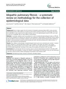
Figure 1. A: Chest radiograph on admission shows diffuse bilateral ground-glass opacity, particularly in the right lung field. B: Chest radiograph on the 11th hospital day shows progression of volume loss of both lungs, mainly in the right lower lobe.
farin (2.25 mg daily) for 2 years because of atrial fibrillation. He had no medical history of autoimmune disease or pulmonary disease. The patient had a 1-pack-day history of cigarette smoking, but had quit smoking more than 25 years before his presentation. He had no occupational or environmental cause for pulmonary fibrosis and DAH (4, 5). On admission, his temperature was 38.2℃ and respiratory rate was 24/min. Chest auscultation revealed diffuse bilateral mild coarse crackles, but there were no third heart sounds and no murmur. His fingers were not clubbed, and the joints of his extremities were not tender or swollen. There were no skin lesions or lower limb edema. The results of all neurologic examinations were normal. Other physical findings and urine microscopy were unremarkable. An ultrasoniccardiogram indicated that left ventricular and tissue valve function was normal. Arterial blood gas at room air showed type 1 respiratory failure (PaO2 of 59 mmHg and PaCO2 of 41 mmHg). Laboratory values showed a hemoglobin value of 12.6 g/dL, leukocyte count was 9.3×10⁹/L, and platelet count was 231×109/L. The international normalized ratio was 2.9 and D-dimer levels were within normal limits. The serum C-reactive protein level was 8.0 mg/dL. Hepatitis C virus antigen was negative, and liver and renal function tests were normal. Serological findings of autoantibodies were as follows: antinuclear antibody, with a titer of 1:40 and a homogeneous pattern; anti-Ro/SS-A Ab, 21.4 Index; and antiLa/SS-B Ab, 41.9 Index. Other autoantibodies, including IgM rheumatoid factor, anti-double-stranded DNA, anticardiolipin, anti-RNP, anti-Scl-70, anti-Jo-1, anti-centromere, and anti-glomerular basement membrane antibodies, were all negative. An enzyme-linked immunosorbent assay for myeloperoxidase anti-neutrophil cytoplasmic antibodies (MPO-ANCAs) and proteinase 3-ANCAs were also negative. No cryoglobulin was detected in the patient’s serum. KL-6 serum levels were within normal levels but surfactant protein D (SP-D) was slightly increased (226.3 ng/mL). The patient was diagnosed with SjS, and this was based
on the following findings: a recurrent sensation of sand in the eyes, frequently drinking liquids to aid in swallowing dry food, positive Schirmer’s test, positive Rose Bengal test, salivary scintigraphy showing delayed uptake and excretion of tracer, and the presence of anti-La/SS-B Ab. Therefore, his disease fulfilled five criteria listed in the international classification criteria for primary SjS revised by the American-European Consensus Group (6). Chest radiograph on admission showed diffuse bilateral ground-glass opacity (GGO), particularly in the right lung field (Fig. 1A). Chest computed tomography (CT) on admission (Fig. 2A) demonstrated diffuse bilateral GGO, thickening of interlobular septa and patchy areas of consolidation, but there was no advanced interstitial fibrotic changes such as volume loss, honeycombing or traction bronchiectasis. Bronchoalveolar lavage (BAL) was performed on the 2nd hospital day. There were no bloody endotracheal secretions, but bronchoscopy yielded progressively bloodier BAL return from the right middle lobe. Bronchoalveolar lavage fluid (BALF) showed a total cell count of 6.45×10⁵/mL, of which 64.5% were lymphocytes (CD4/CD8 ratio was 1.96), 12.8% were neutrophils, and 7.1% were eosinophils. There was no evidence of infection or malignant cells. The percentage of iron-positive alveolar macrophages in BALF was 24% of total macrophages, which confirmed alveolar hemorrhage. Warfarin therapy was stopped after admission and we started steroid pulse therapy (intravenous injection of methylprednisolone, 1 g three times daily) on the 2nd hospital day, followed by 125 mg/day intravenous injection of methylprednisolone, which was slowly tapered. The patient’s hemoptysis disappeared, but on the 6th hospital day, PaO2 was acutely decreased. We administered cyclophosphamide pulse therapy (intravenous injection of cyclophosphamide, 0.5 g monthly). Despite this treatment, on the 7th hospital day, KL-6 and SP-D serum levels were markedly increased (KL6, 1237 U/mL; SP-D, 987 ng/mL) and a chest radiograph on the 11th hospital day also showed progression of volume
296
Intern Med 51: 295-299, 2012
DOI: 10.2169/internalmedicine.51.6288
A
B
Figure 2. A: Chest computed tomography on admission shows extensive bilateral ground-glass opacity, thickening of the interlobular septa and patchy areas of consolidation, but absence of chronic pulmonary fibrosis such as volume loss, traction bronchiectasis and honeycombing. B: A follow-up high resolution computed tomography of the chest 2 months after admission shows bilateral ground-glass opacity, irregular reticular opacity, consolidation, and marked traction bronchiectasis (arrows), with a small volume of right pleural effusion.
loss of both lungs, mainly in the right lower lobe compared with that taken on admission (Fig. 1B). Pulmonary function testing on the 29th hospital day, which was consistent with the result of the chest radiograph, demonstrated a restrictive pattern of lung volume (total lung capacity of 2.36 L, 45% of predicted; forced vital capacity was 1.26 L, 42.7% of predicted). Follow-up BAL performed 1 month after the institution of intravenous immunosuppressive therapy revealed pale bloody BAL return from the right middle lobe, although lymphocytic alveolitis was not detected, which indicated that alveolar hemorrhage had continued (7). Follow-up high resolution CT revealed bilateral GGO, marked traction bronchiectasis, and volume loss in the lower lobes confirming progressive pulmonary fibrosis, but there were no findings of honeycombing (Fig. 1B and 2B). Both KL-6 and SP-D elevation persisted throughout the entire clinical course. Despite early and aggressive immunosuppressive therapy, the patient did not show clinical and radiological improvement, and suddenly died on the 101st hospital day. We performed an autopsy on the patient. The lungs showed diffuse fibrotic changes with a temporally uniform interstitial fibrosing process predominantly in the lower lobes, but fibroblastic foci was absent. These pathological findings were compatible with an f-NSIP pattern. Autopsy also revealed marked diffuse alveolar hemorrhage throughout bilateral lungs. Old and fresh alveolar hemorrhage were mixed, but vasculitis was not detected (Fig. 3). In the kidney, specific findings, which occasionally develop in Sjögren’s syndrome or systemic vasculitis, were not detected. The direct cause of death was finally unclear, but lethal arrhythmia was suspected.
Discussion In SjS, only one case of DAH has been previously reported as a result of pulmonary vasculitis with cryoglobulinemia (8). In contrast, in the present case, multiple tests of
cryoglobulin were all negative in the patient’s serum during the clinical course of disease. We also did not find any underlying causes of DAH such as systemic vasculitis, rheumatoid arthritis, SLE and mixed connective tissue disease (1). Therefore, we speculate that DAH developed as a pulmonary manifestation of SjS. To our knowledge, this is the first case of DAH in SjS without cryoglobulinemia. Recently, Ito et al showed that NSIP was the most frequently observed histological pattern (20 of 33 cases; 61%; 19 f-NSIP and 1 cellular NSIP) in primary SjS. They also showed that lymphocyte counts were elevated in 64% patients in cell differentials of BALF (2). The present patient developed interstitial pneumonia with an f-NSIP pattern, and BALF showed lymphocytic alveolitis (2, 3). Therefore, we consider that interstitial pneumonia also developed as a pulmonary manifestation of SjS. In the majority of disorders associated with DAH, the underlying histopathology shows capillaritis, bland pulmonary hemorrhage, or diffuse alveolar damage (DAD) (9). Pulmonary capillaritis is the most common histologic pattern associated with alveolar hemorrhage syndromes and it is characterized by the presence of fibrinoid necrosis of the alveolar structures, and neutrophil infiltration of the interstitium and adjacent blood vessels. DAD is characterized by the presence of alveolar septal edema, capillary microthrombi, and intra-alveolar hyaline membrane formation. However, the present case did not show the histologic findings of capillaritis or DAD. Bland pulmonary hemorrhage is characterized by the absence of inflammation, edema, or necrosis in the alveolar interstitium and the most common causes of bland pulmonary hemorrhage include idiopathic pulmonary hemosiderosis (IPH). In this case, bland pulmonary hemorrhage pattern was partially observed at autopsy (Fig. 3D). The pathogenesis of bland pulmonary hemorrhage remains unknown, however, studies in IPH have shown disruption of the integrity of the alveolar-capillary membrane at the ultrastructual level (10, 11). Taking these findings into considera-
297
Intern Med 51: 295-299, 2012
DOI: 10.2169/internalmedicine.51.6288
A
B
C
D
Figure 3. A: Photomicrograph of pathology shows a temporally uniform interstitial fibrosing process. (fibrotic nonspecific interstitial pneumonia pattern). Original magnification, ×10; Hematoxylin and Eosin staining. B: Photomicrograph of pathology shows alveolar spaces occupied by red blood cells, which indicates relatively fresh alveolar hemorrhage, and interstitial fibrosis. The fibrosis is composed of mature collagen containing mild inflammatory cells. Original magnification, ×40; Hematoxylin and Eosin staining. C: Photomicrograph of pathology shows alveolar spaces occupied by marked hemosiderin-laden macrophages. The alveolar walls are diffusely thickened by dense collagen with deposition of hemosiderin. Original magnification, ×40; Berlin blue stain. D: Photomicrograph of pathology shows an area of lung parenchyma with the alveolar spaces occupied by red blood cells and the absence of inflammation, edema, and necrosis in the alveolar interstitium (bland pulmonary hemorrhage). Original magnification, ×100; Hematoxylin and Eosin staining.
cyclophosphamide 0.5 g
PSL 60 mg mPSL 1g mPSL 125mg
1
7
15
25
31
PSL 40 mg
38
47
54
72
82
92
99
Hospital days
Figure 4. Clinical course of the patient. mPSL: methylprednisolone, PSL: prednisolone
298
Intern Med 51: 295-299, 2012
DOI: 10.2169/internalmedicine.51.6288
tions, we speculated that these mechanism related to IPH may cause alveolar hemorrhage in this patient. IPH is also known to cause pulmonary fibrosis secondary to recurrent alveolar hemorrhage with deposition of hemosiderin in the interstitium (5, 12). In the present case, follow-up BAL showed persistent alveolar bleeding, and autopsy findings revealed that the alveolar walls were diffusely thickened by dense collagen with deposition of hemosiderin (Fig. 3C). In addition, f-NSIP and alveolar bleeding took place simultaneously in our patient (Fig. 3B). Therefore, recurrent alveolar hemorrhage of this case appeared to cause pulmonary fibrosis of f-NSIP pattern. In conclusion, the findings in the present case suggest that DAH can occur as a pulmonary manifestation of SjS. Clinicians may have to consider SjS as one of the predisposing conditions that cause DAH. The authors state that they have no Conflict of Interest (COI). Acknowledgement We thank Dr. Sayuri Hirooka (Department of Respiratory Medicine, Kumamoto City Hospital, Kumamoto, Japan) for providing us with clinical data.
References 1. Specks U. Diffuse alveolar hemorrhage syndrome. Curr Opin Rheumatol 13: 12-17, 2001. 2. Ito I, Nagai S, Kitaich M, et al. Pulmonary manifestations of primary Sjögren’s syndrome. Am J Respir Crit Care Med 171: 632-
638, 2005. 3. Parambil JG, Myers JL, Lindell RM, Matteson EL, Ryu JH. Interstitial lung disease in primary Sjögren’s syndrome. Chest 130: 1489-1495, 2006. 4. American Thoracic Society,European Respiratory Society. American Thoracic Society/European Respiratory Society International Multidisciplinary Consensus Classification of Idiopathic Interstitial Pneumonias. Am J Respir Crit Care Med 165: 277-304, 2002. 5. Buschman DL, Ballard R. Progressive massive fibrosis associated with idiopathic pulmonary hemosiderosis. Chest 104: 293-295, 1993. 6. Vitali C, Bombardieri S, Jonsson R, et al. American-European Consensus Group. Classification criteria for Sjögren’s syndrome: a revised version of the European criteria proposed by the American-European Consensus Group. Ann Rheum Dis 61: 554558, 2002. 7. Schnabel A, Reuter M, Csernok E, Richter C, Gross WL. Subclinical alveolar bleeding in pulmonary vasculitides: correlation with indices of disease activity. Eur Respir J 14: 118-124, 1999. 8. Johnston SL, Dudley CRK, Unsworth DJ, Lock RJ. Lifethreatening acute pulmonary hemorrhage in primary Sjögren’s syndrome with cryoglobulinaemia. Scand J Rheumatol 34: 404407, 2005. 9. Frankel SK, Cosgrove GP, Fischer A, Meehan RT, Brown KK. Update in the diagnosis and management of pulmonary vasculitis. Chest 129: 452-465, 2006. 10. Gonzalez-Crussi F, Hull MT, Grosfeld JL. Idiopathic pulmonary hemosiderosis: evidence of capillary basement membrane abnormality. Am Rev Respir Dis 114: 689-698, 1976. 11. Corrin B, Jagusch M, Dewar A, et al. Fine structural changes in idiopathic pulmonary hemosiderosis. J Pathol 153: 249-256, 1987. 12. Travis WD, Colby TV, Lombard C, Carpenter HA. A clinicopathologic study of 34 cases of diffuse pulmonary hemorrhage with lung biopsy confirmation. Am J Surg Pathol 14: 1112-1125, 1990.
Ⓒ 2012 The Japanese Society of Internal Medicine http://www.naika.or.jp/imindex.html
299