CASE
REPORT
Rapidly Progressive Glomerulonephritis Associated with Nontuberculous Mycobacteria Shwu-Jiuan Chen1, Yao-Ko Wen1*, Mei-Ling Chen2 1
Division of Nephrology, Department of Medicine and 2Department of Pathology, Changhua Christian Hospital, Changhua, Taiwan, R.O.C.
A 72-year-old woman with a past medical history of nontuberculous mycobacteria (NTM) pulmonary disease was admitted because of hemoptysis and acute renal failure. A chest X-ray showed interstitial infiltration over bilateral lung fields. Kidney biopsy showed immune complex-mediated acute diffuse proliferative glomerulonephritis with 48% crescents and glomerular endocapillary hypercellularity with exudative neutrophils suggestive of infection-related glomerulonephritis. Reactivated NTM infection of the lungs was suspected when mycobacterial cultures of the sputum repeatedly yielded Mycobacterium avium. A lung biopsy revealed chronic inflammation without evidence of alveolar capillaritis. A diagnosis of NTM pulmonary disease was further confirmed by tissue culture of the lung biopsy specimens. Antituberculous drugs in combination with clarithromycin were given for the treatment of NTM infection. Pulmonary symptoms promptly responded to the treatments. Furthermore, renal function steadily improved after initiation of anti-NTM therapy. To our knowledge, this is the first report of rapidly progressive glomerulonephritis associated with NTM infection. [J Chin Med Assoc 2007;70(9):396–399] Key Words: glomerulonephritis, nontuberculous mycobacteria
Introduction Over the centuries, 2 well-known mycobacterial species, namely, Mycobacterium tuberculosis and M. leprae have been the known causes of immense human suffering. Many other mycobacteria are ubiquitous in the environment with low virulence, but can be opportunistic and at times deadly pathogens. They are referred to as atypical mycobacteria, mycobacteria other than tuberculosis, or nontuberculous mycobacteria (NTM). Among the more than 90 known species of NTM, about 1 third have been associated with human diseases. Today, the prevalence of NTM infection is increasing worldwide. It is partly explained by the increase in the number of immunocompromised individuals, such as those suffering from acquired immunodeficiency syndrome, and is also due to an increasing awareness of NTM as human pathogens and improvements in methods of mycobacterial culture. Infection has been well documented to cause glomerulonephritis, with Streptococcus being the most common pathogen and
predominantly affecting children. The pattern of the disease has changed over recent decades. Not only Streptococcus but also other bacterial, viral, fungal and parasitic infections can trigger the disease in both children and adults. Glomerulonephritis caused by tuberculosis infection has rarely been reported in the literature.1–6 To our knowledge, the association of NTM infection with glomerulonephritis has not been described before. Herein, we report a case of acute diffuse proliferative glomerulonephritis with the characteristics of infection-related glomerulonephritis in which M. avium was the most possible causal pathogen.
Case Report A 72-year-old woman was admitted with a 2-week history of cough productive of yellowish, occasionally blood-streaked sputum. She had also noticed profound malaise, decreased appetite and a weight loss of 5 kg within this time period. There was no complaint
*Correspondence to: Dr Yao-Ko Wen, Division of Nephrology, Department of Medicine, Changhua Christian Hospital, 135, Nanhsiao Street, Changhua 500, Taiwan, R.O.C. E-mail:
[email protected] Received: December 12, 2006 Accepted: July 24, 2007 ●
396
●
J Chin Med Assoc • September 2007 • Vol 70 • No 9 © 2007 Elsevier. All rights reserved.
Glomerulonephritis and nontuberculous mycobacteria
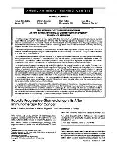
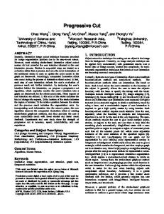
of fever, epistaxis, or arthralgia. She had been diagnosed to have NTM pulmonary disease approximately 5 years before and had received a 12-month course of antituberculous therapy. Her past medical history was otherwise unremarkable. On admission, physical examination showed a body temperature of 36.5°C, regular pulse rate of 92 beats/ minute, respiratory rate of 18 breaths/minute, and blood pressure of 128/76 mmHg. Head and neck examinations were unremarkable. Chest auscultation revealed diffuse crackles over bilateral lung fields. Examinations of the heart and abdomen were normal. She had mild lower extremity pitting edema (+). No skin lesion was found. A chest X-ray demonstrated interstitial infiltration over bilateral lung fields and fibrous change of the right upper lung. Laboratory investigations showed hemoglobin 9.5 g/dL, white blood cell count 15,400/µL, and platelet count 256,000/µL. A blood chemistry panel showed urea nitrogen 32 mg/ dL, creatinine 2.2 mg/dL, sodium 136 mmol/L, potassium 3.8 mmol/L, calcium 8.1 mg/dL, glucose 93 mg/ dL, uric acid 8.4 mg/dL, albumin 3.0 g/dL, and globulin 3.2 g/dL. Urinalysis revealed occult blood (++++) and protein (++), with numerous red blood cells and 35–40 white blood cells per high-power field in the urinary sediment. Daily urinary protein excretion was 1.9 g. Renal ultrasonography showed normal-sized kidneys with increased parenchymal echogenicity suggestive of acute renal parenchymal disease. Owing to acute renal failure and urinalysis findings suggestive of glomerulonephritis, rapidly progressive glomerulonephritis (RPGN) was highly suspected. Serologic studies showed negative findings for antinuclear antibody, antibasement membrane antibody, antineutrophil cytoplasmic antibody, hepatitis B surface antigen, hepatitis C antibody, and human immunodeficiency virus antibody. Antistreptolysin O titer was < 200 IU/mL. Serum complement levels were normal. Rheumatoid factor was negative. Kidney biopsy showed diffuse proliferative glomerulonephritis, with cellular crescents affecting 12 of the 25 examined glomeruli (Figure 1). The glomeruli displayed endocapillary hypercellularity with capillaries full of neutrophils. No thickening or double contouring of capillary loop was seen by periodic acid-Schiff or periodic acid-methenamine silver stain. Mild interstitial infiltrate of inflammatory cells was noted. Interstitial fibrosis and tubular atrophy were present only focally and minimally. No glomerular tuft necrosis or vasculitis was seen. Immunofluorescent studies showed granular staining for IgG (++), IgM (+), C3 (+++), and C1q (+) along the mesangium and capillary loops (Figure 2). There was no IgA deposition. Subendothelial and
J Chin Med Assoc • September 2007 • Vol 70 • No 9
Figure 1. Renal biopsy showing diffuse endocapillary proliferative glomerulonephritis with cellular crescent formation (arrows) (hematoxylin & eosin, 100×).
Figure 2. Immunofluorescence showing strong granular deposits of C3 in the glomerular basement membrane and mesangium.
Figure 3. Electron microscopy showing electron-dense deposits in the subendothelial region (arrows).
mesangial electron-dense deposits were noted on electron microscopy (Figure 3). No subepithelial hump-like deposit was found. There was no evidence of cryoglobulin deposit.
397
S.J. Chen, et al
Because of histologic resemblance to infectionrelated glomerulonephritis, a comprehensive workup for infection was performed. Echocardiography showed no evidence of infective endocarditis. Bronchoscopy revealed no endobronchial lesion. Sputum, blood and urine bacterial cultures were negative. However, sputum smears for acid-fast stain were positive. Furthermore, mycobacterial cultures of the sputum specimens collected by either spontaneous expectoration or bronchoscopic wash repeatedly yielded M. avium. To clarify the lung lesions, a thoracoscopic lung biopsy was performed. The specimens demonstrated dilated alveoli full of mucus, macrophages, and leukocytes. The alveolar septum was widened by lymphoid aggregation, fibrosis, and smooth muscle cell proliferation. No alveolar capillaritis or granuloma was found. NTM pulmonary disease was further confirmed when the mycobacterial culture of the lung biopsy specimens also grew M. avium. Antituberculous agents comprising rifampicin, isoniazide and ethambutol, in combination with clarithromycin were administered for the treatment of pulmonary NTM infection. Pulmonary symptoms promptly responded to the treatments. Repeated sputum smears for acid-fast stain and mycobacterial cultures became negative 1 month later. Follow-up chest X-ray also showed resolution of the pulmonary infiltrates. Furthermore, renal function steadily improved, with serum creatinine level falling to 1.1 mg/dL 3 months after the initiation of anti-NTM therapy.
Discussion The combination of acute glomerulonephritis, as manifested by hematuria and renal insufficiency, and pulmonary hemorrhage, as manifested by hemoptysis and pulmonary infiltrates, is characteristic of pulmonary– renal syndrome. However, these manifestations can be seen in acute glomerulonephritis that is complicated by other pulmonary disorders such as pulmonary edema, pulmonary embolism, or pulmonary infection. There is no doubt that it is important to exclude pulmonary infectious processes before starting immunosuppressive therapy when pulmonary–renal syndrome is suspected. However, definite diagnosis of pulmonary NTM infection remains difficult. First, NTM requires special media and growth conditions. Secondly, the diagnosis of pulmonary NTM infection is complicated by the variability in clinical and radiologic manifestations and the frequent presence of significant prior pulmonary disease. Finally, in contrast to M. tuberculosis, NTM are ubiquitous in the environment. Isolation of NTM from the sputum may represent harmless colonization
398
of the respiratory tract and does not constitute proof of the existence of pulmonary NTM infection. In an attempt to address these difficulties, the American Thoracic Society has established specific criteria that emphasize compatible clinical symptoms/signs, reasonable exclusion of other disease and characteristic imaging findings such as persistent (> 2 months) or progressive pulmonary infiltrates. Bacteriologic criteria include positive cultures of at least 3 sputum/bronchial wash samples within 1 year or any growth from bronchopulmonary tissue biopsy.7 In our patient, the clinical, radiographic and bacteriologic criteria were all satisfied and indicated NTM pulmonary disease. Although the potential virulence of NTM has been increasingly recognized in humans, NTM infectionrelated glomerulonephritis has not yet been described. Nguyen et al reported a patient with RPGN who developed severe lower gastrointestinal bleeding 2 weeks after initiation of aggressive immunosuppressive treatments and was diagnosed to have ulcerative colitis due to infection with M. avium.8 The intestinal bleeding stopped promptly after administration of anti-NTM therapy. However, the renal function did not recover, and the patient was maintained on hemodialysis. The report emphasized an aggravation of gastrointestinal NTM infection after immunosuppressive therapy but did not address the association of NTM infection with RPGN. In our case, arguments may arise as to whether the RPGN was an independent disease or was causally associated with NTM infection. In light of several observations, we strongly considered that NTM infection was responsible for the development of RPGN. First, the kidney biopsy revealed diffuse endocapillary proliferation with neutrophil exudates as a characteristic of infection-related glomerulonephritis. Although subepithelial hump-like deposits were not seen in our case, they are often but not always found in cases of infection-associated glomerulonephritis. Secondly, immunofluorescence studies showed a granular glomerular labeling pattern indicative of immune complex-mediated glomerulonephritis. In the absence of other discernible causes such as a variety of primary glomerulonephritis, systemic lupus erythematosus, Henoch-Schönlein purpura, and mixed cryoblobulinemia, infection-related glomerulonephritis was the most possible clinicopathologic diagnosis. Most importantly, recovery of renal function after treatment of NTM infection further suggested a causal relationship between NTM infection and RPGN. As a result, our case fulfills the following diagnostic criteria for infection-related glomerulonephritis proposed by Montseny et al: (1) evidence of infection preceding the glomerulonephritis; (2) close temporal relationship between the infection
J Chin Med Assoc • September 2007 • Vol 70 • No 9
Glomerulonephritis and nontuberculous mycobacteria
and glomerulonephritis; (3) glomerular change consistent with an infectious glomerulonephritis; (4) absence of systemic disease that may cause the glomerulonephritis; (5) exclude IgA nephropathy.9 When comparing with tuberculosis-associated glomerulonephritis, the glomerular changes with predominantly diffuse endocapillary proliferation observed in our case are nonspecific. On the contrary, tuberculosis-associated glomerulonephritis has more specific glomerular changes, and membranoproliferative glomerulonephritis and IgA nephropathy are the 2 most common histologic findings.1–6 This case also presented a therapeutic dilemma regarding the use of immunosuppressive agents. Because of the generally grave prognosis of RPGN and the known usefulness of immunosuppressive agents in other types of RPGN, corticosteroids, cytotoxic agents, and plasmapheresis, alone or in combination, have been tried in infection-related RPGN.10–15 Although the limited evidence supports the use of immunosuppressive agents in this setting, serious consideration must be given to the fact that renal disease might respond to antimicrobial therapy alone. Furthermore, the risk of overwhelming underlying infection precipitated by immunosuppression might abrogate the benefit for renal function. In conclusion, RPGN could be associated with M. avium infection. Although the prognosis of RPGN is generally thought to be poor, our case showed a favorable prognosis when anti-NTM therapy was administered in a timely manner. The natural history of infection-related glomerulonephritis perhaps contributed to the excellent outcome.
References 1. Cohen AJ, Rosenstein ED. IgA nephropathy associated with disseminated tuberculosis. Arch Intern Med 1985;145:554–6.
J Chin Med Assoc • September 2007 • Vol 70 • No 9
2. Teruel JL, Matesanz R, Mampaso F, Lamas S, Herrero JA, Ortuno J. Pulmonary tuberculosis, cryoglobulinemia and immune complex glomerulonephritis. Nephron 1987;27:48–9. 3. Meyrier A, Valensi P, Sebaoum J. Mesangio-capillary glomerulonephritis and the nephrotic syndrome in the course of disseminated tuberculosis. Nephron 1988;49:341–2. 4. Sopeña B, Sobrado J, Pérez AJ, Oliver J, Courel M, Palomares L, González L. Rapidly progressive glomerulonephritis and pulmonary tuberculosis. Nephron 1991;57:251–2. 5. Pecchini F, Bufano G, Ghiringhelli P. Membranoproliferative glomerulonephritis secondary to tuberculosis. Clin Nephrol 1997;47:63–4. 6. Matsuzawa N, Nakabayashi K, Nagasawa T, Nakamoto Y. Nephrotic IgA nephropathy associated with disseminated tuberculosis. Clin Nephrol 2002;57:63–8. 7. Wallace RJ Jr, O’Brien R, Glassroth J, Raleigh J, Dutta A. Diagnosis and treatment of disease caused by nontuberculous mycobacteria. Am Rev Respir Dis 1990;142:940–53. 8. Nguyen HN, Frank D, Handt S, Rieband HC, Maurin N, Sieberth HG, Matern S. Severe gastrointestinal hemorrhage due to Mycobacterium avium complex in a patient receiving immunosuppressive therapy. Am J Gastroenterol 1999;94: 232–5. 9. Montseny JJ, Meyrier A, Kleinknecht D, Callard P. The current spectrum of infectious glomerulonephritis: experience with 76 patients and review of the literature. Medicine (Baltimore) 1995;74:63–73. 10. Rovzar MA, Logan J, Ogden D, Graham AR. Immunosuppressive therapy and plasmapheresis in rapidly progressive glomerulonephritis associated with bacterial endocarditis. Am J Kidney Dis 1986;7:428–34. 11. Daimon S, Mizuno Y, Fujii S, Mukai K, Hanakawa H, Otsuki N, Yasuhara S, et al. Infective endocarditis-induced crescentic glomerulonephritis dramatically improved by plasmapheresis. Am J Kidney Dis 1998;32:309–13. 12. Kannan S, Mattoo TK. Diffuse crescentic glomerulonephritis in bacterial endocarditis. Pediatr Nephrol 2001;16:423–8. 13. Koya D, Shibuya K, Kikkawa R, Haneda M. Successful recovery of infective endocarditis-induced rapidly progressive glomerulonephritis by steroid therapy combined with antibiotics: a case report. BMC Nephrol 2004;5:18. 14. Couzi L, Morel D, Deminiere C, Merville P. An unusual endocarditis-induced crescentic glomerulonephritis treated by plasmapheresis. Clin Nephrol 2004;62:461–4. 15. Sadikoglu B, Bilge I, Kilicaslan I, Gokce MG, Emre S, Ertuqrul T. Crescentic glomerulonephritis in a child with infective endocarditis. Pediatr Nephrol 2006;21:867–9.
399