REAL-TIME MONITORING OF CELLULAR DYNAMICS USING A MICROFLUIDIC CELL CULTURE SYSTEM WITH INTEGRATED ELECTRODE ARRAY AND POTENTIOSTAT
K. Zόr1, 2*, M. Vergani3, A. Heiskanen2, E. Landini4, M. Carminati3, V. Coman2, I. Vedarethinam2, P. Skafte-Pedersen2, M. Skolimowski2, A. Martinez Serrano5, M. Kokaia6, T. Ramos Moreno5, A. Ghio4, W.E. Svendsen2, M. Dimaki2, Zs. Keresztes7, M. Adamovski8, U. Wollenberger8, D. Sabourin2, G. Ferrari3, R. Raiteri4, M. Sampietro3, M. Dufva2 and J. Emnéus2 1
Lund University, SWEDEN, 2Technical University of Denmark, DENMARK, 3Politecnico di Milano, ITALY, 4 University of Genova, ITALY, 5 University Autonomous of Madrid, SPAIN, 6 Wallenberg Neuroscience Center, Lund University, SWEDEN, 7Hungarian Academy of Sciences, HUNGARY and 8 University of Potsdam , GERMANY
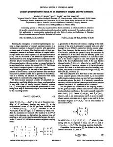
ABSTRACT A versatile microfluidic, multichamber cell culture and analysis system with an integrated electrode array and potentiostat suitable for electrochemical detection and microscopic imaging is presented in this paper. The system, which allows on-line electrode cleaning and modification, was developed for real-time monitoring of cellular dynamics, exemplified in this work by monitoring of redox metabolism inside living yeast cells and dopamine release from PC12 cells. KEYWORDS: Microfluidic Cell Culture System, On-line Measurement, Cellular Dynamics Monitoring, Dopamine Exocytosis INTRODUCTION To conduct cell based experiments, facilitating in situ monitoring of cellular dynamics in real-time, requires novel tools not usually employed in biology. The EU-funded FP7 project “Exploring the cellular dynamics at nanoscale” (EXCELL) is a multidisciplinary collaboration to design and fabricate systems suitable for complete cell-based assays. Electrochemical techniques have proved to be suitable for integration in microsystems since the electronic elements, e.g. electrode arrays and potentiostat can be miniaturized and multiplexed [1].Electrochemical sensors are considered to be sensitive [2] allowing detection without destroying cellular integrity. Integration of an electrode array with a microfluidic cell culture device enables dynamic real-time monitoring of cellular dynamics in terms of e.g. cell differentiation, neurotransmitter release and cellular redox metabolism. This type of analytical system, aside from being a basic research tool in biology and chemistry, can be used as a high throughput monitoring device for exploring cellular dynamics in relation to neurodegenerative disorders and drug screening. THEORY Monitoring of cellular redox metabolism can provide important information about cellular functions, since imbalance in the intracellular redox state can be one of the factors causing neurodegenerative diseases, e.g. Parkinson’s and Alzheimer’s disease [3] and can affect cell signaling [4]. Mediated amperometry, using the ferricyanide-menadione double mediator system, enables monitoring of cellular redox metabolism as previously presented for yeast cells [5]. Detection and quantification of neurotransmitters using electrochemical techniques has been the focus of research for more than three decades. Dopamine, a key analyte in neurodegenerative diseases, successfully quantified by amperometric detection on a thiol-modified Au microelectrode chips [6], plays an important role in evaluation of the neuronal stem cell differentiation. EXPERIMENTAL The developed system comprises two main components, i) microfluidics (cell culture device integrated with a microelectrode chip and multichannel peristaltic pumps operated by step motors from LEGO®) and ii) electronics (miniaturized 24-channel potentiostat operated with a custom-made acquisition and analysis software) (Figure 1). The microfluidic cell culture device (A) consists of 3 micromilled 500 µm thick PMMA layers bonded together using a UV-assisted process (1 min UV exposure, 88 ºC, 60 bar for 20 min). A silicon adhesive gasket (B) is utilized to form a fluidtight sealing between the microfluidic cell culture device and the microelectrode chip (C), supported by a 5 mm thick PMMA holder (D). The microelectrode chips were fabricated using a lift-off process on a Si/SiO2 substrate. A 5 nm thick Ti layer was used as an adhesion layer for the 150 nm thick Au layer. The structure was passivated by depositing a 500 nm silicon nitride layer, which was locally etched with a reactive ion process to define the 12 electrode arrays, each consisting of a working (WE), 978-0-9798064-4-5/µTAS 2011/$20©11CBMS-0001
1532
15th International Conference on Miniaturized Systems for Chemistry and Life Sciences October 2-6, 2011, Seattle, Washington, USA
counter (CE) and reference (RE) electrode. The WEs are 500 µm long interdigitated electrodes (IDE) with 10 µm width and gap. The CEs and REs are Au disks with 700 µm and 50 µm diameter, respectively (c). The assembled microfluidic device and microelectrode chip is placed on the pump platform (E) and coupled to the potentiostat (F) to form the fully assembled system (G) which is controlled by the custom-made software (H). Microscopic imaging trough a window (f) can be performed simultaneously with electrochemical measurements. The potentiostat facilitates experiments using different electrochemical techniques, such as low noise amperometric recordings down to low pA level, cyclic voltammetry (CV) up to 2.5 kV/s sweep rate and electrochemical impedance spectroscopy (EIS) from 1 mHz to 100 kHz.
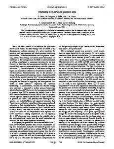
Figure 1: Structure of the analytical system for cell-based assays Prior to integrating the microfluidic device with the microelectrode chip, the latter was treated for 5 min with a mixture of 25% H202/ 50 mM KOH to remove residual organic contaminants [7]. To ensure good electrochemical reproducibility, the IDEs of the microelectrode chip were cleaned on-line by applying a potential sweep in 50 mM KOH (-0.2 to -1.2 V at 50 mV/s). To adapt the IDEs for different cell-based assays, different on-line electrode modifications (self-assembled monolayers (SAM) of thiols) were used: i) cysteamine (200 mM; 2 h) for the experiments with yeast cells and ii) mercaptopropionic acid (MPA) (200mM; 2 h) followed by laminin (20 μg/mL; 4 h) for the experiments with PC12 cells. Baker’s yeast (Saccharomyces cerevisiae) from Jästbolaget AB, Sweden, suspended in cell culture tested PBS (pH 7.4) containing 10 mM K3Fe(CN)6 (F) and K4Fe(CN)6 (Fo) was introduced into the microfluidic chambers. After seeding, the sedimentation of the yeast cells was monitored by EIS (Vrms = 1 mV, 1 Hz to 100 kHz frequency range, 1 s averaging time). To monitor redox metabolism, the cells were first perfused with cell culture tested PBS (pH 7.4) to obtain a baseline followed by a sequential addition of 2 mM F, 100 μM menadione (M) and 10 mM glucose (G) all prepared in PBS. The formed Fo was amperometrically detected as previously described [5] at 350 mV vs. Au RE. PC12 cells were cultivated for 48 h in the microfluidic chambers. Prior to exocytosis measurement the cells were incubated with 100 μM L-dopa in cell culture medium, followed by perfusion with a HEPES buffered solution, according to the previously presented protocol [6]. Dopamine exocytosis was triggered using a HEPES buffered solution with elevated potassium concentration (150 mM KCl) [6] and detected at 550 mV vs. Au RE. Microscopic images were acquired using a ZFL Video Fluorescence Scope (Navitar Imaging Solutions, Rochester, NY) equipped with an Infinity 2-3 CCD camera (Lumenera Corporation, Ottawa, ON). RESULTS AND DISCUSSION The electrochemical performance of the IDEs was followed during the on-line cleaning and modification process as presented in Figure 2A showing the CVs of an IDE after steps of on-line cleaning and thiol (cysteamine) modification. After thiol modification the IDEs show very good reproducibility, which is significant for assay performance. Figure 2B shows 16 CVs of 8 IDEs (for each IDE the both sides were individually used as a WE). The cell culture chambers were designed to be suitable for cell seeding, perfusion culture and performing electrochemical assays as shown in inset b of Figure 3A. The assay inlet (lower part) is in a close proximity of the WEs, ensuring a fast delivery of compounds to the cells growing on the electrodes resulting in an immediate electrochemical response, whereas the perfusion culture inlet (upper part) enables optimal flow for long-term culture eliminating shear stress. To monitor the redox 1533
metabolism (Figure 3A) of yeast (inset a), the electrodes were modified with cysteamine. The interaction between the positively charged SAM and the negatively charged cell wall retains the cells effectively on the electrode surface.
Figure 2: (A) K3Fe(CN)6 CVs of an IDE following on-line cleaning and thiol (cysteamine) modification steps. (B) 16 CVs of 8 IDEs (each side of an IDE was used separately as a WE) after on-line electrode cleaning and modification.
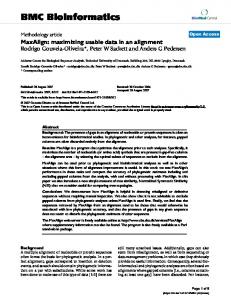
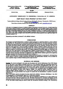
Figure 3: (A) Monitoring of intracellular redox metabolism in living yeast cells on cysteamine modified electrodes (flow rate:10 μL/min) using the double mediator system (Eappl: 350mV vs. Au RE). (B) Cell sedimentation monitoring using EIS. (Inset a)Image of yeast cells on IDE and inset b) a schematic view of a chamber, route for cell seeding and assays) The mechanism of the redox metabolism monitoring is based on the oxidation of the formed K4Fe(CN)6 [3]. Sedimentation of yeast cells was monitored prior the start of the redox metabolism monitoring by means of EIS as presented in Figure 3B. A clear increase in the charge transfer resistance can be observed as a result that the increased yeast cell coverage on the electrode surface. This platform is generally suitable EIS-based monitoring of cellular adhesion [8,9]. PC12 cells, cultivated on MPA and laminin modified electrodes for 48 h, were treated with 150 mM KCl to induce dopamine exocytosis. Figure 4 shows a recorded current-time trace (red line) with 40 s duration resulting from oxidation of the released dopamine from the whole cell population covering the depicted IDE (upper inset). A corresponding current-time trace for a bare IDE (lower inset of Figure 4) is indicated by the black line. CONCLUSION The presented system allows several simultaneous measurements under flow conditions with a potentiostat and software specially developed for cell-based assays. These are significant advancements from the previously reported systems [9]. To highlight the versatility of the platform, the usability of the presented system is demonstrated with two cell-based applications: monitoring of i) redox metabolism and sedimentation of yeast cells (Figure 3) and ii) dopamine exocytosis from cultured PC12 cells (Figure 4). The recorded signals are in good agreement with those previously reported for yeast cells [5] 1534
and a population of PC12 cells [9]. The presented system is capable of parallel microfluidic cell-based assays to study complex biological phenomena, such as stem cell differentiation.
Figure 4: Current-time trace for dopamine exocytosis from PC12 cells (upper inset) grown on MPA and laminin modified IDEs (red line) and a plane IDE (lower inset) without cells (black line) (Eappl: 500mV vs. Au RE). ACKNOWLEDGEMENTS We gratefully acknowledge all the partners of EXCELL consortium. The EU FP7 NMP project EXCELL (NMP4-SL-2008214706) is kindly acknowledged for financial support. REFERENCES [1] X. Xu, S. Zhang, H. Chen and J. Kong, "Integration of electrochemistry in micro-total analysis systems for biochemical assays: Recent developments," Talanta, vol. 80, pp. 8–18, Nov. 2009. [2] J. Wang, "Portable electrochemical systems," Trends Anal. Chem., vol. 21, pp.226 - 232, Apr. 2002. [3] T.G. Hastings and M.J. Zigmond, Neurogenerative disease and oxidative stress: Insights from an animal model of Parkinsonism, Molecular and cellular mechanisms and therapeutic advances, Ed. G. Fiskum, Plenum Press: New York, pp. 37-46, (1996). [4] Y.J. Suzuki, H.J. Forman and A. Sevanian, "Oxidants as stimulators of signal transduction," Free Rad. Biol. Med., vol. 22, pp. 269-285, Jan. 1997. [5] A. Heiskanen, J. Yakovleva, C. Spégel, R. Taboryski, M. Koudelka-Hep, J. Emnéus and T. Ruzgas, "Amperometric monitoring of redox activity in living yeast cells: comparison of menadione and manadione sodium bisulfate as electron transfer mediators," Electrochem. Commun., vol. 6, pp. 219 – 224, Jan. 2004. [6] C. Spégel, A. Heiskanen, S. Pederson, J. Emnéus, T. Ruzgas and R. Taboryski, "Fully Automated Microchip System for the Detection of Quantal Exocytosis from Single and Small Ensembles of Cells," Lab Chip, vol. 8, pp. 323-32, Feb. 2008. [7] L. M. Fischer, M. Tenje, A. R. Heiskanen, N. Masuda, J, Castillo, A. Bentien, J. Émneus, M. H. Jakobsen and A. Boisen, "Gold cleaning methods for electrochemical detection applications," Microelectron. Eng., vol.86, pp. 1282– 1285, Apr. 2009. [8] C. Spégel, A. Heiskanen, L.H. Dæhli Skjolding and J. Emnéus, "Chip based electrochemical systems for cell analysis," Electroanalysis, vol. 20, pp. 680-702, Mar. 2008. [9] A. Heiskanen, C. Spégel, J.Tønnesen, Z. Fohlerova, L. Goulart, J. Hansen, M. Kokaia,,M. Dufva and J. Emnéus, Development of a microfluidic on-line culture system for combined electrochemical and optical real-time detection of cellular processes, Proc. 12th International Conference of Miniaturized Systems for Chemistry and Life Sciences (µTAS 2008), vol. 2, pp. 1168-1171, (2008). CONTACT *K. Zόr, tel: +45256839;
[email protected]
1535