Received February 1, 2013, accepted April 2, 2013, published May 10, 2013. Digital Object Identifier 10.1109/ACCESS.2013.2260791
Real-World Neuroimaging Technologies KALEB MCDOWELL (Senior Member, IEEE)1 , CHIN-TENG LIN (Fellow, IEEE)2 , KELVIN S. OIE1 , TZYY-PING JUNG (Senior Member, IEEE)3 , STEPHEN GORDON (Member, IEEE)4 , KEITH W. WHITAKER1 , SHIH-YU LI2 , SHAO-WEI LU2 , and W. DAVID HAIRSTON1 1 Translational
Neuroscience Branch, U.S. Army Research Laboratory, Aberdeen Proving Ground, MD 21005, USA of Electrical Engineering and the Brain Research Center, National Chiao Tung University, Hsinchu 30010, Taiwan 3 Swartz Center for Computational Neuroscience, Institute for Neural Computation, University of California at San Diego, La Jolla, CA 92093, USA 4 DCS Corporation, Alexandria, VA 22310, USA 2 Department
Corresponding author: K. McDowell (
[email protected]) This work was supported by the U.S. Army Research Laboratory and, in part, under Cooperative Agreement W911NF-10-2-0022.
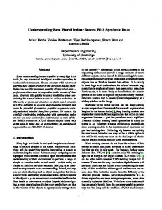
ABSTRACT Decades of heavy investment in laboratory-based brain imaging and neuroscience have led to foundational insights into how humans sense, perceive, and interact with the external world. However, it is argued that fundamental differences between laboratory-based and naturalistic human behavior may exist. Thus, it remains unclear how well the current knowledge of human brain function translates into the highly dynamic real world. While some demonstrated successes in real-world neurotechnologies are observed, particularly in the area of brain-computer interaction technologies, innovations and developments to date are limited to a small science and technology community. We posit that advancements in realworld neuroimaging tools for use by a broad-based workforce will dramatically enhance neurotechnology applications that have the potential to radically alter human–system interactions across all aspects of everyday life. We discuss the efforts of a joint government-academic-industry team to take an integrative, interdisciplinary, and multi-aspect approach to translate current technologies into devices that are truly fieldable across a range of environments. Results from initial work, described here, show promise for dramatic advances in the field that will rapidly enhance our ability to assess brain activity in real-world scenarios. INDEX TERMS Behavioral science, biomarkers, body sensor networks, brain computer interfaces, brain computer interaction, data acquisition, electroencephalography, monitoring, translational research, wearable sensors.
I. INTRODUCTION
Understanding human behavior and, at its core, the human brain has long been considered critical to creating revolutionary technologies. Over the past three decades, significant developments in novel laboratory-based experimental neurotechnologies have led to meaningful and nuanced insights into the connection between human experience and nervous system structure and function; a connection that is considered to be the foundation for understanding how we sense, perceive, and interact with the external world [1], [2]. However, both theoretical and experimental evidence suggests that there may be fundamental differences in how the human brain functions to control behavior when it is situated in ecologically-valid environments (i.e., situated cognition) versus that observed in highly-controlled laboratory environments. This generalizability issue defines the need to expand the capability of neuroimaging VOLUME 1, 2013
technologies beyond the laboratory and into real-world environments where natural human, task, and environmental interactions can be studied. Further, such an extension of neurotechnologies is expected to have a dramatic impact on the rise of novel brain-computer interaction technologies (BCITs; see for discussion: [3], [4]). BCITs are potentially revolutionary technologies that are moving human-systems communication beyond the mere transmission of information between man and machine and toward the capability for mutually-derived data analysis, interpretation, and prediction. The latter is a core capability that has the potential to radically alter human–system interactions across all aspects of everyday life. This article discusses the current state of neuroimaging technologies and the lack of any laboratory-grade system specifically designed for real-world neuroimaging. In large part, real-world neurotechnology development is limited by
2169-3536/$31.00 2013 IEEE
131
K. McDowell et al.: Real-World Neuroimaging Technologies
the fact that only a relatively small circle of scientists and technology developers are focused on these issues and, related to that, the relative inaccessibility of current neuroimaging tools to the broad-based workforce. Here we discuss efforts undertaken by the Army Research Laboratory’s (ARL) Translational Neuroscience Branch in forming a governmentacademic-industry team focused on developing real-world neuroimaging tools that support broad-based scientific and applied pursuits. We highlight the specific translational goals of this effort and numerous advancements of this unique, multidisciplinary team. II. BACKGROUND
‘‘Brain imaging . . . will become even more powerful when high-resolution neural data are gathered from participants moving freely and interacting in natural ways.’’ - Executive Office of the President, National Science and Technology Council [1] Neuroimaging techniques (e.g., electroencephalography [EEG], functional magnetic resonance imaging, functional near-infrared spectroscopy, magnetoencephalography, and positron emission tomography) have unveiled deep insights into brain structures and functions and how mental representations and behavior are generated. However, many of the recent insights into the human brain have been gained from research conducted only with highly-controlled laboratory tasks and environments that are not representative of the real world. Although this approach has advanced our basic understanding of the brain, the extent to which controlled laboratory-based research generalizes to brain function under the complex and dynamic environments of the real world is not well understood. In fact, it has been argued that the consequences of failing to extend neuroimaging approaches to more realistic environments may be far worse than failing to generalize to natural environments; they may generate fundamental misunderstandings of real-world brain function [5]. For example, it has been argued both experimentally and theoretically that the complexity of the human brain’s estimated quadrillion neural connections allows (or even requires) not only different individuals to process information differently, but also the same individuals may engage different brain regions to cognitively process similar information, particularly in response to contextual changes [2], [6]. These concepts suggest that there may be fundamental differences in situated cognition relative to that observed in highly controlled laboratory environments, supporting the need for more ecological approaches [7] focusing on human, task, and environmental interactions [8]. Our primary motivation for attempting neuroimaging in real-world environments is based on the potential impact that answering these open fundamental scientific questions would have on translating basic neuroscience to technology development. Initial attempts at real-world neuroimaging were focused on controlling and replicating aspects of naturally occurring human behaviors within the laboratory [9]–[14]. 132
More recently, attempts have been made to extend neuroimaging to more complex environments previously thought to be unapproachable even with state-of-the-art methodologies [15]–[19]. These efforts have generally adapted laboratorybased neuroimaging technologies to suit specific and unique measurement requirements. While they have made significant steps forward, the overall field is still quite limited; researchers are forced to balance between studying wellcontrolled, potentially unnatural, behavior and performing studies that generalize to ecologically relevant situations. In large part, the research is limited by the technological gap between the neuroimaging tools that currently exist and the tools needed to appropriately conduct neuroimaging research in real-world environments. The development of such tools will require more than a mere extension of current laboratorybased technologies due to the complex dynamics and uncertainties inherent to real-world environments combined with an overwhelming number of influences that limit signalto-noise ratio [15], [19]–[21]. Recently, significant progress has been made in novel technologies that have the potential to radically advance real-world neuroimaging (for reviews, see [4], [22]). While these advancements are enabling preliminary system designs, true real-world neuroimaging will require further development in four critical technology areas: Neuroimaging Hardware including multi-aspect sensors arrays, power, and on-board processing capabilities that meet the wearability and usability requirements for real-world research and application; Algorithms addressing issues ranging from signal-to-noise ratios to multi-aspect sensor integration enabling the joint interpretation of neural signals, human behavior, and environmental context; Interfaces that both facilitate highly multidisciplinary research amongst scientists from varying fields of study and enable effective data collection by naïve participants; and Experimentation, Testing, and Validation including novel paradigms for neuroimaging under uncertainty and technologies for validating new hardware and algorithms. III. TRANSLATIONAL RESEARCH TOOLS
‘‘Agencies should continue to develop new tools and techniques for research and ensure the broadest dissemination possible for those tools.’’ - Executive Office of the President, National Science and Technology Council [1] ARL’s Translational Neuroscience Branch has formed a government-academic-industry team to develop a wide variety of novel tools to enable real-world neuroimaging systems across a wide variety of operationally-relevant environments and in support of neurotechnologies, such as BCITs. Such tools will directly support enhancing human-system integration with the potential for broad-based impacts across almost all aspects of everyday life. However, traditional neuroscience approaches to tool development have the potential for creating the same translational risks that have hindered the transition of neuroscience research in the past. For example, the broad variability in technical system requirements for VOLUME 1, 2013
K. McDowell et al.: Real-World Neuroimaging Technologies
different applications (e.g., temporal and spatial resolution; amounts, types, and sources of data; acceptable signal-tonoise ratios) combined with the sheer complexity of the nervous system and the uncertainty of the signals being detected can lead to an abundance of research results and developed tools that, while important in and of themselves, ultimately cannot be effectively combined into singular functional systems. Furthermore, the development and application of neurotechnologies remains limited to a relatively small circle of scientists and technology developers and has not achieved a broader focus within the greater science and technology (S&T) development community. Complicating this, the large majority of existing tools are expensive and/or require scientists with an extensive knowledge base to use them appropriately. As long as the tools and knowledge needed to develop neurotechnologies remains inaccessible to the broad-based workforce within the S&T community, innovation and development will remain limited, especially with regards to BCITs. To mitigate these issues, it is critical to take a multidisciplinary approach in which scientists and engineers from a wide-range of backgrounds work together to push neuroimaging systems into the real world in both scientific and applied pursuits. We focus on coupling the creation of functional systems targeted at narrow specific aims along with the development of tools targeted at enabling a multidisciplinary team to bridge the scientific and technological gaps between neuroscientists, signal processing experts, and technology innovators, among others [23]. We posit that the results from these kinds of functional systems, even when narrowly focused on specific questions and applications, have the potential to dramatically change how we conceive of human brain functioning and its interactions with real-world technologies, providing insights that will directly influence the types and functionality of the real-world neuroimaging tools being developed. With this in mind, ARL has developed two translational research objectives, described below, that target developing capabilities to answer fundamental scientific questions in real-world environments and applying that knowledge to proof-of-principle neurotechnologies. Both objectives will require wireless, high-resolution, ruggedized, wearable hardware and software tools that disentangle and differentiate brain signal, human behavior, environmental events, and artifacts. However, the relative requirements for each objective are very different. Pursuing these objectives is a team comprised of members from different collaborative programs (e.g., Cognition and Neuroergonomics Collaborative Technology Alliance (CaN CTA; [24]), the Institute for Collaborative Biotechnologies (ICB), Small Business Innovation Research Awards (SBIR), and internal research programs, among others). A. OBJECTIVE 1: LABORATORY GRADE REAL-WORLD NEUROIMAGING
Our first objective is the creation of a toolkit that can support basic research on fundamental questions underlying neural VOLUME 1, 2013
MINDO64 (MINDO) Electroencephalography
BioHarness 3 (Zephyr)
Electrocardiogram (ECG), Respiration rate, Posture, Movement
Q Sensor 2 (Affectiva) Electrodermal Activity (EDA), Movement, Skin Temperature TM
Custom Android Software
FIGURE 1. Prototype Real-World Neuroimaging System. Four components are illustrated, an Android device for data collection and integration, a laboratory grade, dry electrode, wirelesss, high resolution EEG system, a Bioharness 3, and an Affectiva Qsensor. Android screenshots depict some examples of activity logging components, such as a start/stop logger (left), two self-assessment surveys (middle), and a visuomotor continuous tracking task (right).
functions within real-world environments (e.g., see Figure 1). In the near-term, the development of this hardware and software toolkit targets specific questions regarding how the interrelated factors of stress, fatigue, and circadian rhythms impact persons within real-world office environments. As compared to state-of-the-art laboratory neuroimaging tools, the characteristics of the target system include: similar temporal and spatial resolution neural data; the capacity for extensive multi-aspect sensor integration, including physiological, behavioral, and environmental sensing to help build a rich characterization of context; extended duration of data acquisition system wearability; greater ruggedization; limited set-up time requirements; usability by both subject matter experts and trained participants; functionality in office type environments; efficient data storage, transfer, and handling; and complex, often computationally-intensive offline data 133
K. McDowell et al.: Real-World Neuroimaging Technologies
analysis by scientists from a variety of neuroscience and engineering backgrounds. B. OBJECTIVE 2: REAL-TIME NEUROIMAGING FOR BCIT
The second objective is to develop a hardware and software toolkit that can support the development, assessment, and application of BCITs in real-world settings. Near-term objectives focus on designing a system specifically in support of the development and assessment of fatigue-based performance decrements and target-identification brain-computer interaction technologies in real-time. Compared to state-ofthe-art laboratory neuroimaging tools, in the near-term the development of this hardware and software toolkit will target: more narrowly defined neural signals to support application functionality; the capability for multi-aspect sensor integration to help build context; greatly extended duration of data acquisition system wearability to meet expected application use; full ruggedization; limited to no set-up time requirements; usability by technicians with limited experience and naïve participants; functionality across the wide variety of environments in which the applications are expected to be deployed; and capability for real-time analysis with efficient storage, transfer, and handling of raw data to enable further application R&D. IV. REAL-WORLD NEUROIMAGING SYSTEMS
The conceptual and theoretical foundations for the realworld neuroimaging approach we are advocating here can be found in scientific disciplines ranging from artificial intelligence and robotics [25] to embedded and embodied or situated cognition [26] to ecological psychology [27]–[29]. The growing importance of these concepts is reflected in an increasing appreciation that human mental processes are intimately bound with our interactions with the world [30], [31]. Considering this perspective, ARL and its partners advocate an approach to real-world neuroimaging that leverages technological advancements across a broad array of sensors that sample not only the human brain, but human physiology and behavior, in addition to task and environmental conditions [20], [32], [33]. A fundamental element of this multi-aspect approach to neuroimaging is to place information about brain function into the context of when it occurs. In typical laboratory settings, this is accomplished through control of the participant’s actions and environment; however, such control is infeasible in real-world environments and situations. Over the past five years, there have been tremendous advancements in sensor and communication technologies that are enabling our vision of real-world neuroimaging. Perhaps the most significant, direct advancements have occurred in the area of dry and wireless EEG systems. Of the array of existing classes of neuroimaging technologies, EEG has shown the most promise as a near-term solution in terms of quality, mobility, and wearability within real-world environments and, as such, has seen the most commercial development in this domain. A second area of advancement has 134
been in EEG analysis software that is much more broadly available, ranging from commercial ventures (e.g., BESA, MidWare Technologies, Neuroscan) to freely available toolboxes available from academic research groups (e.g. FieldTrip, BrainStorm, EEGLab, ERPLab), to academic software which contains real-time analysis and classification capabilities (e.g., BCILAB toolbox developed by the University of California, San Diego (UCSD); [34], [35] or BCI2000 from the Wadsworth Center of the New York State Department of Health; [36]), as well as sophisticated approaches to extracting neural signals in complex and noisy environments (e.g., see DETECT and EEGVIS toolboxes developed by the University of Texas at San Antonio (UTSA) in collaboration with ARL; [37]–[39]). These advancements in neuroimaging technologies provide a strong basis for leveraging the broad ranging sensor advancements that have occurred in other domains to create multi-aspect systems that meet our team’s objectives. A. LABORATORY GRADE REAL-WORLD NEUROIMAGING
Laboratory-grade systems designed specifically for realworld neuroimaging do not exist to our knowledge. Current laboratory-grade hardware solutions are bulky and not designed for comfort or ease-of-use. For example, wired systems with as many as 256 sensor leads are awkward to wear, take long set-up times of up to an hour, and can cause participant discomfort when worn over extended durations (Figure 2). The typical wet electrodes found in laboratory equipment can begin to dry out within 30-minutes to 2 hours, which directly influences signal quality [40]. High-bandwidth data transmission requirements typically force participants to be tethered to computing systems or to carry relatively heavy hardware, such as batteries, amplifiers, and laptop computers [16]. These hardware constraints limit the naturalistic behaviors that can be observed, as well as the types of contexts that may be investigated. Despite these limitations, there have been several recent efforts to understanding brain function in real-world conditions using actual laboratory systems. One approach has been to take laboratory-grade EEG systems into relatively wellcontrolled, real-world task environments, such as on-road vehicle driving [41], learning marksmanship [21], [42] or even flying a plane [43]. These types of research efforts allow the study of the rich task dynamics and behavioral contexts of real-world conditions, while taking advantage of behaviors that do not generally require a lot of bodily movements that would induce excessive motion and/or muscle-related signal artifacts that negatively affect the quality of the recorded raw EEG signals. However, the constraints imposed by the hardware necessarily limit this approach to a very narrow scope of tasks and experimental durations. Further, even in these real-world environments, participants are so constrained by the systems that it is unclear how effective the results will be in generalizing to other types of naturalistic human behavior. An alternative approach has been to take laboratory-grade measurement and tasks into environments that reflect releVOLUME 1, 2013
K. McDowell et al.: Real-World Neuroimaging Technologies
and depth of real-world neuroimaging, they demonstrate the plausibility of extracting traditional neural signatures in the face of significant artifact. Starting from these initial studies, our group has continued to build towards more fully realized real-world neuroimaging capabilities. First, by extending our efforts into more complex task environments through the use of complex and behaviorally-sophisticated virtual reality simulations (cf., [45]–[47]), we have shown that it is possible to successfully extend current neuroscience research paradigms to better capture and reflect the subtle complexities of realworld, embedded, interaction. Second, by developing more mobile and user-friendly measurement systems we hope to both minimize the burden on the experimenter and to alleviate intrusiveness on the participant, thereby encouraging more naturalistic behavior. 1) OVERVIEW: STRESS AND FATIGUE NEUROIMAGING SYSTEM V1.0
FIGURE 2. State-of-the Art Laboratory Grade EEG System made ‘‘Mobile.’’ Participant wears a high-density wet-electrode cap which is wired to a laptop and amplifiers in a backpack. This system takes approximately 1 hour to set-up, and can become uncomfortable in minutes.
vant aspects of real-world settings. For example, ARL studied standard auditory event-related potentials (ERP) while participants stood, walked, or ran on a treadmill or sat on a six-degree-of-freedom ride motion simulator (RMS) during increasingly dynamic ride-motion conditions [19]. The motion conditions of this study extended the behavioral environments where ERP responses were previously observed far beyond typical laboratory conditions, while still structuring the environment in ways consistent with controlled-laboratory research: the treadmill allowed the control of walking or running speed; the RMS ensured that all participants experienced the exact same ride motions. Except under the most extreme motion conditions, the results demonstrated that even when large body movements induced significant motion- and muscle-related signal artifacts, wellknown temporal (e.g., latencies of N100 and P300 peaks) and spatial (e.g., increased P300 amplitudes from anterior to posterior electrode sites) patterns of ERP responses seen in the laboratory can be recovered. In a similar fashion, team partners at the University of Michigan (UM) and UCSD also demonstrated that visual ERPs could similarly be recovered during treadmill walking using Independent Component Analysis (ICA) techniques [15]. UM further collaborated with ARL to show that visual event-related activity could be observed using artifact-resistant functional connectivity metrics [44]. Though these experiments lack the richness VOLUME 1, 2013
Currently, ARL and its partners, National Chiao Tung University (NCTU) in Taiwan and DCS Corporation (DCS) are collaborating to implement a first-of-its-kind, wearable, high-density, real-world neuroimaging system for studying stress and fatigue in the workplace [48]. This laboratorygrade Stress and Fatigue Neuroimaging (SFN) System V1.0 has been specifically designed to overcome several of the limiting issues for real-world neuroimaging. The prototype is comprised of an Android device, a Samsung Galaxy S3 in the current implementation, which monitors, records, and synchronizes data streamed from three physiological monitoring devices: a high-density MINDO EEG system (Hsinchu, Taiwan), a multifunction Zephyr Bioharness 3 lightweight chest strap (Annapolis, MD, USA), and a multifunction Affectiva QSensor wrist-watch style device (Waltham, MA, USA); as well as obtaining user inputs (Figure 1). All three of these physiological systems use drytype electrodes for faster set-up and longer-term wearability. The complete system weighs approximately 406 grams (0.9 pounds; MINDO-64: 200g, Bioharness 3: 50g, QSensor: 22.7g, Samsung Galaxy S3: 133g). The numerous components of the prototype system have been integrated and the system is undergoing initial phases of use-case testing. As this system is designed for scientific pursuits, the primary analysis software will be offline, where the Android based physiological data and behavioral data can be combined with contextual information from additional sources, such as the user’s calendar, user annotations, or questionnaire responses (see below for more detailed description). State-ofthe-art offline-analyses programs, such as EEGLAB, will be used for data processing. 2) LONG-TERM, WEARABLE NEURAL DATA ACQUISITION SYSTEM
The centerpiece of SFN System V1.0 is the NCTU-developed 64-channel wireless EEG system (MINDO-64), which is designed to address issues of: high-resolution laboratory135
K. McDowell et al.: Real-World Neuroimaging Technologies
grade data acquisition, long-term comfortable wear, quick user set-up, and high portability. The use of high-bandwidth wireless data transmission overcomes the constraints of physically tethering participants to computing systems or having them carry the equipment with them during the experiment. The MINDO-64 system uses both Bluetooth and WiFi modules to transmit EEG signal during recording, offering a maximum 512Hz sampling rate with 24-bit resolution and is the first wireless EEG system integrated into a form factor with a flexible printed circuit board inside, a novel head circumference adaptable mechanical design for improved stability, and active dry sensors that amplify signals at a very early stage to improve signal-to-noise ratios and avoid the need for skin preparation and gel application. The system also stores raw data on-board for use in off-line processing to help mitigate critical issues surrounding wireless transmission. Through the integration of active sensor and power control on the main circuit, the system enables long-term wear of up to 10 consecutive hours of operation time. The system’s wireless technologies, light weight (200g), and dry sensor designs also support comfort, fast set-up, and portability. 3) UNDERSTANDING CONTEXT
One of the most critical assumptions in our vision of real-world neuroimaging is that developing a rich multidimensional characterization of context will be critical to the accurate and meaningful interpretation of measured neural activity. As outlined above, most previous attempts at neuroimaging, both traditional and mobile, have relied on controlling context through environmental, behavioral, and task constraints, which can sacrifice naturalistic human behavior. By contrast, we posit that meaningful interpretation of observed real-world neural signals will result from the characterization and understanding of statistical relationships between broadly defined contextual events (to include behavioral, physiological, task, and environmental events across multiple timescales) and the observed neural activity that supports our behavior. Working in collaboration, ARL and DCS developed a range of applications on the Android platform to enable an observational, multi-aspect measurement approach targeted at building a context to interpret the neural activity related to stress and fatigue. In building the context of the internal state of the user, the system integrates the Bioharness for ECG and respiration rate; the QSensor to provide skin conductance (a form of electrodermal activity) and skin temperature data; and subjective reports related to stress and fatigue. To build a context of the user’s activities, the system integrates 3-axis accelerometers from both the Bioharness and QSensor with incident reports (e.g., voluntary annotation of a stressful event), start and end times of defined user activities (eating, drinking, meetings, exercise, etc.) questionnaire responses, and other user annotations. Off-line software will be used to further integrate the Android-based data with information from the user’s calendar and other information gathered by the experimenters. EEG 136
analysis software has also been used to isolate and interpret EEG activity that is traditionally discarded as artifact (e.g., muscle activity; see description of artifact classifiers by Lawhern and colleagues in Section IV). Our multi-aspect approach advocates integrating these different sources of information, such as artifact, to complete a contextual picture that is sufficient to frame the moment-to-moment dynamics of the user’s brain activity. 4) OTHER IMPORTANT ISSUES
Several other features have been designed into SFN System V1.0 to address issues critical to real-world neuroimaging. For example, the wearer of a real-world EEG system is going to have more responsibility for the system and for the data collection than in typical experiments where the experimenter is present. Towards this end, novel user interfaces have and continue to be designed to enable trained users to set-up the system and troubleshoot bad sensors and periods of ineffective data collection. A second critical issue is the need to reference observational findings to the extant literature on stress and fatigue. To address this issue, our team developed Android-based applications that represent laboratory tasks including a continuous tracking task [49] and can be triggered through a master scheduler. A third issue is the observer effect; the concept that the act of measurement inherently changes the phenomena being measured. While no system can avoid the observer effect, the new technology solutions described here help to minimize some of the most obvious and intrusive effects that have undoubtedly affected the measurement of human behavior in previous research. Finally, SFN System V1.0 has been designed to be highly flexible, enabling the researcher to choose nearly any combination of components and applications, and thus weigh the costs and benefits of the system to measure true naturalistic behavior in addressing their research questions. B. REAL-TIME NEUROIMAGING FOR BCIT
Many of the critical issues for real-world neuroimaging are discussed above and are being addressed in the design of SFN System V1.0. However, there are critical differences in designing such a system for applied as opposed to scientific pursuits. Recently, there have been tremendous advances in commercial EEG technologies, and many systems start to address the issues specific to applied systems. In this section we discuss some of those advances in light of four critical issues for applied systems: 1) BUILT FOR A NARROW PURPOSE
Scientific systems are generally intended for broad-ranging research, where many of the specifics of the signals elicited through their use are unknown to the system designers a priori. As a result, systems are generally designed to maximize data availability for the user. Neuroimaging for BCIT purposes, however, is almost exclusively framed around a previously established target signal, or at least a specific realm of signal types. As a result, BCIT systems can be designed VOLUME 1, 2013
K. McDowell et al.: Real-World Neuroimaging Technologies
efficiently to detect the target signal, at the cost of flexibility or maximal data availability. For example, systems could be designed to use small numbers of electrodes placed in strategic locations, as opposed to the high-density arrays found in laboratory-grade systems. Likewise, designers of specific BCIT systems will have to weigh the costs and benefits of addressing many constraints such as: a) the additional complexity required by high-precision timing accuracy or timing integration with several other devices, b) the ability to flexibly change the devices to which they are connected, c) the need for additional sensory modalities to supplement EEG, and d) the cost and effectiveness of manufacturing processes.
Mindo FIGURE 3. An early prototype system for BCITs. The NCTU-developed MINDO-4 system for potential BCITs that detect and mitigate fatigue-related performance decrements during driving. System uses soft pads (U.S. penny and pen shown for scale) that snap into a headband, and streams data via Bluetooth for easy system integration.
How to optimally create tools that both enable the development of such narrowly focused systems and assist in such cost/benefit analyses remains an open question. Our team’s current research efforts focus on the development of cost-effective, dedicated, integrated components for sensing specific signals and developing prototypes suitable for assessing fatigue-based performance decrements, while also maintaining enough flexibility to assure easy, rapid integration between the neuroimaging component and potential BCIT systems. For example, one of ARL’s first partnerships in neurotechnologies with NCTU resulted in the MINDO-4 (see Figure 3; [50], [51]). 2) REAL-TIME PROCESSING
Perhaps the most critical constraint on BCITs is the requirement for real-time or near-real time processing. Despite the many approaches and advances in EEG analysis, real-time signal processing for online applications is one area that still receives relatively little attention. Currently, the most significant work in this area is coming from the groups developing actual BCITs [52]. Our team’s efforts, described primarily in section V, attempt to integrate and extend these efforts into a systems-level view. VOLUME 1, 2013
3) USABLE BY ANYONE
Future BCITs are envisioned as a core technology that can permeate all aspects of everyday life [3]. As such, it will be critical that the systems are usable by anyone. Consequently, the systems must be simple to setup and troubleshoot and they likely will have to be relatively transparent or even invisible to the operator for compliance purposes. For example, games such as the Star Wars Force Trainer (Uncle Milton) and the Mindflex (Mattel) marked incredible advances in making BCITs usable; however, they are still far from being a reliable neuroimaging technology that people will wear regularly and for long durations in everyday environments. One of the critical elements is the lack of systems and tools to enable non-specialists to develop and assess BCITs [23]. An important step towards this goal was the release of the commercial product, the Emotiv EPOC (San Francisco, CA, USA). This system is relatively easy to set-up and can allow non-specialists to use classifiers to train the system to control keyboard keys among other things. However, the ease of use of the system without sufficient software for basic developer education and training can lead to misinterpretation of system outputs and is, potentially, a significant limiter on BCIT development [23]; current systems allow developers to make basic mistakes that will ultimately inhibit the BCIT’s applicability. Team partners at USCD have also released an open-source BCIT toolkit for MATLAB that aids in bridging the gaps between specialists from different backgrounds (e.g., neuroscientists, engineers, psychologists), BCILAB [53]. Further significant advancements are necessary to fully bridge the gap to non-specialists.
4) SAFE, ROBUST, AND COST EFFECTIVE
The real-world neuroimaging systems developed for BCITs will need to be cost-effective, as well as safe and robust across a wide variety of environments, including some that are highly dynamic and potentially hazardous. While these are issues for the scientific systems described above, as well, there are several factors that emphasize their importance for applied systems: Safety is a critical issue for any system with sensors, electronics, and other components directly above the scalp. Health and cleanliness issues must be addressed by all system designs. In the scientific domain, constraints are placed on experimentation to limit the risk to study participants, such as those emplaced by Institutional Review Boards. However, as applied systems become more functional and available, they are likely to be found in situations with the potential for trauma-inducing head impact. As discussed in section V, ARL and NCTU are investigating solutions to enable safe system designs for all environments. Systems will also have to be robust and cost-effective. Robust systems will need to operate consistently and reliably within an environment that could be physically damaging to a sensitive unit, as well as being able to properly deal with the noise and artifacts that will occur within the BCIT application. For example, a system designed for use 137
K. McDowell et al.: Real-World Neuroimaging Technologies
A. NEUROIMAGING HARDWARE
Recent technological advances have greatly increased our capabilities for data acquisition and measurement of all manner of variables outside of the laboratory. For neuroimaging, a number of commercially-available devices are starting to make fieldable recording of EEG data more reality than science fiction (e.g. ABM X-series (Carlsbad, CA, USA), Emotiv Epoc, Quasar DSI (San Diego, CA, USA)). However, there remain several interrelated technical hurdles that still must be overcome for real-world neuroimaging systems to truly provide either laboratory-grade real-world measurement capabilities or commercially-viable end-user applications. In the neurotechnologies field, one of the most active, current research and development areas to meet either of these objectives is in dry sensor designs that can provide comparable signal quality as standard passive or active sensor designs that require conductive gels and extended application time [54], [55]. Generally, conductive gels serve two primary purposes. The first is to increase the conductive nature of the skin, by creating a moistened conductive bridge with the electrode tip, lowering the overall impedance between the internal biological source and the recording system components. 138
0 -200 -400 -1
0
1
2
3
4
5
Time (sec)
6
7
8
9
1
20
30
40
50
40
50
Frequency (Hz) Power (dB)
200
1
40 20 0 -20 -40 0
10
1
120 100 80 60 40 20 0 0
10
0 -200
0
1
2
3
4
5
Time (sec)
6
7
8
9
400 200 0 -200 -400 -1
10
100 80 60
400
-400 -1
Dry Wet
Power (dB)
200
20
30
Frequency (Hz)
Power (dB)
Voltage (uV) Voltage (uV)
Normal
100 80 60 40 20 0 -20 -40 0
400
Voltage (uV)
Jaw Clench Eye Blink
V. ENABLING TECHNOLOGIES FOR FUTURE SYSTEMS
As discussed above, major technological, scientific, and conceptual strides have been made towards developing realworld neuroimaging as a viable and legitimate approach to research and technology application development. Still, the creation of integrated, fieldable, wearable, multi-aspect measurement systems and the attendant capabilities to enable the efficient handling, analysis, and modeling of real-world neuroimaging data, and the use of such data in systems designs, will require further advancements across a wide range of technology domains to enable a fully realized capability. This section delineates major challenges and targeted enabling technology capabilities for each of the four critical technical areas identified previously: Neuroimaging Hardware, Algorithms, Interfaces, and Experimentation, and discusses recent research and development efforts within our programs that have started to address these challenges.
This problem, however, has generally been alleviated by the development of ultra-high impedance amplifiers capable of matching the conductance of dry skin [56], which can be in excess of megaohms. Second, it provides a physical bridge to easily penetrate hair and dead skin, ensuring a strong, reliable connection. One approach to dry sensor designs is to use a soft, pliable pad which conforms to the contours of the skin, providing a large surface area over which to collect the electrical potential and ensuring a reliable connection. Partners at NCTU have shown that using a conductive fabric wrapped around a pliable foam polymer (see Figure 3 for an example) allows a consistent contact, even under motion. This design also alleviates concerns about long-term comfort, due to the soft nature of the material, and poses minimal injury risk (see below; [40]). As a validation of this approach, an example comparison between a traditional gelled wet electrode and
0
1
2
3
4
5
Time (sec)
6
7
8
9
20
30
Frequency (Hz)
40
50
Impedance Changes During Extended Wear Impedance (Kohm)
within an off-road vehicle must be capable of functioning for extremely long time periods when subject to vibration (e.g., see [19]), as well as electrical noise from the engine and other sources. Further, the systems must be available at a cost that is affordable to a wide range of potential users and BCIT developers. Alternative approaches to cost-effective robust EEG measurement range from systems with very low-cost disposable sensors to higher-quality, highly-reconfigurable systems with lower sensor density. ARL is currently working with the Defense Advanced Research Projects Agency (DARPA) on its efforts to advance portable, inexpensive, easy-to-use hardware solutions to EEG (e.g., see DARPA proposed SBIR SB131-002). In section V below, we highlight several specific algorithmic approaches enabling robust system design.
20
10
0 0
1
2
3
4
5
Measurement Times (Hours)
FIGURE 4. Data quality for dry EEG systems are comparable to that of conventional ‘‘wet’’ gel-based approaches for both brain and muscle activity (top panels). Further, dry electrodes have much more stable impedance over time as compared to gel electrodes, which tend to dry out, increasing the contact impedance over time (bottom, adapted from [48]).
a dry electrode pad is shown in Figure 4. The two different kinds of electrodes were placed in contact with a subject’s forehead, and the contact impedance was measured and monitored over an extended period of time. Aside from having highly correlated signals, as shown both in time and frequency plots (Figure 4, top panels), the results showed that, although impedance for the dry electrode was substantially greater than for the wet electrode, it was still in a reasonable range for EEG measurements. However, in contrast to the wet electrode, which tended to dry out such that the impedance became higher over time, the dry electrode remained stable after several hours of recording. Note that in this comparison, we did not perform any skin preparation for either style of electrodes. Due to the large surface contact area, the dry pads are able to achieve relatively low contact impedance similar VOLUME 1, 2013
K. McDowell et al.: Real-World Neuroimaging Technologies
to that of traditional gel approaches. Importantly, this electrode style requires minimal preparation, is a cost-effective solution, and in conjunction with a high-impedance amplifier, provides signal quality approximately equivalent to that of the classic ‘‘wet’’ electrode after preparation, and shows even better performance than the wet style over moderate and long time periods (Figure 4). This technique provides comparable performance to laboratory-grade wet electrodes on glabrous skin, with decreases in performance when attached to hairy, nonglabrous skin [57]. To penetrate hair, several variations of spring-loaded multi-pin style electrodes have been developed with signal characteristics similar to that of the foam pad electrode and equivalent functionality as gelled methods. In this case, an array of tiny pins penetrates the hair, distributing the connection across several individual contact points [58]. In the near-term, this approach may be more feasible for systems designed for scientific study than for fielded applications due to cost, the potential for injury risk in some settings, and the potential for participant discomfort over long time periods. Additionally, because dry approaches rely solely on mechanical connection with the scalp, there are open questions about whether this will be a reliable, consistent medium over time, especially in high-movement scenarios. Given this and the potential for discomfort from the mechanical connection, NCTU, with ARL partners, are currently performing use-case evaluations and exploring novel materials (see below) and sensor designs. Form factor design will continue to be a critical development area, because, in order for any system to be truly fieldable, user comfort and safety must be a primary consideration. For example, the system must be comfortable for long-term wear; designs will need to avoid the possibility of ‘‘hot spots’’ of discomfort that can form as a result of constant pressure on the same locations. This likely means avoiding designs that involve semi-rigid or stiff spring-based mechanisms for holding electrodes in place, such as the Emotiv EPOC or Quasar DSI models. These approaches require pressure behind each electrode and could be particularly problematic for larger head sizes, especially if used in combination with multi-pin style dry electrodes that could place pressure on very small contact points [59]. Similarly, systems must be as lightweight as possible to minimize muscle fatigue, while remaining as unobtrusive to the user as possible so as not to inhibit their inclination to wear the unit on a regular basis. This includes social issues, as well as usability, in order to ensure consistent compliance across a potentially diverse user base. Considering that these systems are worn on the head, safety becomes critical to putting neurotechnology applications into task environments where risks from impact (e.g. an automobile or bicycle crash) are present. Protective helmets utilize deformable materials, which are designed to absorb energy by deformation, thus protecting the head from trauma. This deformation requires both time and space, and poses at least two problems for any design involving hard components, such as communications gear or EEG sensor mounted directly VOLUME 1, 2013
above the scalp, which compromises the space between it and the helmet shell. First, these components become potential sources of direct ballistic injury to the user by transferring the energy directly into the scalp. Secondly, rigid structures will inhibit the intended deformation of the material, thus decreasing the helmet’s effectiveness in absorbing energy and allowing more to be transferred on to the wearer. For electronic components to be seamlessly integrated with embedded protection devices there must be significant advances in the area of deformable electronics in order to minimize risk to the user. To this end, ARL has been investigating the fabrication of pliable, stretchable conductive polymer substrates as a basis for electronics platforms that may serve as the foundation for biological sensing [60]. While recent advances in material sciences have allowed the development of deformable conductive polymers, one primary challenge with the current commercially available solutions is that the conductance changes dramatically as the material deforms. This poses a severe challenge for EEG, which targets extremely small fluctuations in voltage against a high-noise background. However, collaborators in ARL’s Weapons & Materials Research Directorate have developed a unique process that yields soft meso-scale polymer composites capable of maintaining low levels of signal attenuation (< 3 dB) while subjected to biaxial hyperelastic stretching (See Figure 5). Particularly, by adjusting the manufacturing process to adjust the orientation behavior of carbon-nanotube particulates within elastomer substrates, structures can be formed that maintain conductivity even while being stretched by large amounts. Meanwhile, sophisticated and unique new electromechanical characterization techniques were recently developed by collaborators in ARL’s Vehicle Technology Directorate that allow us, for the first time, to make accurate measurements of the influence that very large biaxial strains, frequency, temperature, and material type have on stretchable electronic signal attenuation and filtering characteristics [61]. Ongoing efforts at ARL, funded by the ARL Director’s Research Initiative [62], combine the expertise across three of ARL’s directorates in materials science and neurotechnology to exploit this technology for EEG by exploring the use of such conductive, deformable materials as a replacement for the fixed-conductor metallic wire leads connecting electrode tips with the primary electronics of the systems. For example, an early prototype sample (shown in Figure 5) replicates a typical 9-electrode 10/20 style layout, but is capable of stretching to match different head sizes while causing minimal signal attenuation. Further iterations focus on refining the fabrication techniques to tune the material conductance profile, as well as identifying the appropriate methods for characterizing and validating the efficacy of this technology when used for the EEG domain. While one goal is integration with the reconfigurable suspension pads of a helmet, thus alleviating the concerns mentioned above during hazardous situations, stretchable or compressive polymers could additionally be an ideal replacement for rigid conductors when using a multi-pin or bristle style electrode, alleviating 139
K. McDowell et al.: Real-World Neuroimaging Technologies
the concerns about long-term comfort and safety discussed above.
FIGURE 5. Stretchable, pliable conductors for biosensing. Example prototype 9-sensor strip of stretchable, pliable materials usable for EEG data collection can stretch beyond 33%. This technology allows it to easily conform for differing head sizes (left), while also providing comfort and safety of the user when built into head protection devices.
At the systems level, some of the concerns found at the sensor level are also relevant (e.g., weight, comfort, ruggedization), while several additional technology advances will be needed, such as reducing power consumption and increasing onboard processing power and data transmission bandwidth. All of these pervasive challenges are amplified by the implementation of multi-aspect measurement approaches that capture not only EEG data to measure brain activity, but also other sensors to capture additional data that characterize non-brain related physiological, behavioral, and contextual variables. The necessity for the use of multiple measures stems from concerns that are both conceptual and pragmatic. Conceptually, the highly multi-dimensional nature of human behavior, in general, and human brain behavior, specifically, strongly suggests that no single available measure alone can provide the information needed to fully characterize human cognitive performance (cf. [20]). Moreover, given the highly integrated nature of many of the objective signals typically employed, such as measures of central or peripheral physiological function, specific, behaviorally-relevant states are not uniquely mapped to specific values of these measures. This indicates that information from other simultaneously varying measures of state or context is needed to disambiguate the meaning of the recorded signals. Practically, in either real-world experimental paradigms or neurotechnology application environments, performance of a given task at any given time is not typically known, making it difficult, if not impossible, to assign often task-dependent meanings to patterns of observed measurements. External variables are needed for the segmentation of continuous data streams into task-relevant epochs, which are typically well-defined in the laboratory. Choosing the right combination of sensor technologies to adequately characterize the relevant aspects of real-world behavior will be a critical challenge, which is complicated for real-world neuroimaging systems by the need to integrate and adequately synchronize these data streams under the constraints of weight, power, data transmission, and cost. 140
B. ALGORITHMS
Once neuroimaging data, real-world or otherwise, are acquired, extensive and often computationally-intensive processing is necessary before the data are in a form that is easily usable and suitable for the analysis, modeling, and interpretation of brain behavior or for the transformation into metrics of user state or performance in brain-based human-systems interactions. Classically, the processing and analysis of EEG data in research settings requires extensive effort by trained experts who must first visually inspect data and identify non-brain-related signal artifacts or other, often irrelevant, sources of signal variance. This process is time consuming even in highly-controlled laboratory experiments where task conditions are held constant for task durations that are relatively short, the size of datasets is relatively small, and task performance is highly constrained and well annotated in the data streams (e.g., time-stamped event markers indicating task, stimulus, or response onsets and offsets). However, in a approach to real-world neuroimaging research, many of these controls that serve to constrain the data processing and analysis problem in laboratory-based research are inapplicable. From a scientific perspective, one possible solution to this issue entails recording over much longer time periods than the typical laboratory study, with concomitant increases in the size of experimental datasets and the costs of data processing. From an application development perspective, the emphasis for real-time or near real-time system interactions that are central to many neurotechnology concepts obviates the possibility to depend upon the extensive, expert-driven, post-hoc data processing typical of the research laboratory. However, a common need in real-world neuroimaging research and neurotechnology development is the ability to detect and identify salient events whose occurrence during realistic behavior is not known a priori in either case. Consider the ubiquitous example of events that have long posed significant challenges in EEG analysis: the presence of motion- and muscle-related signal artifacts (for a review, see [63]). In laboratory experimentation, the presence of artifact components is often reduced by asking participants to minimize their body movements, though the persistent necessity for time-consuming visual inspection and data rejection would suggest that the effectiveness of such instructions is far from perfect. Moreover, during real-world neuroimaging experiments, such constraints would be counter-productive to the observation and study of naturalistic behavior. For user applications, it would be impractical to constrain user movements in order to achieve reliable functionality, leading to a potential lack of user acceptance. In either case, an increase in the likelihood and frequency of motion- and muscle-related artifacts in these data are expected. Our team has achieved several advancements in algorithm development to enhance real-world neuroimaging for both scientific and applied pursuits. UTSA and ARL partners have used autoregressive (AR) modeling techniques to create type-specific artifact models. This enables both automatic artifact detection and classificaVOLUME 1, 2013
K. McDowell et al.: Real-World Neuroimaging Technologies
VOLUME 1, 2013
Artifact Classification Accuracy 100
A
Original Stimuli
Jaw Clench Jaw Movement Move Eyebrows Eyeblink Eyes Left Eyes Up Rotate Head
B
None
Rotate Head
Eyes Up
Eyes Left
Eyeblink
Jaw Clench Jaw Movement Move Eyebrows
Classified Stimuli
0
None
PCA Weights
-2 -10
0
ensi
0
on 3
2
-2 -8
-6
-4
Dimension 2
-2
Dim
Dimension 1
tion [64] and offers a possible avenue for cleaning the data ahead of subsequent analysis or system integration. To derive type-specific artifact models it is necessary to obtain labeled training instances for each artifact type. The results of this research effort indicate that this can be done by having the research subject undergo a quick battery (< 10 min) of calibration tasks designed to elicit standard artifacts (e.g. eye blinking, jaw clenching, and head turning) prior to system use. Once the type-specific AR models have been created, the parameters of the model are then used as features to train a multi-class support vector machine (SVM) [64]. This approach has yielded remarkable average classification results > 95% across the following artifact categories: eye blink, jaw clench, jaw movement, head rotation, eyebrow movement, left eye movements, upward eye movements, and none (Figure 6). This work represents an important step in objective artifact detection and extends previous research in artifact classification, thus enabling artifact to be considered as markers of salient events and a critical component of developing the context of the neural data. Further, this is a relatively fast computational approach that is suitable to realtime processing and applications. The AR approach above has shown promise when exemplar artifacts are known. However, it is unrealistic to assume that all artifacts can be captured using that approach. An alternative method is to utilize measures that account for differences in the dynamic nature of brain versus non-brain signal sources and are therefore less affected by the presence of artifacts in the EEG signal. Recently, ARL has collaborated with UM and DCS on work assessing phase synchronization that has shown it is possible to detect statistically significant changes in neural dynamics during activities that would have traditionally been ‘‘off-limits’’ due to artifact severity [65], [44], [66]. For example, Lau, et al. [44] demonstrated that a P300-like cognitive response to a visual oddball discrimination task could be obtained during walking using the weighted phase lag index (WPLI), which is a measure describing the phase synchronization between disparate neural sources recorded at the scalp [67]. Phase synchronization is used to refer to the tendency of two (or more) oscillators to exhibit consistent phase locking or entrainment, in response to specific conditions, such as the establishment of a causal pathway or the presence of a common, third-party influence [68]. By limiting analysis to only the imaginary portion of the cross-spectrum between two electrodes, phase analysis methods such as the WPLI are able to strongly discount volume conduction effects, which result in the mixture of the signals produced by different neural and non-neural sources before they reach the sensors located on the scalp, giving the researcher a clearer picture of true neural dynamics. Gordon, et al. [66] extended this approach by using the AR methods described earlier to first model the data prior to computing phase synchronization statistics. Using this technique, the authors were able to demonstrate further resistance to EMG artifacts by analyzing a combination of simulated and real neural data.
-4 0
FIGURE 6. Performance of AR-based artifact identification methods. Discrimination accuracy is extremely high for subject-specific models of 7 different type of movement artifacts (A). Independent features are easily visualized when separated using Principle Component Analysis (first 3 components shown, B).
Importantly, the approach described here does not rely on a priori knowledge of what the artifacts may be and are also applicable to the low-density electrode montages expected in many BCIT applications. These are approaches that operate in ‘‘scalp space.’’ Over the past several years, methods for blind source separation (BSS) have become relatively standard for the separation of brain and non-brain related signal components in EEG data. Blind source separation methods, specifically, ICA approaches developed by UCSD collaborators, work by projecting the scalp recordings, which are assumed to be a linear mixture of a set of latent source variables, into a new source space by maximizing statistical independence between the source variables [69]. This approach has been shown to be useful for both scientific and applied pursuits; however, there are a number of practical issues when performing ICA on EEG data. For starters, ICA requires that the number of sources be equal to the number of electrodes. This assumption is unlikely to be true in practice and thus the source separation problem is typically not well-posed and is often under-determined. The sources must also be stationary and at 141
K. McDowell et al.: Real-World Neuroimaging Technologies
most one source can be Gaussian. The assumption of source stationarity is equally unlikely, at least over extended periods of time and/or when multiple complex tasks are being performed. Finally, to capture the source statistics, the researcher must know a priori neural event boundaries and must possess enough data to adequately represent the underlying processes of interest. Despite these issues ICA has proven to be an effective tool for data cleaning and pre-processing [70] and has received attention with respect to isolating various neural processes of interest [71]. UCSD collaborators are working on a number of extensions proposed to improve the performance of standard ICA techniques, similar to what has been advocated by other groups [72]. These include adding the capacity for multiple mixture [73] and adaptive mixture models [74] that use methods, such as maximum likelihood estimation, to produce the most likely decomposition at different time points. In addition, clustering methods and machine learning approaches have been used to identify and group relatively stable components across task conditions and recording sessions [75], [76]. In this way, rather than assuming all components from a specific decomposition are unique and equally informative, researchers are able to better identify relationships between changes in context and changes occurring in seemingly independent neural processes. To further combat some of the many issues related to processing EEG, our multi-aspect approach looks beyond just EEG. By simultaneously recording multi-aspect physiological data, researchers can better understand the subtleties of the brain-body-behavior relationship [16]. This also facilitates the development of more robust tools by providing secondary sources of information that can be leveraged when and as needed to develop the context for interpreting measured neural activity. Currently, collaboration between DCS and ARL is underway to simultaneously record high-density EEG, EMG of the face, neck and shoulders, eye tracking, facial expression tracking, and upper limb motion tracking. Similar efforts are underway at partner locations in UCSD, UM, and NCTU. The goal of these efforts is to derive relationships that can be used to fuel the systems-level development of neurotechnologies that then can be reliably situated in real-world contexts. We are also looking towards the development of more real-time processing techniques. Low-pass and high-pass filters can be implemented in hardware or through specific software applications. AR methods such as those previously described can be applied quickly and efficiently and many phase synchronization approaches have online extensions that do not require epoching of the data, but rather use the Hilbert transform to derive a more continuous notion of phase [77]. Our partners have also worked on hardware-based ICA applications [78]. Significant progress has been made by BCI developers, who have viewed standard EEG processing as forms of feature extraction that then could be used to feed stateclassification tools. These tools range from relatively stock 142
approaches, such as Linear Discriminant Analysis (LDA), Common Spatial Patterns (CSP), or Support-Vector Machines (SVM), that operate on minimally processed data, to more in-depth designs, such as those from our collaborators [79], [80], in which complex, multi-tiered pre-processing techniques and/or event-related analyses are leveraged in conjunction with standard machine learning approaches. These latter approaches represent a significant step towards the types of capabilities needed for current BCIT development. Rather than taking an agnostic view of the information contained in the EEG signals and attempting to perform state classification on the raw or minimally processed data, these neurotechnology developers are taking a systems-level view in which multiple components each enable the extraction of different information from the imaged neural data. To support this development, engineers and scientists working at UCSD have been developing a tool, known as BCILAB, that enables rapid deployment of a number of EEG processing pipelines coupled with state-of-the-art classification systems [81]. C. INTERFACES
One significant, if perhaps insufficiently appreciated, challenge to the real-world neuroimaging approach will be the research into and the design and development of interfaces that can enable minimally-intrusive sampling of participant behaviors while they are observed in natural conditions, and the effective interaction of researchers and developers with the large and complex datasets that will result from these research efforts. Thus, the requirements for user interfaces will be significantly altered for a real world neuroimaging system compared to conventional neuroimaging systems. One of the main changes will be the shift in responsibility for monitoring the hardware and data collection from an expert who is not wearing the system to a wearer with limited or potentially unknown level of expertise. In SFN System V1.0, ARL and DCS have designed a range of applications for the Android platform on cell phones and tablet computers. The main feature of the system is a set of widgets on the home screen that allow the user to self-report the start and end of pre-defined activities (eating, drinking, meeting, conversations, etc.), as well as unexpected events (startling sounds, forgot to log the start or end of an event, adjusted the fit or positioning of measurement equipment, etc.). There is also a master scheduler application, which is nearly invisible to the user. This application can trigger the start of self-report surveys and experimental tasks based on a preset schedule. We have converted a number of published self-report surveys, such as the Karolinska Sleepiness Survey [82], the task induced fatigue scale [83], the NASA TLX [84], [85], and visual analog scales of stress and fatigue [86], [87], into touch-screen versions that can be completed quickly with minimal impact on the user’s work or everyday tasks and behavior. In the near future, we plan to enable automatic processing of the incoming data in order to trigger and aid in troubleshooting the physiological systems when they start to fail. These applications, which are undergoing use-case VOLUME 1, 2013
K. McDowell et al.: Real-World Neuroimaging Technologies
evaluations, all assist in transitioning data collection and monitoring capabilities from an experimenter to the user. Data collected with SFN System V1.0 will aid our development of future versions of the system in which we aim to implement automatic processing of the physiological data streams to further remove the need to intrusively sample subjective data. A second critical challenge is enabling an effective interaction between scientists, engineers, and technology innovators to analyze, interpret, and ultimately create novel technologies from the complex multi-aspect datasets created with these technologies. UCSD continues to evolve their EEGLAB graphical-user interface to incorporate many of the aforementioned advances in algorithms and to enable EEG specialists to more readily access advanced signal processing techniques. Similarly, UTSA and ARL have partnered to implement the DETECT toolbox for MATLAB that offers the ability for EEG specialists to access the AR models described above [37], [38]. Finally, as described above, UCSD’s BCILAB was specifically developed for nonEEG specialists (Christian Kothe, personal communications). While important progress has been made, the area of interface development for the broader community remains a significant challenge. D. EXPERIMENTATION, TESTING, AND VALIDATION
Above we highlighted three different initial approaches that have been taken to extending neuroimaging into real-world settings: examining human behavior in relatively well-controlled real-world task environments; examining real-world tasks under controlled laboratory conditions; and examining laboratory tasks in more realistic task conditions. All of these approaches enable significant advances, both scientifically and technically, towards the real-world neuroimaging vision laid out here. However, the maturation of enabling capabilities discussed here, as well as the growing ubiquity of sensor-based user technologies in our everyday lives, is opening up new possibilities for the study of human neural behavior in its natural state. In particular, these technologies are envisioned to enable more ecological approaches utilized in other disciplines in the human and behavioral sciences-approaches that, by contrast, have not been prevalent in the cognitive neurosciences-to both the observation and the experimental study of real-world brain behavior. From both the scientific and applied perspectives, our current efforts focus on understanding and developing technologies that are tailored to individual users. This approach is feasible in large part due to the recent re-emergence of a focus on individual differences in the field of cognitive neuroscience (e.g., see [88], [89]) coupled with the remarkable advances observed in the application of machine learning and classifications approaches to individualizing BCIT applications over the past decade. These advances are making subtle but critical shifts in the paradigms researchers are employing to understand brain function. To this end, we are employing paradigms of large data collection efforts that capture both sufficient real-world behaviors and large participant popuVOLUME 1, 2013
lations. Like many within the cognitive neuroscience community, our focus is not only on understanding similarities between people, but also on uncovering the differences that make people unique. We aim to further this paradigm and focus on understanding the differences in neural processing between events within individuals. Our primary focus is on developing capabilities for neuroimaging in actual real-world settings; however, one of the bridging technologies that may further our team’s scientific objectives and enable the development of future systems is virtual reality (VR). Our general approach outlined here, when merged with VR, will introduce new and potentially insightful cognitive neuroscience experimental paradigms. Still, further tool development in VR is needed to instantiate this vision. To meet these needs, ARL has been developing a novel software framework for the intelligent control of dynamic, complex, high-resolution VR environments. The Graph-based Interactive Narrative (GIN) system [46] utilizes a three-layered hierarchical graph framework that models the physical topography of the environment as a set of nodes (i.e., path intersections) and edges (i.e., paths the user can traverse between nodes), and allows the monitoring of the participant’s movement through the graph and the manipulating of stimulus presentations relative to the participant’s chosen path. The GIN system aims to: 1) provide experimental participants with an increased ability to freely control their navigation throughout the virtual world (i.e., increasing perceptions of agency [90]); 2) maintaining experimental control by adapting the presentation of stimulus events to the participants chosen path while adhering to the requirements of the experimental design; and 3) improving the tractability of the development of VR scenarios for experimentation by providing capabilities for automatically controlling the placement and occurrence of stimulus events. The primary rationale for addressing this last point stems from the time- and labor-intensity inherent to designing, developing, and implementing experimental VR paradigms that capture the relevant contextual effects on human tasks, actions, and environmental actions (cf. [45]). As a complimentary approach, ARL partners at UCSD have developed a new software framework, the Simulation and Neuroscience Application Platform (SNAP) (https://github.com/sccn/SNAP), that aims to provide new rapid prototyping, testing, and real-time control of complex tasks and paradigms for implementation in real-world neuroimaging experimental designs. The SNAP framework is integrated with a commercial-grade C++ game engine, Panda3D, to enable the efficient transition from controlling simple 2-D psychological stimuli to running complex experimental tasks involving full, immersive 3-D visual displays. Similar capabilities, for example implemented on portable interface platforms (see ‘‘Interfaces,’’ above), could support the implementation of experimental paradigms in real-world environments. In addition to novel paradigms, as fieldable neuroimaging expands and new hardware approaches are developed, it will be necessary to continually evaluate and compare the efficacy 143
K. McDowell et al.: Real-World Neuroimaging Technologies
of new technologies. For example, recent ARL tests of the timing accuracy and variance in trigger integration associated with several different commercial EEG systems has revealed strong timing errors in certain products [88], [91]. Similarly, comparisons of system design features that directly affect overall usability, such as average application time, adaptability for different head sizes, user comfort, or electrode stability over time, have led to a stronger understanding of which elements are most critical for application to systems targeted for real-world use and are aiding future technology development [59]. One of the critical issues with real-world neurotechnology testing and evaluation (T&E) is the lack of current formal standards or procedures for EEG sensors and systems (i.e., no T&E standards have been adopted by IEEE or ASTM International). The most common approaches to T&E rely on humans to provide the native signals (e.g., see Figure 4), which is sub-optimal due to the inherent variability and inconsistency of human EEG signals. Further, while this approach may be acceptable for evaluating certain aspects of sensor design, it is more problematic for evaluating entire systems. Our team has addressed this particular problem by attempting to develop and validate an EEG ‘‘phantom’’ device, which could be used to replace the human head as an objective fixture to generate reference signals for these kinds of tests (Figure 7). Specifically, the phantom serves as a standard, known-quantity signal that can be used with a putative system, allowing an experimenter to quantify any differences in the outputs of the recording system. The physical properties of the phantom are designed to emulate the conductive properties of brain matter, skull, and skin. And while there currently are a number of computational models that simulate these properties of the head, no physical models are commercially available that also have analogous conductive properties. Likewise, the few physical, conductive models or ‘‘phantoms’’ discussed in the academic literature (e.g. [95]–[98]) have not been created in an appropriate form factor for use with complete conventional EEG systems. Being structured as such would enable a host of applications, including: use as a benchmark for typical off-the-shelf cap-based EEG systems, assessing the impact of particular environments on signal recording quality, and validating the efficacy of putative signal detection or discrimination algorithms. In order to quickly advance the development of this capability, ARL proposed and awarded an SBIR project topic (‘‘Neurological Simulator for Applied Data Collection,’’ Army SBIR A10-066) for a phantom model that has physical and electrical properties analogous to the human head with multiple sources for creating EEG-like signals from distinct, interior points. Initial prototypes developed under this program have ranged from use of simple conductive paints over a plastic shell (Physical Optics Corp (Torrence, CA, USA); CFD Research Corp. (Huntsville, AL, USA)), to varying mixtures of carbon nanotube polymers doped to match the mean conductance of the human head (Physical Optics Corp [99]; Creare, Inc (Hanover, NH, USA)), and even a 3-layer realistic model based on an MRI of an actual head with embedded 144
dipole source points (Creare, Inc, [100]). Even though the initial attempts fall short of the necessary conductive realism to serve as a universal standard, models developed thus far have already been found useful for evaluating system latency [89]. Ongoing efforts focus on improving fabrication techniques to ensure reproducibility of the device while achieving conductive realism across the entire ‘‘head’’ structure.
FIGURE 7. Sample prototype ‘‘phantom head’’ devices developed under the SBIR program. Models provided by CFDRC (left), POC (center), and Creare (right) are designed to be conductively analogous to the human head, with embedded sources to create realistic electrical dipoles detectable at the surface.
VI. MOVING TOWARD APPLICATIONS
One of the main goals of developing real-world neuroimaging techniques is to enable the development of BCITs that can be implemented in realistic scenarios. An example of such efforts is an SAIC-University of California, Santa Barbara (UCSB)-ARL collaboration that aims to demonstrate the effective extension of a rapid serial visual presentation (RSVP) based-BCIT to an operationally-relevant context [47]. Recent advances in the application of signal processing and classification algorithms and techniques to EEG signals have enabled the development of an RSVP BCIT, which lets operators rapidly review or triage visual imagery without requiring a manual response to each image [101]. Various groups have shown that this approach can be used to make the process of identifying targets in large ensembles of images an order of magnitude faster than a manual search (e.g., see efforts by ARL partners, [47]). However, initial applications typically used obvious target objects and the only task required of the operator was to focus on the image presentation. In order to quickly advance the development of this capability, ARL successfully proposed a SBIR topic (‘‘Neurological Simulator for applied Data Collection,’’ Army SBIR A10066), with three Phase I projects awarded, and one subsequent Phase II project awarded. This project is aimed at developing a phantom model that has physical and electrical properVOLUME 1, 2013
K. McDowell et al.: Real-World Neuroimaging Technologies
ties analogous to the human head, with multiple sources for creating EEG-like signals from distinct, interior points. The RSVP BCIT was incorporated into the simulation by having an RSVP window pop-up at various times throughout the mission. Operators had to switch from what they were doing and engage the BCIT. During the RSVP presentation, each image was sorted based on an interest score derived from the evoked brain response. The operator was then presented with the images that generated the highest interest score and they manually verified which of the top scoring image(s) contained targets. The initial development of this simulation environment was focused on identifying the optimal parameters and approach for classification of the neural response in an RSVP paradigm. This included presentation properties such as size, eccentricity, and rate [104], as well as the effects of changes in attentional state on classification accuracy [105]. In addition, early studies examined the effect of operator multitasking on system performance [106]. After the optimal design parameters were identified, a simulation environment was developed to quantify the benefit of incorporating neural processing techniques into a relevant operational context. Preliminary results indicate that the RSVP neural classification scheme can dramatically reduce the target search time while maintaining the accuracy level achieved by traditional manual search methods. Currently, the simulation environment (called RSVP-based Adaptive Virtual Environment with Neural-processing or RAVEN) is undergoing final implementation and testing that focuses on the effect of multitasking and task difficulty on overall system performance. RAVEN System
Operator Screen Sensor Banner
fatigue-based monitoring neurotechnologies demonstrated that high correlations could be obtained between EEG activity and lane deviation performance on a simplified driving task under static and motion conditions [107]–[109]. Our team further extended this effort to demonstrate that auditory mitigations could be used to aid performance in this scenario [110], [111]. Currently, we are systematically extending these efforts to more complex and realistic scenarios including adding: road, traffic, and environmental complexity; multitasking; and vehicle convoy operations. Efforts such as the applications illustrated above aid in understanding the practical aspects of real-world neuroimaging implementation and give insights into many of the conceptual issues discussed herein. They also just hint at the possible brain-based technologies that will allow computers, for the first time, to generate and leverage sophisticated analyses of the emotional and cognitive states and processes of the people using them. These analyses have the potential to radically change our current insights into basic human brain function and to revolutionize the basic interactions people have, not only with the systems they use, but also with each other. ACKNOWLEDGMENT
The authors would like to thank all of our numerous research partners who have supported this effort over the past seven years. Special thanks to J. Touryan for his assistance in describing the efforts on the ICB RAVEN project, R. Mrozek and G. Slipher for their efforts on the stretchable, pliable conductors, B. Kellihan, J. Canady, and T.J. Doty for their efforts in developing SFN System V1.0, S. Kerick for aiding the description of several aspects of the paper, T. Lee (DCS Corporation) and S. Fish (The University of Texas at Austin), who graciously spoke on our behalf in the online videos accompanying this document, and the public affairs offices at The University of Texas at Austin and ARL for their assistance with video production. REFERENCES
Mode1: Driving
Mode 2: Search
RSVP Window
Mode 3: RSVP
Sensor Portal
Mission Map
FIGURE 8. RSVP-based Adaptive Virtual Environment with Neural-processing (RAVEN) system. RAVEN system main operator screen and RSVP window. The primary task is identification of dismounts while the vehicle is navigating a simulated environment. Secondary tasks include identification of potential IED locations, monitoring and responding to communications (audio and text). The BCI component is engaged during RSVP search (Mode 3) and top scoring images are presented on the main operator screen.
ARL, and its partners at Teledyne Scientific & Imaging, SAIC, DCS, NCTU, UCSD, and the U.S. Army Tank Automotive Research, Development, and Engineering Center are attempting a similar effort to extend NCTU- and UCSD-developed driving performance prediction and mitigation technologies into realistic driving scenarios. Early VOLUME 1, 2013
[1] Social, Behavioral, and Economic Research in the Federal Context, National Science and Technology Council, Subcommittee on Social, Behavioral, and Economic Research, Executive Office of the President, Washington DC, USA, 2009. [2] Committee on Opportunities in Neuroscience for Future Army Applications, National Research Council, Opportunities in Neuroscience for Future Army Applications. Washington, DC, USA: National Academies Press, 2009. [3] B. J. Lance, S. E. Kerick, A. J. Ries, K. S. Oie, and K. McDowell, ‘‘Brain– computer interface technologies in the coming decades,’’ Proc. IEEE, vol. 100, no. 5, pp. 1585–1599, May 2012. [4] L.-D. Liao, C.-T. Lin, K. McDowell, A. E. Wickenden, K. Gramann, T.-P. Jung, L.-W. Ko, and J.-Y. Chang, ‘‘Biosensor technologies for augmented brain–computer interfaces in the next decades,’’ Proc. IEEE, vol. 100, no. 5, pp. 1553–1566, May 2012. [5] A. Kingstone, D. Smilek, J. Ristic, C. K. Friesen, and J. D. Eastwood, ‘‘Attention, researchers! It is time to take a look at the real world,’’ Current Directions Psychol. Sci., vol. 12, no. 5, pp. 176–180, Oct. 2003. [6] G. M. Edelman and J. A. Gally, ‘‘Degeneracy and complexity in biological systems,’’ Proc. Nat. Acad. Sci., vol. 98, no. 24, pp. 13763–13768, Nov. 2001. [7] J. J. Gibson, ‘‘The theory of affordances,’’ in Perceiving, Acting, and Knowing, Toward an Ecological Psychology, R. Shaw and J. Bransford, Eds. New York, NY, USA: Wiley, 1977. 145
K. McDowell et al.: Real-World Neuroimaging Technologies
[8] A. Gevins, H. Leong, R. Du, M. E. Smith, J. Le, D. DuRousseau, J. Zhang, and J. Libove, ‘‘Towards measurement of brain function in operational environments,’’ Biol. Psychol., vol. 40, nos. 1–2, pp. 169–186, May 1995. [9] C. Babiloni, C. Del Percio, M. Iacoboni, F. Infarinato, R. Lizio, N. Marzano, G. Crespi, F. Dassù, M. Pirritano, M. Gallamini, and F. Eusebi, ‘‘Golf putt outcomes are predicted by sensorimotor cerebral EEG rhythms,’’ J. Physiol., vol. 586, no. 1, pp. 131–139, Jan. 2008. [10] S. F. Liang, C. T. Lin, R. C. Wu, Y. C. Chen, T. Y. Huang, and T. P. Jung, ‘‘Monitoring driver’s alertness based on the driving performance estimation and the EEG power spectrum analysis,’’ in Proc. IEEE 27th Annu. Int. Conf. Eng. Med. Biol. Soc., vol. 6. Jun. 2006, pp. 5738–5741. [11] G. F. Wilson and C. A. Russell, ‘‘Performance enhancement in an uninhabited air vehicle task using psychophysiologically determined adaptive aiding,’’ Human Factors, vol. 49, no. 6, pp. 1005–1018, Dec. 2007. [12] G. F. Wilson, ‘‘An analysis of mental workload in pilots during flight using multiple psychophysiological measures,’’ Int. J. Aviation Psychol., vol. 12, no. 1, pp. 3–18, Jan. 2002. [13] A. J. Haufler, T. W. Spalding, D. L. Santa Maria, and B. D. Hatfield, ‘‘Neuro-cognitive activity during a self-paced visuospatial task: Comparative EEG profiles in marksmen and novice shooters,’’ Biol. Psychol., vol. 53, nos. 2–3, pp. 131–160, Jul. 2000. [14] S. E. Kerick, B. D. Hatfield, and L. E. Allender, ‘‘Event-related cortical dynamics of soldiers during shooting as a function of varied task demand,’’ Aviation Space Environ. Med., vol. 78, no. 5 pp. 153–164, May 2007. [15] J. T. Gwin, K. Gramann, S. Makeig, and D. P. Ferris, ‘‘Removal of movement artifact from high–density EEG recorded during walking and running,’’ J. Neurophysiol., vol. 103, no. 6, pp. 3526–3534, Apr. 2010. [16] K. Gramann, J. T. Gwin, D. P. Ferris, K. Oie, T.-P. Jung, C.-T. Lin, L.-D. Liao, and S. Makeig, ‘‘Cognition in action: Imaging brain/body dynamics in mobile humans,’’ Rev. Neurosci., vol. 22, no. 6, pp. 593–608, Nov. 2011. [17] K. R. Dixon, K. Hagemann, J. Basilico, C. Forsythe, S. Rothe, M. Schrauf, and W. E. Kincses, ‘‘Improved team performance using EEG-and contextbased cognitive-state classifications for a vehicle crew,’’ in Foundations of Augmented Cognition. Neuroergonomics and Operational Neuroscience, D. D. Schmorrow, I. V. Estabrooke, and M. Grootjen, Eds. Berlin, Germany: Springer-Verlag, 2009, pp. 365–372. [18] M. Simon, E. A. Schmidt, W. E. Kincses, M. Fritzsche, A. Bruns, C. Aufmuth, M. Bogdan, W. Rosenstiel, and M. Schrauf, ‘‘EEG alpha spindle measures as indicators of driver fatigue under real traffic conditions,’’ Clinical Neurophysiol., vol. 122, no. 6, pp. 1168–1178, Jun. 2011. [19] S. E. Kerick, K. S. Oie, and K. McDowell, ‘‘Assessment of EEG signal quality in motion environments,’’ US Dept. Army, Army Res. Lab., Aberdeen Proving Ground, Aberdeen, MD, USA, Tech. Rep. ARL-TR4866, Jun. 2009. [20] K. S. Oie, S. Gordon, and K. McDowell, ‘‘The Multi-aspect measurement approach: Rationale, technologies, tools, and challenges for systems design,’’ in Designing Soldier Systems, Current Issues in Human Factors. Farnham, U.K.: Ashgate, 2012. [21] S. E. Kerick and K. McDowell, ‘‘Understanding brain, cognition, and behavior in complex dynamic environments,’’ in Proc. 5th Int. Conf. Found. Augmented Cognit., 2007, pp. 35–41. [22] S. Makeig, C. Kothe, T. Mullen, N. Bigdely-Shamlo, Z. Zhang, and K. Kreutz-Delgado, ‘‘Evolving signal processing for brain–computer interfaces,’’ Proc. IEEE, vol. 100, no. 5, pp. 1567–1584, May 2012. [23] K. McDowell, K. Whitaker, W. D. Hairston, and K. S. Oie, ‘‘Concepts for developing and utilizing crowdsourcing for neurotechnology advancement,’’ US Dept. Army, Army Res. Lab., Aberdeen Proving Ground, Aberdeen, MD, USA, Tech. Rep. ARL-SR-088, Jan. 2013. [24] K. S. Oie, K. McDowell, J. Metcalfe, W. D. Hairston, S. Kerick, T. Lee, and S. Makeig, ‘‘The cognition and neuroergonomics (CaN) collaborative technology alliance (CTA): Scientific vision, approach, and translational paths,’’ US Dept. Army, Army Res. Lab., Aberdeen Proving Ground, Aberdeen, MD, USA, Tech. Rep. ARL-SR-0252, 2012. [25] R. A. Brooks, ‘‘New approaches to robotics,’’ Science, vol. 253, no. 5025, pp. 1227–1232, 1991. [26] M. L. Anderson, ‘‘Embodied cognition: A field guide,’’ Artif. Intell., vol. 149, no. 1, pp. 91–130, Sep. 2003. [27] E. Brymer and K. Davids, ‘‘Ecological dynamics as a theoretical framework for development of sustainable behaviours towards the environment,’’ Environ. Educ. Res., vol. 19, no. 1, pp. 45–63, May 2012.
146
[28] E. K. Morris, ‘‘Behavior analysis and ecological psychology: Past, present, and future. A review of harry heft’s ecological psychology in context,’’ J. Exp. Anal. Behavior, vol. 92, no. 2, pp. 275–304, Sep. 2009. [29] J. J. Gibson, ‘‘The theory of affordances,’’ in Perceiving, Acting, and Knowing, Toward an Ecological Psychology, R. Shaw and J. Bransford, Eds. New York, NY, USA: Wiley, 1977. [30] D. H. Ballard, ‘‘Animate vision,’’ Artif. Intell., vol. 48, no. 1, pp. 57–86, Feb. 1991. [31] W. H. Warren, ‘‘The dynamics of perception and action,’’ Psychol. Rev., vol. 113, no. 2, pp. 358–389, Apr. 2006. [32] K. Oie and K. McDowell, ‘‘Neurocognitive engineering for systems development,’’ Synesis, J. Sci., Technol., Ethics, Policy, vol. 2, no. 1, pp. 26–37, 2011. [33] L. D. Liao, C. Y. Chen, I. J. Wang, S. F. Chen, S. Y. Li, B. W. Chen, J. Y. Chang, and C. T. Lin, ‘‘Gaming control using a wearable and wireless EEG-based brain-computer interface device with novel dry foam-based sensors,’’ J. Neuroeng. Rehabil., vol. 9, no. 1, pp. 1–5, 2012. [34] A. Delorme, C. Kothe, A. Vankov, N. Bigdely-Shamlo, R. Oostenveld, T. O. Zander, and S. Makeig, ‘‘MATLAB-based tools for BCI research,’’ in Proc. Brain-Comput. Inter. Conf., 2010, pp. 241–259. [35] A. Delorme, T. Mullen, C. Kothe, Z. A. Acar, N. Bigdely-Shamlo, A. Vankov, and S. Makeig, ‘‘EEGLAB, SIFT, NFT, BCILAB, and ERICA: New tools for advanced EEG processing,’’ Comput. Intell. Neurosci., vol. 2011, pp. 1–10, Feb. 2011. [36] G. Schalk, D. J. McFarland, T. Hinterberger, N. Birbaumer, and J. R. Wolpaw, ‘‘BCI2000: A general-purpose brain-computer interface (BCI) system,’’ IEEE Trans. Biomed. Eng., vol. 51, no. 6, pp. 1034–1043, Jun. 2004. [37] V. Lawhern, W. D. Hairston, and K. Robbins, ‘‘DETECT: Detection of events in continuous time toolbox: User’s guide, examples and function reference documentation,’’ US Dept. Army, Army Res. Lab., Aberdeen Proving Ground, Aberdeen, MD, USA, to be published. [38] V. Lawhern, W. D. Hairston, and K. Robbins, ‘‘A MATLAB toolbox for event detection and identification in time series, with applications to artifact detection in EEG signals,’’ PLoS ONE, vol. 8, no. 4, p. e62944, Apr. 2013. [39] K. A. Robbins, ‘‘EEGVIS: A MATLAB toolbox for browsing, exploring, and viewing large datasets,’’ Front. Neuroinf., vol. 6, 2012. [40] C. T. Lin, L. D. Liao, Y. H. Liu, I. J. Wang, B. S. Lin, and J. Y. Chang, ‘‘Novel dry polymer foam electrodes for long-term EEG measurement,’’ IEEE Trans. Biomed. Eng., vol. 58, no. 5, pp. 1200–1207, May 2011. [41] C. Papadelis, C. Kourtidou-Papadeli, P. D. Bamidis, I. Chouvarda, D. Koufogiannis, E. Bekiaris, and N. Maglaveras, ‘‘Indicators of Sleepiness in an ambulatory EEG study of night driving,’’ IEEE Eng. Med. Biol. Soc., vol. 1, no. 1, pp. 6201–6204, Jan. 2006. [42] S. E. Kerick, L. W. Douglass, and B. D. Hatfield, ‘‘Cerebral cortical adaptations associated with visuomotor practice,’’ Med. Sci. Sports Exercise, vol. 36, no. 1, pp. 118–129, Jan. 2004. [43] T. Schnell, A. Postnikov, and N. Hamel, ‘‘Neuroergonomic assessment of simulator fidelity in an aviation centric live virtual constructive (LVC) application,’’ in Proc. 6th Int. Conf., FAC, Held Part HCI Int., Jul. 2011, pp. 221–230. [44] T. M. Lau, J. T. Gwin, K. G. McDowell, and D. P. Ferris, ‘‘Weighted phase lag index stability as an artifact resistant measure to detect cognitive EEG activity during locomotion,’’ J. Neuroeng. Rehabil., vol. 9, no. 1, pp. 1–9, Jul. 2012. [45] J. Vettel, B. Lance, C. Mantueffel, M. Jaswa, M. Cannon, T. Johnson, V. Paul, and K. Oie, ‘‘Mission-based scenario research: Experimental design and analysis,’’ US Dept. Army, Army Res. Lab., Aberdeen Proving Ground, Aberdeen, MD, USA, Tech. Rep. ARL-RP-0352, 2012. [46] B. Lance, J. Canady, and K. Oie, ‘‘Design and functionality of the graphical interacting narrative (Gin) system version 0.2,’’ US Dept. Army, Army Res. Lab., Aberdeen Proving Ground, Aberdeen, MD, USA, Tech. Rep. ARL-TR-6076, 2012. [47] J. Touryan, L. Gibson, J. H. Horne, and P. Weber, ‘‘Real-time measurement of face recognition in rapid serial visual presentation,’’ Frontiers Psychol., vol. 42, no. 2, pp. 1–9, Feb. 2011. [48] B. Kellihan, T. J. Doty, W. D. Hairston, J. Canady, K. W. Whitaker, C.-T. Lin, T.-P. Jung, and K. McDowell, ‘‘A real-world neuroimaging system to evaluate stress. A translational approach to neurotechnology development,’’ in Proc. Trans. Approach Neurotechnol. Develop., Symp. Augmented Cognit., Held Part HCI Int., Apr. 2013, pp. 1–6.
VOLUME 1, 2013
K. McDowell et al.: Real-World Neuroimaging Technologies
[49] S. Makeig, T. P. Jung, and T. J. Sejnowski, ‘‘Awareness during drowsiness: Dynamics and electrophysiological correlates,’’ Can. J. Exp. Psychol., vol. 54, no. 4, pp. 266–273, Dec. 2000. [50] C.-T. Lin and K. McDowell, ‘‘Prolog to the section on neurotechnological systems: The brain–computer interface,’’ Proc. IEEE, vol. 100, no. 5, pp. 1551–1552, May 2012. [51] C. T. Lin, L. W. Ko, C. J. Chang, Y. T. Wang, C. H. Chung, F. S. Yang, J. R. Duann, T. P. Jung, and J. C. Chiou, ‘‘Wearable and wireless brain– computer interface and its applications,’’ in Proc. Found. Augmented Cognit. Neuroergonomics Oper. Neurosci. Conf., 2009, pp. 741–748. [52] S. Makeig, C. Kothe, T. Mullen, N. Bigdely-Shamlo, Z. Zhang, and K. Kreutz-Delgado, ‘‘Evolving signal processing for brain–computer interfaces,’’ Proc. IEEE, vol. 100, no. 5, pp. 1567–1584, May 2012. [53] C. A. Kothe and S. Makeig, ‘‘Estimation of task workload from EEG data: New and current tools and perspectives,’’ in Proc. IEEE Annu. Int. Conf. Eng. Med. Biol. Soc., Sep. 2011, pp. 6547–6551. [54] C. Grozea, C. D. Voinescu, and S. Fazli, ‘‘Bristle-sensors–low-cost flexible passive dry EEG electrodes for neurofeedback and BCI applications,’’ J. Neural Eng., vol. 8, no. 2, pp. 025005-1–025005-3, Apr. 2011. [55] Y. M. Chi, T.-P. Jung, and G. Cauwenberghs, ‘‘Dry-contact and noncontact biopotential electrodes: Methodological review,’’ IEEE Rev. Biomed. Eng., vol. 3, no. 1, pp. 106–119, Jan. 2010. [56] G. Gargiulo, R. A. Calvo, P. Bifulco, M. Cesarelli, C. Jin, A. Mohamed, and A. van Schaik, ‘‘A new EEG recording system for passive dry electrodes,’’ Clinical Neurophysiol., vol. 121, no. 5, pp. 686–693, May 2010. [57] C.-T. Lin, L.-D. Liao, Y.-H. Liu, I.-J. Wang, B.-S. Lin, and J.-Y. Chang, ‘‘Novel dry polymer foam electrodes for long-term EEG measurement,’’ IEEE Trans. Biomed. Eng., vol. 58, no. 5, pp. 1200–1207, May 2011. [58] L. D. Liao, I. J. Wang, S. F. Chen, J. Y. Chang, and C. T. Lin, ‘‘Design, fabrication and experimental validation of a novel dry-contact sensor for measuring electroencephalography signals without skin preparation,’’ Sensors, vol. 11, no. 6, pp. 5819–5834, May 2011. [59] W. D. Hairston, K. W. Whitaker, A. J. Reis, J. M. Vettel, J. C. Bradford, S. E. Kerick, and K. McDowell, ‘‘Usability of four commercially-oriented EEG systems,’’ J. Neural Eng., to be published. [60] B. J. Lance, W. D. Hairston, G. Apker, K. W. Whitaker, G. A. Slipher, R. A. Mrozek, S. E. Kerick, J. S. Metcalfe, C. Mantufel, M. Jaswa, J. Canady, and K. McDowell, ‘‘2012 year-end report on eurotechnologies for in-vehicle applications,’’ US Dept. Army, Army Res. Lab., Aberdeen Proving Ground, Aberdeen, MD, USA, to be published. [61] G. A. Slipher, R. A. Mrozek, and J. A. Shumaker, ‘‘Tunable band-pass filters employing stretchable electronic components,’’ in Proc. ASME Conf. Smart Mater., Adapt. Struct. Intell. Syst., 2012, pp. 1–4. [62] G. Slipher, R. Mrozek, K. Whitaker, W. D. Hairston, and W. Nothwang, ‘‘Stretchable conductive elastomers for soldier bio-sensing applications,’’ Dept. U.S. Army Res. Lab., Univ. Aberdeen Proving Ground, Aberdeen, MD, USA, Tech. Rep. FY13-VTD-009, 2012. [63] R. Dhiman, J. S. Saini, and A. M. Priyanka, ‘‘Artifact removal from EEG recordings—An overview,’’ in Proc. NCCI, 2010, pp. 1–6. [64] V. Lawhern, W. D. Hairston, K. McDowell, M. Westerfield, and K. Robbins, ‘‘Detection and classification of subject-generated artifacts in EEG signals using autoregressive models,’’ J. Neurosci. Methods, vol. 208, no. 2, pp. 181–189, Jul. 2012. [65] J. Vettel, A. Dagro, S. Gordon, S. Kerick, R. Kraft, S. Luo, S. Rawal, M. Vindiola, and K. McDowell, ‘‘Brain structure-function couplings (FY11),’’ US Dept. Army, Army Res. Lab., Aberdeen Proving Ground, Aberdeen, MD, USA, Tech. Rep. ARL-TR-5893, 2012. [66] S. M. Gordon, P. J. Franaszczuk, W. D. Hairston, M. Vindiola, and K. McDowell, ‘‘Comparing parametric and nonparametric methods for detecting phase synchronization in EEG,’’ J. Neurosci. Methods, vol. 212, no. 2, pp. 247–258, Jan. 2013. [67] M. Vinck, R. Oostenveld, M. van Wingerden, F. Battaglia, and C. Pennartz, ‘‘An improved index of phase-synchronization for electrophysiological data in the presence of volume-conduction, noise and sample-size bias,’’ Neuroimage, vol. 55, no. 4, pp. 1548–1565, Apr. 2011. [68] M. G. Rosenblum, A. S. Pikovsky, and J. Kurths, ‘‘Phase synchronization of chaotic oscillators,’’ Phys. Rev. Lett., vol. 76, no. 11, pp. 1804–1807, Mar. 1996. [69] S. Makeig, A. J. Bell, T.-P. Jung, and T. J. Sejnowski, ‘‘Independent component analysis of electroencephalographic data,’’ in Proc. Adv. Neural Inf. Process. Syst. Conf., 1996, pp. 145–151. [70] I. Winkler, S. Haufe, and M. Tangermann, ‘‘Automatic classification of artifactual ICA-components for artifact removal in EEG signals,’’ Behavioral Brain Funct., vol. 7, no. 1, pp. 1–30, Aug. 2011. VOLUME 1, 2013
[71] J. Onton, M. Westerfield, J. Townsend, and S. Makeig, ‘‘Imaging human EEG dynamics using independent component analysis,’’ Neurosci. Biobehavioral Rev., vol. 30, no. 6, pp. 808–822, 2006. [72] F. Raimondo, J. E. Kamienkowski, M. Sigman, and D. F. Slezak, ‘‘CUDAICA: GPU optimization of infomax-ICA EEG analysis,’’ Comput. Intell. Neurosci., vol. 2012, no. 206972, pp. 1–8, 2012. [73] T. W. Lee, M. S. Lewicki, and T. J. Sejnowski, ‘‘ICA mixture models for unsupervised classification of non-Gaussian classes and automatic context switching in blind signal separation,’’ IEEE Trans. Pattern Anal. Mach. Intell., vol. 22, no. 10, pp. 1078–1089, Oct. 2000. [74] J. A. Palmer, S. Makeig, K. K. Delgado, and B. D. Rao, ‘‘Newton method for the ICA mixture model,’’ in Proc. IEEE Int. Conf. Acoust., Speech Signal Process., Apr. 2008, pp. 1805–1808. [75] N. Bigdely-Shamlo, A. Vankov, R. R. Ramirez, and S. Makeig, ‘‘Brain activity-based image classification from rapid serial visual presentation,’’ IEEE Trans. Neural Syst. Rehabil. Eng., vol. 16, no. 5, pp. 432–441, Oct. 2008. [76] R. Grandchamp, C. Braboszcz, S. Makeig, and A. Delorme, ‘‘Stability of ICA decomposition across within-subject EEG datasets,’’ in Proc. IEEE Annu. Int. Conf. Eng. Med. Biol. Soc., Sep. 2012, pp. 6735–6739. [77] C. J. Stam, G. Nolte, and A. Daffertshofer, ‘‘Phase lag index: Assessment of functional connectivity from multi channel EEG and MEG with diminished bias from common sources,’’ Human Brain Mapping, vol. 28, no. 11, pp. 1178–1193, Nov. 2007. [78] C. T. Lin, L. W. Ko, J. C. Chiou, J. R. Duann, R. S. Huang, S. F. Liang, T. W. Chiu, and T. P. Jung, ‘‘Noninvasive neural prostheses using mobile and wireless EEG,’’ Proc. IEEE, vol. 96, no. 7, pp. 1167–1183, Jul. 2008. [79] C.-T. Lin, S.-F. Tsai, H.-C. Lee, H.-L. Huang, S.-Y. Ho, and L.-W. Ko, ‘‘Motion sickness estimation system,’’ in Proc. Int. Joint Conf. Neural Netw., Jun. 2012, pp. 1–6. [80] N. Bigdely-Shamlo, A. Vankov, R. R. Ramirez, and S. Makeig, ‘‘Brain activity-based image classification from rapid serial visual presentation,’’ IEEE Trans. Neural Syst. Rehabil. Eng., vol. 16, no. 5, pp. 432–441, Oct. 2008. [81] C. A. Kothe and A. Delorme, ‘‘BCILAB: A new tool for braincomputer interfacing,’’ in Proc. Frontiers Human Neurosci. Conf., 2011, pp. 1–10. [82] T. Akerstedt and M. Gillberg, ‘‘Subjective and objective sleepiness in the active individual,’’ Int. J. Neurosci., vol. 52, nos. 1–2, pp. 29–37, May 1990. [83] G. Matthews and P. A. Desmond, ‘‘Personality and multiple dimensions of task-induced fatigue: A study of simulated driving,’’ Personality Individual Differences, vol. 25, no. 3, pp. 443–458, Sep. 1998. [84] S. G. Hart and L. E. Staveland, ‘‘Development of NASA-TLX (Task Load Index): Results of empirical and theoretical research,’’ in Advances in Psychology, vol. 52, P. A. Hancock and N. Meshkati, Eds. Amsterdam, The Netherlands: North-Holland, 1988, pp. 139–183. [85] A. C. Trujillo, ‘‘Evaluation of electronic formats of the NASA task load index,’’ NASA, Langley Research Center, Hampton, VA, USA, Tech. Rep. NASA/TM-2011-217166, Aug. 2011. [86] F. X. Lesage and S. Berjot, ‘‘Validity of occupational stress assessment using a visual analogue scale,’’ Occupational Med., vol. 61, no. 6, pp. 434–436, Apr. 2011. [87] T. Rutledge, E. Stucky, A. Dollarhide, M. Shively, S. Jain, T. Wolfson, M. B. Weinger, and T. Dresselhaus, ‘‘A real-time assessment of work stress in physicians and nurses,’’ Health Psychol., vol. 28, no. 2, pp. 194–200, Mar. 2009. [88] W. D. Hairston, ‘‘Accounting for timing drift and variability in contemporary electroencepholography (EEG) systems,’’ US Dept. Army, Army Res. Lab., Aberdeen Proving Ground, Aberdeen, MD, USA, Tech. Rep. ARL-TR-5945, 2012. [89] W. D. Hairston, S. G. Diamond, D. Kynor, and C. Stachowiak, ‘‘Procedure for overcoming time locking variability in low-cost EEG systems and demonstration on a realistic head phantom,’’ in Proc. Cognit. Neurosci. Soc. Conf., Apr. 2012, pp. 1–5. [90] M. Havranek, N. Langer, M. Cheetham, and L. Jäncke, ‘‘Perspective and agency during video gaming influences spatial presence experience and brain activation patterns,’’ Behavioral Brain Functions, vol. 8, no. 1, pp. 1–34, Jul. 2012. [91] W. D. Hairston, S. G. Diamond, D. Kynor, and C. Stachowiak, ‘‘Procedure for overcoming time locking variability in low-cost EEG systems and demonstration on a realistic head phantom,’’ in Proc. Cognit. Neurosci. Soc. Conf., Chicago, IL, Apr. 2012. 147
K. McDowell et al.: Real-World Neuroimaging Technologies
[92] W. D. Hairston, ‘‘Accounting for timing drift and variability in contemporary electroencepholography (EEG) systems,’’ US Dept. Army, Army Res. Lab., Aberdeen Proving Ground, Aberdeen, MD, USA, Tech. Rep. ARL-TR-5945, 2012. [93] K. W. Whitaker and W. D. Hairston, ‘‘Assessing the minimum number of synchronization triggers necessary for temporal variance compensation in commercial electroencephalography (EEG) systems,’’ US Dept. Army, Army Res. Lab., Aberdeen Proving Ground, Aberdeen, MD, USA, Tech. Rep. ARL-TN-0495, 2012. [94] W. D. Hairston and A. J. Ries, ‘‘Adrift in a sea of data: Assessing the effects of timing variability and drift on EEG analyses,’’ Soc. Neurosci. Abstracts, Washington, DC, USA, 2011. [95] L. Gavit, S. Baillet, J. F. Mangin, J. Pescatore, and L. Garnero, ‘‘A multiresolution framework to MEG/EEG source imaging,’’ IEEE Trans. Biomed. Eng., vol. 48, no. 10, pp. 1080–1087, Oct. 2001. [96] K. Miller, K. Chinzei, G. Orssengo, and P. Bednarz, ‘‘Mechanical properties of brain tissue in-vivo: Experiment and computer simulation,’’ J. Biomech., vol. 33, no. 11, pp. 1369–1376, Nov. 2000. [97] C. K. Looi and Z. N. Chen, ‘‘Design of a human-head-equivalent phantom for ISM 2.4-GHz applications,’’ Microw. Opt. Technol. Lett., vol. 47, no. 2, pp. 163–166, Oct. 2005. [98] R. M. Leahy, J. C. Mosher, M. E. Spencer, M. X. Huang, and J. D. Lewine, ‘‘A study of dipole localization accuracy for MEG and EEG using a human skull phantom,’’ Electroencephalogr. Clin Neurophysiol., vol. 107, no. 2, pp. 159–173, Aug. 1998. [99] N. Patnekar, R. Pradhan, B. Campbell, and N. Tun, ‘‘Development of a head-shaped neurological simulator for field EEG characterization,’’ in Proc. Conf., Jun. 2012, pp. 1–5. [100] T. J. Collier, D. B. Kynor, J. Bieszczad, W. E. Audette, E. J. Kobylarz, and S. G. Diamond, ‘‘Creation of a human head phantom for testing of electroencephalography equipment and techniques,’’ IEEE Trans. Biomed. Eng., vol. 59, no. 9, pp. 2628–2634, Sep. 2012. [101] P. Sajda, E. Pohlmeyer, J. Wang, L. C. Parra, C. Christoforou, J. Dmochowski, B. Hanna, C. Bahlmann, M. K. Singh, and S. F. Chang, ‘‘In a blink of an eye and a switch of a transistor: Cortically coupled computer vision,’’ Proc. IEEE, vol. 98, no. 3, pp. 462–478, Mar. 2010. [102] P. Sajda, A. Gerson, and L. Parra, ‘‘High-throughput image search via single-trial event detection in a rapid serial visual presentation task,’’ in Proc. IEEE EMBS Conf. Neural Eng. Int., Mar. 2003, pp. 7–10. [103] K. McDowell, P. Nunez, S. Hutchins, and J. S. Metcalfe, ‘‘Secure mobility and the autonomous driver,’’ IEEE Trans. Robot., vol. 24, no. 3, pp. 688–697, Jun. 2008. [104] L. Gibson, J. Touryan, A. J. Ries, K. McDowell, H. Cecotti, and B. Giesbrecht, ‘‘Adaptive integration and optimization of automated and neural processing systems-establishing neural and behavioral benchmarks of optimized performance,’’ US Dept. Army, Army Res. Lab., Aberdeen Proving Ground, Aberdeen, MD, USA, Tech. Rep. ARL-TR-6055, 2012. [105] B. Giesbrecht, M. P. Eckstein, and C. K. Abbey, ‘‘Neural decoding of semantic processing during the attentional blink,’’ J. Vis., vol. 9, no. 8, pp. 121–124, Aug. 2009. [106] H. Cecotti, R. W. Kasper, J. C. Elliott, M. P. Eckstein, and B. Giesbrecht, ‘‘Multimodal target detection using single trial evoked EEG responses in single and dual-tasks,’’ in Proc. IEEE 27th Annu. Int. Conf., Sep. 2011, pp. 6311–6314. [107] S. F. Liang, C. T. Lin, R. C. Wu, Y. C. Chen, T. Y. Huang, and T. P. Jung, ‘‘Monitoring driver’s alertness based on the driving performance estimation and the EEG power spectrum analysis,’’ in Proc. IEEE 27th Annu. Int. Conf., May 2005, pp. 5738–5741. [108] G. Yang, Y. Lin, and P. Bhattacharya, ‘‘A driver fatigue recognition model using fusion of multiple features,’’ in Proc. IEEE Int. Conf. Syst., Man Cybern., Oct. 2005, pp. 1777–1784. [109] C. T. Lin, Y. C. Chen, R. C. Wu, S. F. Liang, and T. Y. Huang, ‘‘Assessment of driver’s driving performance and alertness using EEG-based fuzzy neural networks,’’ in Proc. IEEE Int. Symp. Circuits Syst., May 2005, pp. 152–155. [110] T. P. Jung, K. C. Huang, C. H. Chuang, J. A. Chen, L. W. Ko, T. W. Chiu, and C. T. Lin, ‘‘Arousing feedback rectifies lapse in performance and corresponding EEG power spectrum,’’ in Proc. IEEE Annu. Int. Conf., May 2010, pp. 1792–1795. [111] C. T. Lin, K. C. Huang, C. F. Chao, J. A. Chen, T. W. Chiu, L. W. Ko, and T. P. Jung, ‘‘Tonic and phasic EEG and behavioral changes induced by arousing feedback,’’ NeuroImage, vol. 52, no. 2, pp. 633–642, Aug. 2010.
148
KALEB MCDOWELL (SM’11) received the B.S. degree in operations research and industrial engineering from Cornell University, Ithaca, NY, USA, in 1992, the M.S. degree in kinesiology and the Ph.D. degree in neuroscience and cognitive science from the University of Maryland, College Park, MD, USA, in 2000 and 2003, respectively. He is currently the Chief of the Translational Neuroscience Branch and Chair of the Neuroscience Strategic Research Area, ARL. Since joining ARL in 2003, he has contributed to over 40 peer-reviewed publications, and has led several major research and development programs focused on translational neuroscience, indirect vision systems and vehicle mobility. His current research interests include translating basic neuroscience into applications for use by healthy populations in everyday, real-world environments. He serves as an Associate Editor on the IEEE Transactions on Neural Systems and Rehabilitation Engineering and the IEEE Transactions on Fuzzy Systems. He was the recipient of the Department of Army Research and Development Achievement Awards for technical excellence in 2007 and 2009 and the ARL Award for Leadership in 2011.
CHIN-TENG LIN (F’05) received the B.S. degree in control engineering from National Chiao-Tung University (NCTU), Hsinchu, Taiwan, in 1986, and the M.S.E.E. and Ph.D. degrees in electrical engineering from Purdue University, West Lafayette, IN, USA, in 1989 and 1992, respectively. Since August 1992, he has been with the College of Electrical and Computer Engineering, NCTU, where he is the Provost and the Chair Professor with the Department of Electrical Engineering. He has authored textbook Neural Fuzzy Systems (Prentice Hall). He has authored over 174 journal papers, including about 86 IEEE transactions papers. His current research interests include translational neuroscience, computational intelligent technologies, brain-computer interface, smart living technology, and robotics. He was elevated to IEEE Fellow for contributions to biologically inspired information systems. He was honored with Outstanding Electrical and Computer Engineer (OECE), Purdue University in 2011. He is the IEEE Distinguished Lecturer from 2003 to 2005. He is currently the Editor-in-Chief (EIC) of the IEEE Transactions on Fuzzy Systems, the Co-EIC of the Journal of Neuroscience and Neuroengineering, and was the Deputy EIC of the IEEE Transactions on Circuits and Systems from 2006 to 2007. He is the General Chair of FUZZ-IEEE 2011 held in Taipei, and was the Program Chair of the 2006 IEEE International Conference on Systems, Man, and Cybernetics held in Taipei. He was the president of Board of Government (BoG) of Asia Pacific Neural Networks Assembly (APNNA) from 2004 to 2005. He has own the Outstanding Research Award granted by National Science Council (NSC), Taiwan, since 1997 to present, the Outstanding Professor Award granted by the Chinese Institute of Engineering (CIE) in 2000, and the Taiwan Outstanding Information-Technology Expert Award in 2002. He was also elected to be one of 38th Ten Outstanding Rising Stars in Taiwan in 2000. He is a member of Tau Beta Pi, Eta Kappa Nu, and Phi Kappa Phi honorary societies.
VOLUME 1, 2013
K. McDowell et al.: Real-World Neuroimaging Technologies
KELVIN S. OIE was born in Washington, D.C., USA, on October 18, 1970. He received the B.S. in kinesiological sciences in 1996, the M.A. in kinesiology in 1999, and the Ph.D. degree in neuroscience and cognitive sciences in 2006 from the University of Maryland, College Park, MD, USA. He is currently serving as a Research Kinesiologist with the US Army Research Laboratory (ARL), which he joined in December 2004 as part of the Human Research and Engineering Directorate (HRED). He has authored over 30 journal articles and other technical publications, and is currently the Lead Scientist and Manager of a multiyear, multi-organization, basic research program, the Cognition and Neuroergonomics Collaborative Technology Alliance, examining a broad range of issues related to the fundamental understanding of human neurocognitive performance in real-world environments. Previously, he has managed over 25M in Army-funded basic and applied research programs and grants, involving over 100 domestic and international scientists, engineers, and students. His previous research examined visual and sensorimotor performance in indirect vision operations, and adaptive processes in human multisensory integration. He was the recipient of the graduate fellowship in 1999 from the University of Maryland.
TZYY-PING JUNG (SM’06) received the B.S. degree in electronics engineering from National Chiao-Tung University, Hsinchu, Taiwan, in 1984, and the M.S. and Ph.D. degrees in electrical engineering from The Ohio State University, Columbus, OH, USA, in 1989 and 1993, respectively. He was a Research Associate with the Computational Neurobiology Laboratory, The Salk Institute, La Jolla, CA, USA. He is currently a Research Scientist and the Co-Director of the Center for Advanced Neurological Engineering, Institute of Engineering in Medicine, University of California at San Diego (UCSD), CA. He is also an Associate Director of the Swartz Center for Computational Neuroscience, Institute for Neural Computation, and an Adjunct Professor of bioengineering at UCSD. In addition, he is a Professor of Department of Computer Science, National Chiao Tung University, Hsinchu. His current research interests include biomedical signal processing, cognitive neuroscience, machine learning, time-frequency analysis of human EEG, functional neuroimaging, and brain computer interfaces and interactions.
STEPHEN GORDON (M’09) received the B.S. degree in mechanical engineering and electrical engineering from Tennessee Technological University, Cookeville, TN, USA, in 2003, and the M.S. and Ph.D. degrees in electrical engineering from Vanderbilt University in Nashville, TN, USA, in 2005 and 2009, respectively. He is currently a Researcher for DCS Corporation, Alexandria, VA, USA. His current work includes signal processing methods for EEG data as well as methods for deriving and classifying operator state using both eye tracking and EEG measures. His current research interests include signal processing and machine learning with a focus on state classification for use in planning and decision-making.
VOLUME 1, 2013
KEITH W. WHITAKER received the B.S. degree in neurobiological sciences from the University of Florida, Gainesville, FL, USA, in 2005. He was a Lab Manager and Research Assistant at Scripps Florida before received the Ph.D. degree in neuroscience from the Institute for Neuroscience at The University of Texas at Austin, TX, USA. He is currently a Biologist with the Translational Neuroscience Branch, US Army Research Laboratory (ARL), Aberdeen Proving Grounds, MD, USA. He has been supported by the US Department of Defense’s Science, Mathematics and Research for Transformation (SMART) Program since 2007. His current research interests include biomarkers related to human performance.
SHIH-YU LI was born in Taipei, Taiwan, in 1980. He received the B.S. degree in mechanical and mechatronic engineering from National Taiwan Ocean University (NTOU), Keelung, Taiwan, in 2004, the M.S. degree in aeronautics and astronautics engineering from National Cheng Kung University (NCKU), Tainan, Taiwan, in 2006, and the Ph.D. degree from National Chiao Tung University (NCTU), Hsinchu, Taiwan, in 2010. He is currently a Post-Doctoral Fellow with the Brain Research Center, National Chiao Tung University (NCTU-BRC), Hsinchu. He has authored 22 SCI journal papers, including one IEEE transactions papers and 18 Q1 ranking papers. His current interests include biomedical signal processing, computational intelligence, intelligent fuzzy logic control, chaos analysis and brain computer interface.
SHAO-WEI LU received the B.S. degree in physics and the M.S. degree from the Institute of Molecular Medicine, National Tsing-Hua University, Hsinchu, Taiwan, in 2007. He is currently an Assistant Researcher with the Brain Research Center, National Chiao-Tung University, Hsinchu, where he is involved in electroneurophysiology and biomedical signal processing.
W. DAVID HAIRSTON received the B.S. degree in experimental psychology from Appalachian State University, Boone, NC, USA, in 1999, and the Masters degree in experimental psychology from Wake Forest University, Winston-Salem, NC, in 2001, while working as a Technician in neurobiology, where he received the Ph.D. degree in neurobiology and anatomy in 2006. After serving as a Post-Doctoral Fellow in WFU Radiology and the US Army Research Laboratory, he joined the ARL Human Research and Engineering Directorate in 2009. He is currently serving as a Neuroscientist for the Translational Neuroscience Branch at ARL. His current research interests include multisensory integration, developing novel methods for validating neurophysiological tools, and developing technologies allowing the translation of novel neuroimaging tools into usable formats for real-world environments.
149


![[PDF] Download Entrepreneurship A RealWorld Full Books.pdf ...](https://m.moam.info/img/260x300/pdf-download-entrepreneurship-a-realworld-full-boo_64780a0d097c4737708c5da5.jpg)