Biomolecules 2014, 4, 160-180; doi:10.3390/biom4010160 OPEN ACCESS
biomolecules ISSN 2218-273X www.mdpi.com/journal/biomolecules Article
Reconstructing Protein Structures by Neural Network Pairwise Interaction Fields and Iterative Decoy Set Construction Claudio Mirabello 1,2 , Alessandro Adelfio 1,2 and Gianluca Pollastri 1,2, * 1
School of Computer Science and Informatics, University College Dublin, Belfield, Dublin 4, Ireland 2 Complex and Adaptive Systems Laboratory, University College Dublin, Belfield, Dublin 4, Ireland
* Author to whom correspondence should be addressed; E-Mail:
[email protected]; Tel.: +353-1-716-5382. 24 December 2013; in revised form: 22 January 2014; Accepted: 30 January 2014; Published: 10 February 2014
Abstract: Predicting the fold of a protein from its amino acid sequence is one of the grand problems in computational biology. While there has been progress towards a solution, especially when a protein can be modelled based on one or more known structures (templates), in the absence of templates, even the best predictions are generally much less reliable. In this paper, we present an approach for predicting the three-dimensional structure of a protein from the sequence alone, when templates of known structure are not available. This approach relies on a simple reconstruction procedure guided by a novel knowledge-based evaluation function implemented as a class of artificial neural networks that we have designed: Neural Network Pairwise Interaction Fields (NNPIF). This evaluation function takes into account the contextual information for each residue and is trained to identify native-like conformations from non-native-like ones by using large sets of decoys as a training set. The training set is generated and then iteratively expanded during successive folding simulations. As NNPIF are fast at evaluating conformations, thousands of models can be processed in a short amount of time, and clustering techniques can be adopted for model selection. Although the results we present here are very preliminary, we consider them to be promising, with predictions being generated at state-of-the-art levels in some of the cases. Keywords: protein folding; protein structure prediction; artificial neural networks
Biomolecules 2014, 4
161
1. Introduction Predicting the fold of a protein from its amino acid sequence is one of the grand open problems in computational biology [1], called protein structure prediction or protein folding prediction, with the former having a slightly broader connotation. In the scheme of protein structure prediction, two main groups of techniques have long being identified, whose applicability depends on the characteristics of the protein itself. If this belongs to a fold that has already been observed, it is often possible to use a Homology Modelingor Template-Based(TB) approach. If the right fold can be identified, by a variety of techniques ranging from simple sequence-sequence alignments to more complex approaches relying on structural information, knowledge- or physics-based potentials and, sometimes, elaborate statistical learning algorithms [2–9], it is generally possible to predict at least the core of a structure with good approximation by using one or more known structures belonging to the fold as “templates”. Some domains at the latest CASPcompetitions were predicted by TB techniques with very high accuracy [10–12] (up ˚ to 95% of the protein within one Angstrom of the native). There is, however, the case of proteins whose fold has not been observed thus far, also known as Novel Folds. In this case, predicting via TB is not an option, as there are no known templates. Moreover, even when a protein does in fact belong to a known fold, it is possible that current techniques may not be able to identify it. This is often the case when the relationship between the protein and the known instances of the correct fold is one of very remote homology or of analogy. In order to predict the structure of such proteins, models have to be built only starting from the amino acid chain, without any help from the templates. This is the case of de novo, or ab initio (AI) modeling. When modelling is attempted by AI techniques, the results are generally much poorer than in the TB case [12,13], as we are not fully capable of mimicking nature in a reliable manner. Normally, methods attempting to predict proteins ab initio try to fold a protein conformation under the guidance of an energy function, be this physics-based or knowledge-based (or, most often, a combination of the two). Both the manner of folding, the energy function and their interplay come in a dazzling variety of forms [14–22], although it is clear that full atomistic treatment of a protein comes at a tremendous cost and is feasible only in very limited circumstances on currently available hardware [23]. Although the fraction of proteins for which TB techniques are applicable has steadily grown for years, partly because of the growth of the Protein Data Bank [24], partly because of the impact of structural genomics initiatives [25], a sizeable fraction of known sequences still stubbornly belongs to the AI category, and improved AI techniques are going to be necessary for the foreseeable future. Folding proteins by AI techniques remains both interesting and challenging. A comprehensive solution to the problem would give a better understanding of how proteins actually work and behave in vivo and may lead to the ability to design proteins in silico. The challenges of this approach, however, are highlighted by the fact that a solution has remained elusive in spite of decades of efforts. The core of the problem is that replicating the way nature works is computationally costly, way beyond what we can conceivably afford, and as a consequence, the AI problem has to be solved by simplified models, which trade off accuracy for computational tractability. In this paper, we present the prototype of a new approach to AI protein folding: a Protein Model Reconstructorthat uses a potential function based on a class of artificial neural networks we have
Biomolecules 2014, 4
162
designed [26]. This potential function, which measures the native-likeness of a protein conformation, is based on a very simple (hence, computationally affordable) geometrical model of a protein, but is also capable of evaluating the local context of each of the residues in the protein and, especially, is adaptive and requires minimal human intervention: rather than starting from a physical model and shedding complexity or designing a complex feature set to base the evaluation of a conformation upon, we simply train the model to identify native-like or partially native-like conformations from non-native-like ones by using a large set of decoys as a training set. We model this potential function by adapting a fast, easily scalable Protein Model Quality Assessment Program(MQAP) based on Neural Network-Pairwise Interaction Fields (NNPIF) [26]. This model inputs the conformation as a set of coordinates and amino acid identities and takes care of automatically extracting a feature set that guides its evaluation. We have implemented and describe in this work an original protocol in which training decoys are iteratively generated by the NNPIF themselves during successive folding simulations, in what is a mix of supervised and self-taught learning. NNPIF is capable of evaluating a protein model in a fraction of a second by only relying on its alpha carbon atoms (or Cα trace) as the input, along with a small number of other simple features, and no information from structural templates is needed. As a result of this, thousands of models can be evaluated in a short amount of time, and clustering techniques can be adopted for model selection. Although the results we present here are preliminary, we consider them to be promising, with predictions being generated at state-of-the-art levels in some of the cases. 2. Experimental Section 2.1. Overview This ab initio predictor can be divided in two main blocks: • A protein structure (Cα trace) reconstructor; • A neural network predicting the quality of a model, serving as an evaluation/potential function, that guides the reconstructor. The model reconstructor is a component that manipulates a set of 3D coordinates representing a protein conformation, or model. While a complete protein structure model contains coordinates for every atom in the backbone and side-chains of the protein, this particular model reconstructor only uses a simplified representation of the protein, where each residue is represented by a single point, coinciding with its Cα atom. This minimal set of coordinates is generally called the Cα trace. A model reconstruction is carried over a number of steps, where the Cα trace is progressively shaped in a fold as close as possible to the perfect (native) one. In order to do so, the native-likeness of the Cα trace has to be evaluated by an evaluation function, so that the reconstruction leads to building a good final model. In this case, the evaluation function is a knowledge-based potential based on a parametric, adaptive algorithm, an artificial neural network (ANN) called the “Neural Network-Pairwise Interaction Field” (NNPIF) [26], which we have developed. An NNPIF automatically maps the interaction between each Cα atom with its closest neighbour atoms into a feature vector. This feature vector is hidden, that is, it
Biomolecules 2014, 4
163
is learned with the aim of being most informative to predict the quality of the protein conformation. All hidden features are computed for all of the Cα atoms and added up into a global feature vector for the conformation. This global feature vector is mapped via a second processing stage of the NNPIF into a global target property, which, in this case, is the quality (native-likeness) of the conformation. The reconstructor implements the modelling process as a random search in which local perturbations to the Cα trace are generated, and the resulting conformation is ranked by the evaluation function and kept or abandoned according to a probabilistic schedule. The NNPIF evaluation function needs to be trained on examples (often called “decoys”) for which some meaningful measure of native-likeness is known. In fact, one such measure needs to be adopted: ideally, one that may be easily calculated for a conformation for which the native structure is known and that is, at the same time, meaningful, and that induces a tractable optimisation landscape. In this work, we use a measure of native-likeness that is purely geometrical, and gauge the overall distance between a conformation and its native state essentially by assessing the match between native contacts and contacts in the conformation, where a contact is simply a pair of Cα atoms that are within a predefined threshold in the Euclidean space. Even when a measure of native-likeness can be easily obtained, sampling the vast space of protein conformations (even when simplified as Cα traces) is difficult, especially when one does not know beforehand where the evaluation function will guide the search. In other words, a decoy set may represent very well a part of the conformational space, and the evaluation function trained on this decoy set may, thus, evaluate conformations from that part of the space satisfactorily, but this is not much use if the search leads elsewhere. In order to deal with this problem, instead of using a static training decoy set, new examples are generated and added to the training set as training proceeds. That is, if the evaluation function leads the search in a part of the conformational space, that part is automatically sampled, and the samples are added to the decoy set. More specifically, training is split in a number of different phases. The training set is initialised with native structures from the Protein Data Bank (PDB) [24]. Then, before the n-th training phase starts, more examples are generated by sampling the search pathways followed by the reconstructor using the NNPIF trained in phase n − 1 and added to the training set. The n-th training phase will then employ the new, expanded dataset. The same procedure is iterated until the desired training depth is reached. While this is essentially supervised learning, it borrows some ideas from self-teaching, e.g., [27], and active learning [28]. In order to gauge the accuracy of the NNPIF-based predictor, we perform tests using two different sets containing a number of target proteins (targets). As the predictor can produce a single model in a short amount of time, many different models are generated and grouped in clusters based on their mutual similarity, as the biggest clusters tend to contain models that are closer to the native one [29]. A small number of models is selected from the clusters and compared to the native model for the target. From the comparison, we obtain a score for the quality of the model called the Global Distance Test(GDT) score. The GDT score is described in [30] as: GDT (predicted, native) = (GDT1 + GDT2 + GDT4 + GDT8 )/4
(1)
Biomolecules 2014, 4
164
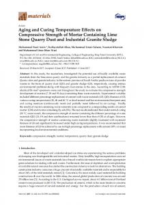
˚ to their where GDTX is the percentage of atoms of the predicted model that are closer than X A homologous in the native model when the two structures are superimposed. 2.2. NNPIF The NNPIF considers the interactions between couples of amino acids (AA) in a model in order to evaluate its quality, or native-likeness. Only the Cα trace of a model is taken into account. A version of this NNPIF has been used in the past as a model quality assessment system (MQAP) [26], which is a similar problem, in that a protein conformation has to be evaluated as a whole and mapped into a measure of its quality. The NNPIF uses two stages, implemented as layered feed-forward neural networks (FFNN), to predict the native-likeness of a model based on its Cα trace. In order to use an FFNN model, the input (in this case, the protein conformation) needs to be represented as a directed acyclic graph (DAG) [26,31–33]). While a conformation can be naturally viewed as an undirected graph in which atoms are nodes and interactions are edges, there are numerous ways to encode this representation in a DAG form [33–36]. In this work, in order to represent a protein structure as a DAG, we consider the interactions between Cα atoms as leaves of a tree where the root is the output of the predictor (Figure 1). Figure 1. A pentapeptide structure is processed by the Neural Network Pairwise Interaction Fields (NNPIF). The structure is represented as a directed acyclic graph (DAG) by considering as leaf nodes the interactions between couples of atoms. Each interaction is modeled by the function, F (), via an NN, N F . The inputs encode characteristics of each of the two interacting atoms (ax , ay ) in the pair and their interaction (dxy ). The outputs are combined in hidden vectors Y . The contribution of each interaction to the quality of the structure is modeled by the function, G(), via an NN, N G , which produces the global output, O.
Biomolecules 2014, 4
165
The interaction between two Cα atoms (i, j) is described as follows: Xij = F (ai , aj , dij )
(2)
where ai and aj are vectors describing the two interacting atoms in position i and j in the sequence and dij describes the nature of the interaction between them. Notice how this representation is by no means limited to describing the atoms’ identity and their distance. For instance, ai can be used to represent an AA’s Cα by the identity of the AA it is in alongside the identities of several AAs that neighbour it in the sequence or in a conformation. Evolutionary information extracted from similar sequences predicted the structural features, such as secondary structure, etc. Similarly, dij is generally a vector containing an encoding of any pairwise property of atoms i and j, which is considered relevant for the prediction. This will naturally include the distance between the atoms, but may also describe the type of covalent bond between the atoms, if any, the possible presence of a hydrogen bond between the AA the atoms are part of, etc. Notice also how, given the flexibility of the input encoding (any unary or binary property of atoms can be represented), more mediated information could easily be added, for instance, correlated mutations, which have proven valuable for the prediction of protein residue contacts [37]. Function F () is implemented by an FFNN, N F , with a single hidden layer and a sigmoidal (hyperbolic tangent) output. This function is computed for each Cα atom, i, considering only the 10 closest neighbours in the Euclidean space with the sequence separation distance >5. On average, a residue ˚ distance from 20 other residues, including the ones with sequence separation distance is within a 20 A ≤ 5 [38]. If we call the set of the 10 neighbours to the i − th atom Ci , we can compute the feature vector for ai : X Yi = K Xij (3) j∈Ci
Where K is a normalization constant. Notice that this feature vector is in fact the sum of the outputs of the hidden layers of 10 neural networks. That is, it has no explicit meaning, and no direct supervision is necessary for it; but, upon success in training, it will be a representation of the complex of all the interactions between an AA and its close neighbours, which is locally optimal for the prediction of the overall target property (the conformation’s native-likeness). The feature vectors for each of the Cα atoms in the trace are computed and then combined (added component by component) to produce a global feature vector. This vector is input to the second layer of the NNPIF, which directly predicts the target property: L X O = G( Yi )
(4)
i=1
where L is the length of the protein. The function, G(), is implemented by a FFNN N G with a single hidden layer and a linear output. The functions, F and G, are considered stationary (the same functions can be used, respectively, for each pair of atoms and for each conformation); so, the same N F is replicated for all of the interactions, and the same N G is used for all of the models. Training of the overall NNPIF is performed by gradient descent, using the squared distance between the predicted and the target evaluation value. The gradient is computed in closed form, via a version of the back-propagation algorithm [39].
Biomolecules 2014, 4
166
To speed up training, we do not compute the gradient of the error on all the training set, but estimate it on a small number of examples. This allows a speedier convergence, as many gradient descent steps are made for each evaluation of the training set instead of only one. Moreover, the stochastic element introduced by this practice (as the error landscape is estimated and not exact, it varies at every learning step) is helpful in avoiding local minima in the optimisation. In our experiments, the gradient is computed once for every two examples that are evaluated. Considering an average of 60 AAs per conformation, N F ’s weights are updated once every 1,200 interactions that are evaluated, while N G ’s weights are updated once every two contributions to its gradient that are calculated. The NNPIF can evaluate a single model in a fraction of a second of CPU time. This means that thousands of models can be generated in a reasonable amount of time. Training, on the other end, can take weeks or months of computing time to complete, depending on the size of the training set used. 2.2.1. Inputs, Output The interactions between couples of neighbouring Cα atoms are considered as the input. For each atom, i, in the Cα trace, an ordered list, Li , of other atoms in the Cα trace is compiled. The order of a given atom, j, in the list Li is given by the Euclidean distance between i and j, Dij ; the closest atom will be the first in the list, the farthest will be the last. All of the atoms that have a sequence separation distance with i