ORIGINAL PAPER
Relationship between immunoexpression of mucin peptide cores MUC1 and MUC2 and Lauren’s histologic subtypes of gastric carcinomas V. Barresi, E. Vitarelli, M. Grosso, G. Tuccari, G. Barresi Department of Human Pathology, University of Messina, Messina, Italy
©2006, European Journal of Histochemistry Laurèn’s system subdivides gastric cancers into an intestinal type and a diffuse type. This histological classification mirrors histogenetic hypotheses according to which the intestinal-type cancer derives from intestinal metaplasia and dysplasia, while the diffuse-type originates directly from gastric mucosa, with or without a preceding non-metaplastic dysplasia. Studies concerning mucins expression in gastric neoplastic and preneoplastic lesions have provided contradictory data concerning such histogenetic relationships. The aim of the present study was to verify whether a correlation between mucins phenotype and Lauren’s classification subsists. 40 gastric adenocarcinomas, subdivided, according to Laurèn’s classification, into 27 intestinal-type, 10 diffusetype and 3 unclassified cases, were examined for MUC1 and MUC2 immunohistochemical expression. Intestinal-type carcinomas displayed a MUC1-positive staining in 23/27 cases and a MUC2-positive immunoreaction in 10/27 cases. Diffuse-type carcinomas expressed MUC1 in 3/10 and MUC2 in 8/10 cases, respectively. According to the mucins expression pattern, three phenotypes were identified: the gastric phenotype (MUC1+/MUC2–); the gastro-intestinal phenotype (MUC1+/MUC2+) and the intestinal phenotype (MUC1–/MUC2+). The gastric phenotype was significantly higherin intestinal-type adenocarcinomas, whereas cases showing an intestinal phenotype were significantly more frequent in diffuse-type adenocarcinomas. These findings provide evidence for a lack of correlation between Lauren’s classification and MUC1 and MUC2 phenotypes. In particular, the term intestinal-type tumour as referred to gland-forming gastric cancer does not seem to reflect an immunohistochemical phenotype. Key words: MUC1, MUC2, gastric carcinoma, Lauren’s classification. Correspondence: Gaetano Barresi, Dipartimento di Patologia Umana, Università degli studi di Messina, Policlinico Universitario Gaetano Martino, 98125 Messina, Italy Tel: +39.90.2212537. Fax:+39.90.2938324. E-mail:
[email protected] Paper accepted on September 12, 2006 European Journal of Histochemistry 2006; vol. 50 issue 4 (October-December):301-310
n Western countries, Lauren’s histologic classification system for gastric carcinomas is largely used (Lauren, 1965). It classifies human gastric carcinomas from a standard haematoxylin and eosin stain, into two main groups, the intestinal and diffuse types, corresponding to differentiated and undifferentiated types (Nakamura et al., 1968; Sugano et al., 1982), respectively. In terms of histogenesis, it is generally accepted that the Lauren’s intestinal-type gastric carcinoma is preceded by the sequential steps of chronic gastritis, intestinal metaplasia, dysplasia, and intramucosal carcinoma (Uchino et al., 1993), whereas diffuse-type gastric carcinoma seems to originate via hyperplastic or de novo changes, with or without concurrent non-metaplastic dysplasia (Correa et al., 1992). Nonetheless, conflicting data have recently emerged regarding such histogenetic relationships from studies concerning mucins expression in gastric cancerous and pre-cancerous lesions (Baldus et al., 1998; Machado et al., 2000; Tsukashita et al., 2001; Gurbuz et al., 2002; Wada et al., 2005). Mucins are high-molecular-weight glycoproteins consisting of a central polypeptidic structure (apomucin) to which numerous carbohydrate chains are attached by an O-glycosidic linkage. They can be divided into two types, secretory and membrane-bound mucins, respectively. At present, 20 different human genes encoding for apomucins have been identified (MUC1, 2, 3A, 3B, 4, 5AC, 5B, 6, 7, 8, 9, 11, 12, 13, 15, 16, 17, 18, 19, 20) (Swallow et al., 1987a; Swallow et al., 1987b; Gum et al., 1989; Gum et al., 1990; Porchet et al., 1991; Toribara et al., 1991; Fox et al., 1992; Bobek et al., 1993; Gross et al., 1993; Shankar et al., 1994; GuyonnetDuperat et al., 1995; Ho et al., 1995a; Lapensèe et al., 1997; Williams et al., 1999; Wu et al., 2001; Williams et al., 2001; Yin et al., 2001; Pallesen et al., 2002; Gum et al., 2002; Chen et al., 2004; Higuchi et al., 2004). MUC1 and MUC2 are the most widely investigated mucins in gastric carcinomas. MUC1 has been detected in mucous cells of the
I
301
V. Barresi et al. Table 1. Clinico-pathological characteristics and mucins expression profile in 40 gastric adenocarcinomas. Case
Age
Gender
Localization
WHO Classif.
Laurèn Classif.
WHO Grade
pTNM
MUC1
MUC2
1 2 3 4 5 6 7 8 9 10 11 12 13 14 15 16 17 18 19 20 21 22 23 24 25 26 27 28 29 30 31 32 33 34 35 36 37 38 39 40
74 73 63 60 73 76 69 77 66 71 72 69 78 54 66 74 70 66 74 77 73 77 71 57 61 66 71 74 77 71 59 70 64 66 59 74 77 65 66 77
F M F F M M F M M F F F M F M F M F F M M M M M M F M F M M M F F M F F M M M M
Cardias Cardias Corpus Corpus Corpus Corpus Corpus Corpus Corpus Antrum Antrum Corpus Cardias Antrum Corpus Corpus Antrum Corpus Corpus Corpus Corpus Antrum Corpus Antrum Corpus Antrum Corpus Corpus Corpus Corpus Antrum Corpus Antrum Corpus Corpus Antrum Corpus Corpus Corpus Antrum
SCR SCR SCR SCR SRC Mucinous Tubular Tubular SCR SCR Tubular Tubular SCR SCR Mucinous Tubular SCR Tubular Tubular Tubular Tubular Tubular Tubular Tubular Tubular Tubular Tubular Papillar Mucinous Tubular Tubular Tubular Tubular Tubular Tubular Tubular Tubular Tubular Tubular Tubular
Diffuse Diffuse Diffuse Diffuse Diffuse Unclassified Intest Intest Diffuse Diffuse Intest Intest Diffuse Diffuse Unclassified Intest Diffuse Intest Intest Intest Intest Intest Intest Intest Intest Intest Intest Intest Unclassified Intest Intest Intest Intest Intest Intest Intest Intest Intest Intest Intest
MD WD WD MD MD WD WD WD WD MD WD MD WD MD WD WD MD WD MD WD WD MD WD WD MD WD MD WD WD MD WD WD WD MD WD WD MD WD MD WD
T2N1Mx T3N1Mx T3N3Mx T2N1Mx T2N1Mx T2N1Mx T3N1Mx T2N0Mx T2N0Mx T2N1Mx T2N0Mx T1N0Mx T2N1Mx T2N3Mx T2N0Mx T1N2Mx T1N0Mx T2N1Mx T2N1Mx T1N0Mx T2N1Mx T1N0Mx T2N1Mx T1N0Mx T2N1Mx T2N0Mx T2N1Mx T2n1Mx T1N0Mx T2N1Mx T2N1Mx T1N0Mx T2N1Mx T2N1Mx T3N1Mx T2N1Mx T1N0Mx T3N1Mx T2N1Mx T2N1Mx
0 0 0 0 0 2 1 2 0 0 0 1 2 1 0 1 1 1 2 2 1 1 2 2 2 1 1 1 2 1 2 0 2 2 1 1 0 1 1 0
2 1 2 1 2 2 0 0 1 2 2 0 0 0 2 0 2 0 0 0 0 0 0 0 0 2 0 0 0 2 2 2 0 1 2 0 1 1 0 2
surface epithelium and neck region of the antrum, as well as in pyloric glands and in oxynthic glands of the body region in normal gastric mucosa (Ho et al., 1993; 1995b; Lesuffleur et al., 1994). During gastric carcinogenesis, expression of underglycosylated forms of MUC1 mucin has been demonstrated (Baldus et al., 1998; Reis et al., 1998) and their presence seems to be closely tied to a poor prognosis (Akyurek N et al., 2002; Kocer et al., 2004). MUC2 mucin, expressed in the colon, small intestine and airways, and not found in normal gastric mucosa, is detected in intestinal metaplasia and in gastric carcinoma (Filipe et al., 1996); it is considered a prognostic factor associated with an unfavourable outcome in patients with gastric cancer (Zhang et al., 2004; Leteurtre et al., 2006). Despite the many studies that have been performed, conflicting data have emerged as to correlations between mucins phenotype and gastric carcinoma 302
classification systems (Machado et al., 2000; Tsukashita et al., 2001; Gurbuz et al., 2002; Roessler et al., 2005; Kabashima et al., 2005; Leturtre et al., 2006). Lauren’s hypothesis, according to which gland-forming gastric cancer is related to intestinal differentiation, remains still to be validated. Indeed, whereas some authors find a higher rate of MUC2 expression in intestinal-type cancer compared to diffuse-type (Baldus et al., 1998; Kocer et al., 2004), others report a low prevalence of this intestinal marker of differentiation in intestinal-type gastric adenocarcinomas (Machado et al., 2000; Tsukashita et al., 2001), in contrast with histogenetic theory according to which gastric intestinal-type cancer would derive via chronic gastritis, intestinal metaplasia and dysplasia. In the present study we determined MUC1 and MUC2 immunohistochemical expression profiles in a series of gastric cancers preliminarily classified according to Laurèn’s system,
Original Paper
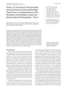
Figure 1. a. Normal gastric glands are not labelled by MUC2 antibody, whereas MUC2 staining is clearly evident in intestinal metaplasia (MUC2 staining; original magnification x100). b. MUC1 intense immunoreaction in mucous neck cells of gastric normal epithelium (MUC1 staining, original magnification x200).
and aimed to investigate whether any correlation exists between MUC1 and MUC2 mucin phenotypes and Lauren’s classification system.
Materials and Methods 40 cases of gastric adenocarcinomas were selected from the files of the Department of Human Pathology, University of Messina, Italy. Tissue samples were obtained from surgical resection; for each case, age and gender of the patient, tumour location, grading and pTNM stage were evaluated by reviewing clinical charts and pathological records. Patients that had undergone pre-operative chemotherapy were excluded from the study. According to the Lauren’s system, cases were subdivided into 27 intestinal-type, 10 diffuse-type, and 3 unclassified adenocarcinomas. Helicobacter Pylori infection was not evidenced in any sample. Immunohistochemical
procedures were performed on 4 µm formalin fixed, paraffin embedded, tissue sections obtained from each representative paraffin block. Briefly, endogenous peroxidase activity was preliminarily blocked with 3% H2O2 in PBS for 30 min at room temperature. Sections were successively incubated at 4°C overnight with the following primary monoclonal antibodies: NCL-MUC1 (Ma695; 1:100, Novocastra, Newcastle, UK) and NCL-MUC2 (Ccp58; 1:100, Novocastra Newcastle, UK). Microwave pre-treatment using 0.01M sodium citrate buffer was employed for each immunoreaction. The bound primary antibody was visualized by the avidin-biotin-peroxidase detection complex (ABC) method with a commercial kit (Vectastain ABC Elite Kit, Vector Laboratories, Burlingame, Calif.,USA), according to the manufacturer's instructions. 3-3' diaminobenzidine (DAB) was used as the chromogen. Sections of breast carcinoma for MUC1 and normal small bowel mucosa for MUC2 were used as positive controls. Negative control slides were also made by omitting the primary antibody. The extent of positivity for MUC1 and MUC2 was scored according to the percentage of stained neoplastic cells: 0=